Clinical Application Value of Ultrasound in the Diagnosis of Acute Appendicitis
-
摘要:
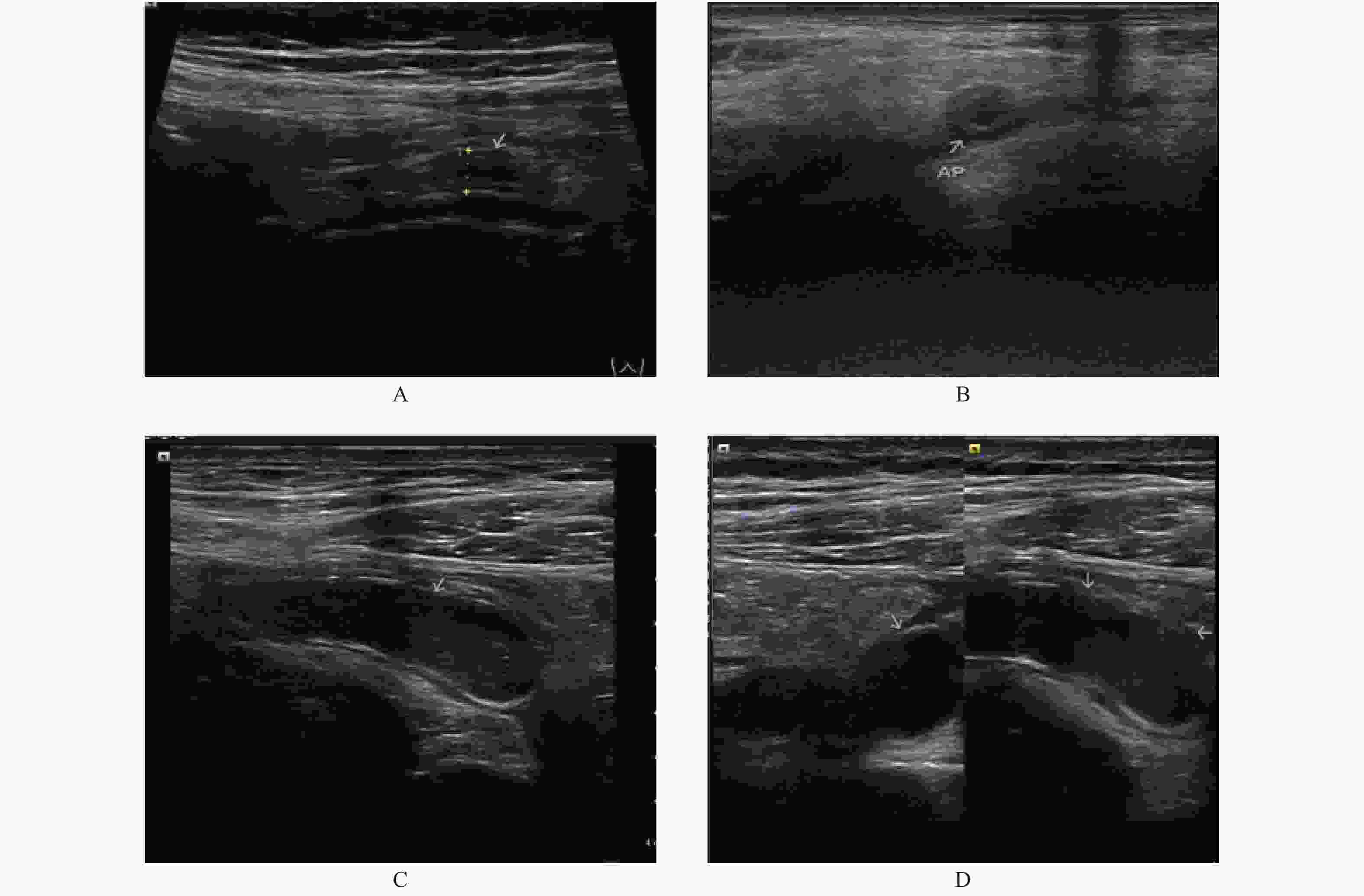
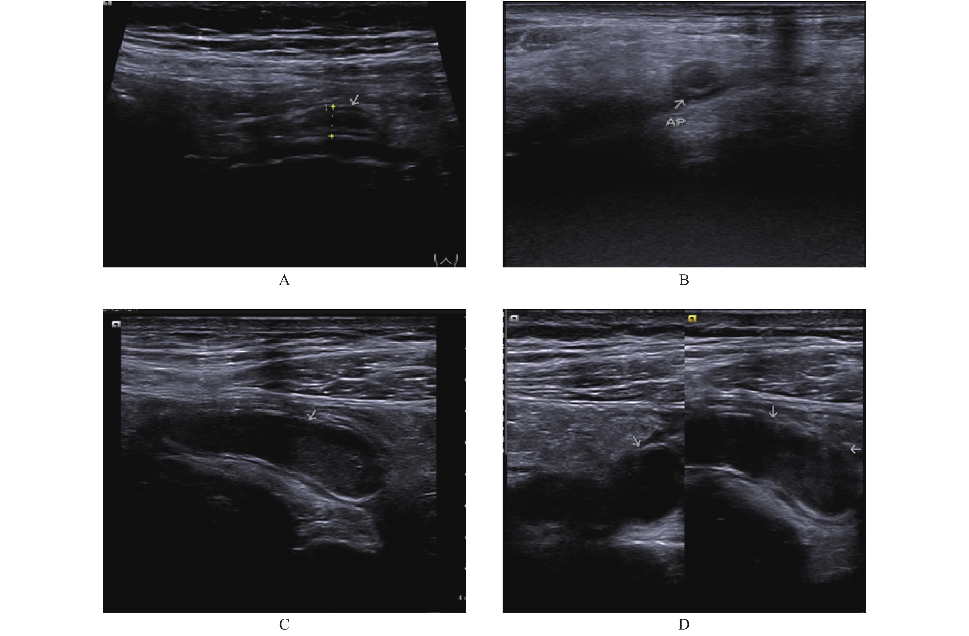
目的 探讨超声在急性阑尾炎诊断中的临床应用价值。 方法 回顾性分析2019年至2020年昆明医科大学第二附属医院所收治的423例急性阑尾炎术后临床资料,评价超声对急性阑尾炎诊断准确率,分析超声对不同类型阑尾炎的管腔直径,管壁厚度结果以及右下腹回盲部管壁增厚,右下腹局部积液,局部强回声对比差异。 结果 超声共检出急性阑尾炎374例,诊断准确率为88.4%(374/423)。急性阑尾炎阑尾管腔直径、管壁厚度(mm)分别为:(7.1±1.3)、(3.2±0.9),急性化脓性阑尾炎阑尾管腔直径、管壁厚度(mm)分别为:(10.0±1.9)、(4.3±1.1),急性坏疽性阑尾炎管腔直径、管壁厚度(mm)分别为:(12.4±2.2)、(5.3±1.3),三组结果比较差异有统计学意义(P < 0.05)。急性坏疽性阑尾炎和急性化脓性阑尾炎合并回盲部管壁增厚,右下腹局部积液,局部强回声,淋巴结肿大的比例均高于急性单纯性阑尾炎,差异有统计学意义(P < 0.05)。 结论 超声能够为不同病理类型急性阑尾炎的诊断提供可靠的参考依据。 Abstract:Objective To explore the clinical value of ultrasound in the diagnosis of acute appendicitis. Methods The clinical data of 423 cases of acute appendicitis treated in our hospital from 2019 to 2020 were retrospectively analyzed to evaluate the diagnostic accuracy of ultrasonography for acute appendicitis. The results of lumen diameter, tube wall thickness, tube wall thickening in the right lower abdominal ileocecal part, local effusion in the right lower abdomen, and local strong echo comparison for different types of appendicitis were analyzed. Results A total of 374 cases of acute appendicitis were detected by ultrasound, and the diagnostic accuracy was 88.4%(374/423). The lumen diameter and wall thickness(mm)of appendicitis with acute appendicitis were(7.1±1.3)and(3.2±0.9), the lumen diameter and wall thickness(mm)of appendicitis with acute suppurative appendicitis were(10.0±1.9)and(4.3±1.1), and the lumen diameter and wall thickness of appendicitis with acute gangrenous appendicitis were(12.4±2.2)and(5.3±1.3), respectively, with statistically significant differences among the three groups(P < 0.05). Acute gangrene appendicitis and acute suppurative appendicitis combined with thickening of ileocecal wall, local effusion in the right lower abdomen, local strong echo and lymph node enlargement were all higher than those in acute simple appendicitis, and the differences were statistically significant(P < 0.05). Conclusion Ultrasonography can provide a clear basis for the diagnosis of different pathological types of acute appendicitis. -
Key words:
- Acute appendicitis /
- Ultrasonography /
- Pathological type
-
表 1 不同病理类型急性阑尾炎超声诊断与术后病理诊断结果的对比(n)
Table 1. Comparison of ultrasonographic diagnosis and postoperative pathological diagnosis of acute appendicitis in all patients(n)
组别 病理诊断 超声诊断 符合率(%) 急性单纯性阑尾炎 143 120 83.9 急性化脓性阑尾炎 180 162 90.0 急性坏疽性阑尾炎 100 92 92.0 χ2 22.74 19.91 19.91 P 0.000 0.000 0.000 表 2 超声在不同病理类型急性阑尾炎中的直接征象对比(
$\bar x \pm s$ )Table 2. Comparison of direct signs of ultrasonography in acute appendicitis of different pathological types(
$\bar x \pm s$ )组别 n 管腔直径(mm) 管壁厚度(mm) 管壁层次 管壁连续性 急性单纯性阑尾炎 120 7.1 ± 1.3 3.2 ± 0.9 清晰 完整 急性化脓性阑尾炎 162 10.0 ± 1.9 4.3 ± 1.1 欠清晰 欠完整 急性坏疽性阑尾炎 92 12.4 ± 2.2 5.3 ± 1.3 不清晰 连续性中断 q 16.52 13.43 P 0.001 0.010 表 3 超声在不同病理类型急性阑尾炎中的间接征象对比[n (%)]
Table 3. Comparison of indirect signs of ultrasonography in acute appendicitis of different pathological types [n (%)]
组别 n 回盲部管壁增厚 右下腹局部积液 右下腹局部强回声 右下腹淋巴结肿大 急性单纯性阑尾炎 120 62(51.7) 24(20.0) 8(6.7) 39(32.5) 急性化脓性阑尾炎 162 130(80.2) 105(64.8) 97(59.9) 81(50.0) 急性坏疽性阑尾炎 92 83(90.2) 86(93.5) 67(72.8) 61(66.3) χ2 26.45 50.07 71.52 14.63 P 0.000 0.000 0.000 0.001 -
[1] 马晓慧. 腹部CT在急性阑尾炎诊断治疗中的积极意义和诊断要点探讨[J].临床医药文献电子杂志,2020,7(20):123-125. [2] 曹勇,王克蓉,苏剑,等. 高频与低频彩超联用诊断急性阑尾炎105例分析[J].现代医用影像学,2017,26(3):790-792. [3] 夏磊. 经腹部浅表超声在急性阑尾炎临床诊断中的价值[J].牡丹江医学院学报,2019,40(3):85-87. [4] 吕晓梅. 不同病理类型急性阑尾炎的超声图像特征观察[J].中国医药指南,2013,11(31):455-456. [5] 蔡志南. 彩色多普勒超声在急性阑尾炎临床诊断中的应用价值[J].中国医药指南,2018,16(1):54-55. [6] 张文婷. 二维及彩色多普勒超声诊断阑尾炎的价值[J].世界最新医学信息文摘,2018,18(54):172-173. [7] Hwang M E. Sonography and Computed Tomography in Diagnosing Acute Appendicitis[J]. Radiol Technol,2018,89(3):224-237. [8] Trout A T,Sanchez R,Ladino-Torres M F,et al. A critical evaluation of US for the diagnosis of pediatric acute appendicitis in a real-life setting:how can we improve the diagnostic value of sonography?[J]. Pediatric Radiology,2012,42(7):813-823. doi: 10.1007/s00247-012-2358-6 [9] 龙安军,项国靓,刘窗,等. 多层螺旋CT在诊断急性阑尾炎中的价值探讨[J].现代医用影像学,2016,25(6):1162-1163. [10] Augustin T,Cagir B,Vandermeer T J. Characteristics of perforated appendicitis:Effect of delay is confounded by age and gender[J]. Journal of Gastrointestinal Surgery,2011,15(7):1223-1231. doi: 10.1007/s11605-011-1486-x [11] 刘波. 超声间接征象对急性阑尾炎的诊断价值研究[J].影像研究与医学应用,2018,2(17):28-29. doi: 10.3969/j.issn.2096-3807.2018.17.015 [12] 宋希根. 急性阑尾炎行CT与超声诊断临床比较[J].淮海医药,2017,35(2):189-190. [13] 苏红,吕开红,杨勇. 彩色多普勒超声诊断小儿急性阑尾炎的临床应用价值[J].临床医学研究与实践,2017,2(33):151-152. [14] 辛洪兵,朱德仓. 高低频超声联合检查对急性阑尾炎诊断价值[J].影像研究与医学应用,2020,4(19):225-226. doi: 10.3969/j.issn.2096-3807.2020.19.129 [15] 李淑杰. 高、低频超声联合检查在急性阑尾炎诊断中的临床价值分析[J].影像研究与医学应用,2020,4(02):223-224. [16] 于英,王宇. 高频超声与低频超声用于诊断急性阑尾炎的价值分析[J].中国处方药,2017,15(8):129-130. doi: 10.3969/j.issn.1671-945X.2017.08.093 [17] 梁华波. 螺旋CT诊断和鉴别诊断急性右下腹疼痛的价值[J].世界最新医学信息文摘,2015,15(95):146-146. [18] 许慧君,王光霞. 高频超声对不同病理类型急性阑尾炎及并发症的诊断价值[J].中国中西医结合外科杂志,2019,25(02):145-150. doi: 10.3969/j.issn.1007-6948.2019.02.006 [19] 寇智勇,王志强,李文亮,等. 胃癌阑尾转移并发急性阑尾炎1例[J].昆明医科大学学报,2015,36(08):137-138. doi: 10.3969/j.issn.1003-4706.2015.08.041 -


 下载:
下载:


