The Application Value of CT Pulmonary Blood Flow Distribution in the Diagnosis of Pulmonary Embolism
-
摘要:
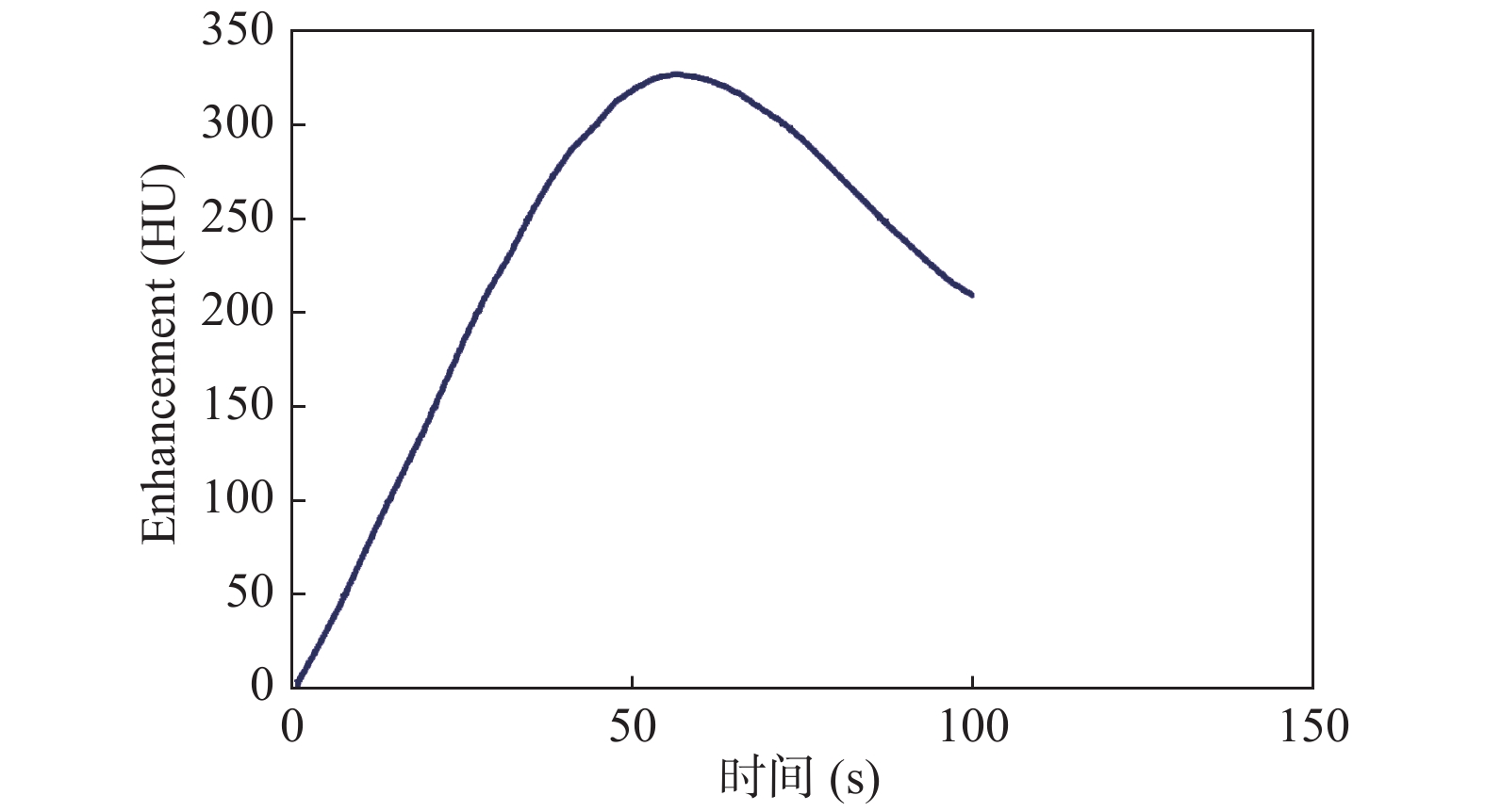
目的 探讨CT肺叶血流分布造影在肺栓塞的诊断的可行性。 方法 利用显影剂的显影效能,采用技巧处理图像,建立对应的映射机制,通过彩色显影法,观察肺叶血流分布,推测肺部血液灌注情况。 结果 在注入显像剂后,显像剂随血液流动并通过其显像效率,导致CT图像上CT值发生变化,通过肺部原始CT图影像,可以找到血管栓塞处。显影后彩色图影像的蓝色区块,会出现在栓塞血管末梢处,直接从彩色图来观察栓塞区域,较CTA和核医学分析更容易,且更有效率。 结论 CT肺叶血流分布造影是通过彩色图影像的分析,描述肺叶血流分布,对肺栓塞的诊断有辅助作用,值得进一步研究及临床应用。 Abstract:Objective To explore the feasibility of CT pulmonary blood flow distribution in the diagnosis of pulmonary embolism. Methods Using the imaging efficiency of the imaging agent, the corresponding mapping mechanism was established by processing the image with skill. The blood flow distribution of the lung lobe was observed by color development method, and the pulmonary blood perfusion was speculated. Results After the imaging agent was injected, the imaging agent flowed with the blood and passed its imaging efficiency, resulting in changes in the CT value on the CT image. Through the original CT image of the lungs, the embolism can be found. After the development, the blue area of the color map image appeared at the end of the embolized blood vessel. Observing the embolized area directly from the color map was easier and more efficient than CTA and nuclear medicine analysis. Conclusion CT pulmonary blood flow distribution angiography is to describe the blood flow distribution of the pulmonary lobes through the analysis of color map images, and has an auxiliary role in the diagnosis of pulmonary embolism. It is worthy of further research and clinical application. -
Key words:
- Distribution of blood flow rate of diagnosis /
- CT /
- Pulmonary embolism
-
表 1 肺叶各区块单位血流量
Table 1. Blood flow unit of every lung lobe
肺叶区块
编号单位血流量
(Vi)肺叶区块
编号单位血流量
(Vi)R1 60(0.60) L1 67(0.67) R2 51(0.51) L2 69(0.69) R3 43(0.43) L3 63(0.63) R4 44(0.44) L4 83(0.83) R5 32(0.32) L5 62(0.62) R6 19(0.19) L6 32(0.32) R7 100(1.00) L7 75(0.75) R8 100(1.00) L8 74(0.74) R9 67(0.67) L9 83(0.83) R10 56(0.56) L10 63(0.63) R11 43(0.43) L11 43(0.43) R12 18(0.18) L12 23(0.23) 注:括号内为Ri值。 -
[1] 王秋桐, 吴爽, 赵瑞. 肺栓塞发病机制及致病因素研究进展[J].临床误诊误治,2020,33(1):108-112. doi: 10.3969/j.issn.1002-3429.2020.01.026 [2] 王海峰. 超声心动图测量肺动脉高 压对急性肺栓塞患者的危险分层及预后的应用价值[J].临床肺科杂 志,2017,22(3):476-478. [3] Mcgee M, Whitehead N, Martin J, et al. Drug-associated pulmonary arterial hypertension[J]. Clinical Toxicology,2018,56(9):1-9. [4] 王建国, 王辰, 郭佑民, 等. CT肺血管成像分析肺栓塞程度及右心功能的价值[J].中国医学影像学杂志,2009,17(02):25-29. [5] 仝春冉, 张中和, 马春梅, 等. 简化肺栓塞严重性指数及生化标志物的联合检测在肺栓塞预后评价中的价值[J].中华结核和呼吸杂志,2014,37(02):34-38. [6] Kratzer L, Noakes P, Baumwol J, et al. Under-utilisation of β blockers in patients with acute coronary syndrome and comorbid chronic obstructive pulmonary disease[J]. Int Med J,2018,48(8):931-936. doi: 10.1111/imj.13795 [7] 吴京兰, 谭四平, 沈比先. 双源CT双能量肺灌注成像技术诊断急性肺动脉栓塞的临床应用[J].临床心血管病杂志,2011,27(5):57-63. [8] Felix G. Meinel,Anita Graef,Fabian Bamberg,et al. Effectiveness of automated quantification of pulmonary perfused blood volume using dual-energy CTPA for the severity assessment of acute pulmonary embolism[J]. Investigative Radiology,2013,48(8):75-87. [9] 李树峰, 张震. 肺栓塞的影像联合诊断价值的探讨[J].医学综述,2020,26(3):554-558. doi: 10.3969/j.issn.1006-2084.2020.03.027 [10] Daniel J. Hou, David K. Tso, Chris Davison, et al. Clinical utility of ultra high pitch dual source thoracic CT imaging of acute pulmonary embolism in the emergency department: Are we one step closer towards a non-gated triple rule out?[J]. European Journal of Radiology,2013,82(10):1793-1798. doi: 10.1016/j.ejrad.2013.05.003 [11] 胡丽霞, 龚静山, 饶梓彬, 等. 多层螺旋CT在肺栓塞检查中的方法与技巧[J].放射学实践,2015,20(01):69-73. [12] 李辉, 李铁一, 郝晓光. 值得注意的急性肺动脉栓塞平扫CT征象[J].中华放射学杂志,2014,38(11):19-27. [13] 孙长青, 徐炳福, 刘兰, 等. 肺通气灌注显像与64 排CT肺动脉造影在肺动脉栓塞诊断中的比较研究[J].中国医学装备,2016,13(3):61-64. doi: 10.3969/J.ISSN.1672-8270.2016.03.017 [14] Joachim Ernst Wildberger, Ernst Klotz, Hendrik Ditt, et al. Multislice computed tomography perfusion imaging for visualization of acute pulmonary embolism: animal experience[J]. European Radiology,2005,15(7):1378-1386. doi: 10.1007/s00330-005-2718-9 [15] 王燕林, 木合拜提·买合苏提, 刘文亚, 等. CT肺动脉造影对大面积肺栓塞严重程度的评估[J].中国医学 影像学杂志,2016,24(1):8-11. -


 下载:
下载:


