Application of Various Sealing Materials and Adhesive Systems in Extracorporeal Pit and Fissure Sealing
-
摘要:
目的 比较树脂型窝沟封闭剂、流动树脂、复合体、玻璃离子等材料结合不同粘结系统行窝沟封闭术后的边缘密闭性,为经济欠发达地区寻找简捷有效的防龋措施。 方法 离体牙随机分五组:全酸蚀技术 + 树脂型窝沟封闭剂组(A组,对照组)、自酸蚀粘结剂 + 树脂型窝沟封闭剂组(B组)、自酸蚀粘结剂 + 流动树脂组(C组)、自酸蚀粘结剂 + 复合体组(D组)及玻璃离子组(E组),体外行窝沟封闭并记录操作时间。行体外温度循环后进行扫描电镜及体视显微镜检测。 结果 (1)各组均存在不同程度的微渗漏;(2) B组及C组与对照组(A组)差异有统计学意义(P < 0.05),操作时间短于对照组;(3) D组及E组微渗漏高于对照组(P < 0.05);(4) D组在试件制备过程中无折断及松脱等现象。 结论 自酸蚀粘结系统结合树脂型窝沟封闭剂及流动树脂实施窝沟封闭术时操作简便、微渗漏较低;复合体抗压强度较大。 Abstract:Objective To evaluate marginal integrity and microleakage of resin-based sealant, flowable composite resin, compomer and glass ionomer cement combined with certain adhesive systems, in order to explore a more convenient, effective and time-saving approach which intends to be applied in less developed areas. Methods 65 maxillary premolar teeth were kept in artificial saliva, pits and fissures were cleaned by ART equipments. The samples were allocated into 5 groups randomly: (A) phosphoric acid etching + Concise (control); (B) AdperTM Easy One + Concise; (C) AdperTM Easy One + FiltekTM Z350XT flowable resin; (D) AdperTM Easy One + Dyract Extra compomer; and (E) GC Fuji VII. The pit and fissure sealant were applied, and the operation time was recorded in vitro. After thermocycling, specimens were evaluated by scanning electron microscope and stereomicroscope. Results i) All the groups showed various level of microleakage; ii) Group B and C showed similar microleakage as compared with the control group, while their operation time were shorter than that of the control group (P < 0.05); iii) Group D and E showed greater microleakage than other groups (P < 0.05); iv) No shedding of sealing materials during teeth cutting in Group D. Conclusions Resin-based sealant and flowable composite resin combined with self-etching adhesive system has advantages of operative convenience and effectiveness. Dyract Extra compomer has better abrasion resistance. -
Key words:
- Pit and fissure sealant /
- Sealing materials /
- Adhesive system /
- Marginal integrity /
- Microleakage
-
表 1 采用不同材料及技术进行窝沟封闭的实验分组
Table 1. Sealing with various materials and techniques in different groups
组别 粘结处理材料 封闭材料 固化方式 A组(对照组) 37%正磷酸 树脂型窝沟封闭剂(Concise) LED光固化 B组 Easy One自酸蚀粘结剂 树脂型窝沟封闭剂(Concise) LED光固化 C组 Easy One自酸蚀粘结剂 流动树脂(Z350XT) LED光固化 D组 Easy One自酸蚀粘结剂 复合体(Dyract) LED光固化 E组 — 玻璃离子(Fuji VII) 自凝固化 表 2 染料渗漏等级评分
Table 2. Crading of dye penetration
分值 渗漏情况描述 0 边缘无染色 1 渗入深度未及封闭材料与牙釉质界面长度的1/2 2 渗入深度超过封闭材料与牙釉质界面长度的1/2,
未及沟底3 染料延伸至沟底 表 3 各组窝沟封闭操作时间比较(
$\bar x \pm s$ )Table 3. Comparison of operation time between different groups (
$\bar x \pm s$ )组别 n 操作时间(s) A组 13 129.69 ± 9.49#^&§ B组 13 108.54 ± 10.00*& C组 13 110.38 ± 12.47*& D组 13 153.85 ± 17.53*#^§ E组 13 95.31 ± 20.63*& 与A组比较,*P < 0.05;与B组比较,# P < 0.05;与C组比较,^P < 0.05;与D组比较,&P < 0.05;与E组比较,§P < 0.05。 表 4 体式显微镜下各组材料渗漏情况[n(%)]
Table 4. Microleakage detected by stereomicroscope [n(%)]
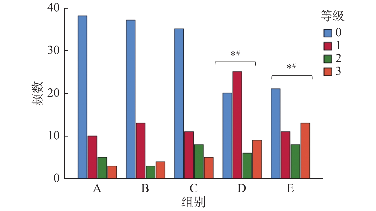
组别 渗漏分值 其他 合计 0 1 2 3 X A 38(63.3) 10(16.7) 5(8.3) 3(5.0) 4(6.7) 60(100) B 37(61.7) 13(21.7) 3(5.0) 4(6.7) 3(5.0) 60(100) C 35(58.3) 11(18.3) 8(13.3) 5(8.3) 1(1.7) 60(100) D 20(33.3) 25(41.7) 6(10.0) 9(15.0) 0(0.0) 60(100)*# E 21(35.0) 11(18.3) 8(13.3) 13(21.7) 7(11.7) 60(100)*# X为试件制备过程中,材料出现折裂、松动及脱落等现象,无法正常观察染料渗透情况。与A组比较,*P < 0.05;与B组比较,#P < 0.05。 -
[1] Seow W K. Early childhood caries[J]. Pediatr Clin North Am,2018,65(5):941-954. doi: 10.1016/j.pcl.2018.05.004 [2] Zhou N,Wong H M,Wen Y F,et. Efficacy of caries and gingivitis prevention strategies among children and adolescents with intellectual disabilities:A systematic review and meta-analysis[J]. J Intellect Disabil Res,2019,63(6):507-518. doi: 10.1111/jir.12576 [3] Carvalho J C,Dige I,Machiulskiene V,et al. Occlusal caries:Biological approach for its diagnosis and management[J]. Caries Res,2016,50(6):527-542. doi: 10.1159/000448662 [4] Naaman R,El-Housseiny A A,Alamoudi N. The use of pit and fissure sealants-a literature review[J]. Dent J(Basel),2017,5(4):34. [5] 魏立梅,刘丽敏,孙婷. 窝沟封闭防龋的循证治疗方案[J]. 中国医疗前沿,2012,7(1):48-49. doi: 10.3969/j.issn.1673-5552.2012.01.0030 [6] 张海亮,赵玉宏,欧晓艳. 3种窝沟封闭剂微渗漏的实验研究[J]. 口腔医学研究,2011,27(12):86-89. [7] Garcia Godoy F,de A raujo F B. Enhancement of fissure sealant penetration and adaptation:the enameloplasty technique[J]. J Clin Ped iatr Den t,1994,19(1):13-18. [8] Frenchen J E,Makoni F,Sithole W D. ART restorations and glass ionomer sealants in Zimbabwe:survival after 3 years[J]. Community dentistry and oral epidemiology,1998,26(6):372-381. doi: 10.1111/j.1600-0528.1998.tb01975.x [9] 翁春城,刘娟,吕长海. 自酸蚀粘结系统及复合体联合应用于ART的实验研究[J]. 昆明医科大学学报,2015,36(6):120-126. doi: 10.3969/j.issn.1003-4706.2015.06.030 [10] Lenzi T L,Guglielmi Cde A,Umakoshi C B,et al. One-step self-etch adhesive bonding to pre-etched primary and permanent enamel[J]. J Dent Child(Chic),2013,80(2):57-61. [11] 计艳,龚玲,王瑜,等. 自酸蚀粘结剂对窝沟封闭微渗漏影响的实验研究[J]. 实用口腔医学杂志,2012,28(4):449-452. doi: 10.3969/j.issn.1001-3733.2012.04.08 [12] Garg D,Mahabala K,Lewis A,et al. Comparative evaluation of sealing ability,penetration and adaptation of a self etching pit and fissure sealant- stereomicroscopic and scanning electron microscopic analyses[J]. J Clin Exp Dent,2019,11(6):e 547-552. [13] 欧晓丽,施春梅,周嫣,等. GC FujiⅦ玻璃离子应用于乳牙窝沟封闭疗效评价[J]. 中国妇幼保健,2013,28(3):544-546. doi: 10.7620/zgfybj.j.issn.1001-4411.2013.03.59 [14] Alsabek L,Al-Nerabieah Z,Bshara N,et al. Retention and remineralization effect of moisture tolerant resin-based sealant and glass ionomer sealant on non-cavitated pit and fissure caries:randomized controlled clinical trial[J]. J Dent,2019,86:69-74. doi: 10.1016/j.jdent.2019.05.027 [15] 吕长海, 凌均棨, 凌征宇. LED固化系统应用于乳牙复合体修复的研究. 中国实用口腔科杂志[J]. 2010, 3(5): 295-297. -


 下载:
下载:


