

Periodontal Ligament Stem Cell BMP-2-PSH Composite Membrane in Repairing Alveolar Bone Defect in New Zealand Rabbits
-
摘要:
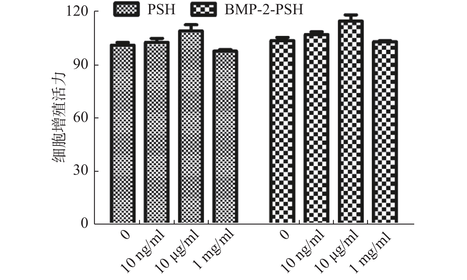
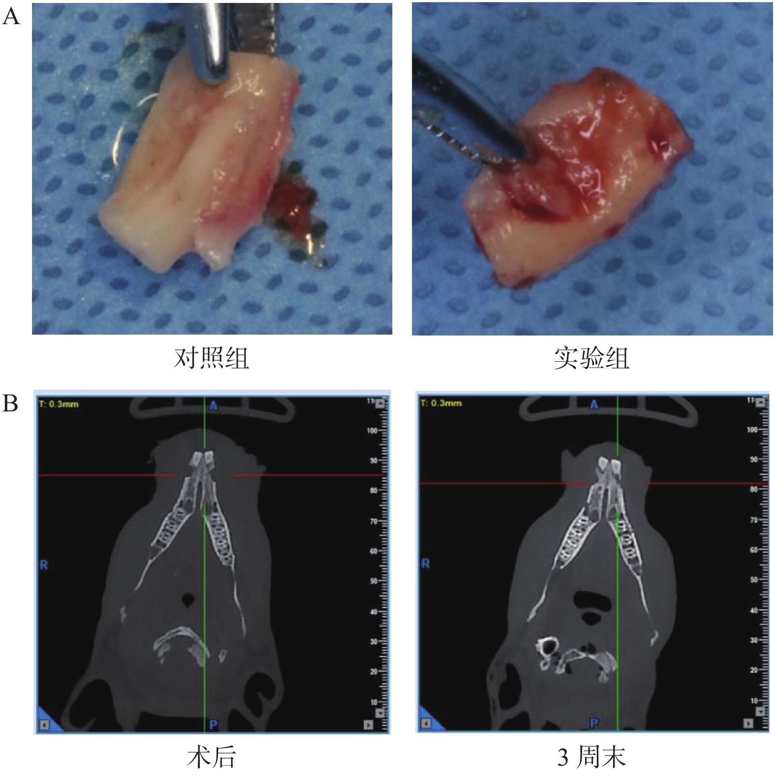
目的 拟构建新西兰兔牙周缺损模型,确定hPDLSCs-BMP-2-PSH膜的生物安全性和其体内成骨修复作用。 方法 在确定BMP-2-PSH膜没有细胞毒性后将培养好的hPDLSCs细胞膜片复合至BMP-2-PSH膜上。分别在每只新西兰兔下颌左中切牙牙槽骨缺损内植入hPDLSCs/PSH复合膜作为实验组,右中切牙牙槽骨缺损内植入对照性多孔纤维膜材料作为对照组(或不植入任何材料作为对照),然后全瓣复位,缝合。术后即刻和每3周(连续观察12周)进行影像学(CT)检查,观察骨缺损愈合情况;同时,分别在术后第4、8、12周观察PDLSCs/BMP-2双膜材料的吸收度和各组样本新骨形成量和新骨形态。 结果 不同浓度PSH膜和BMP-2-PSH膜浸提液对hPDLSCs细胞不具有细胞毒性,hPDLSCs细胞能够成功复合到BMP-2-PSH膜上;CBCT及组织形态学分析显示,hPDLSCs/PSH复合膜能够有效促进牙槽骨缺损修复再生。 结论 hPDLSCs/PSH复合膜具有良好生物安全性,有利于修复新西兰兔牙周缺损。 -
关键词:
- 牙周膜干细胞 /
- BMP-2-PSH复合膜 /
- 牙周再生
Abstract:Objective It is planned to build a New Zealand rabbit periodontal defect model and determine the biological safety of hPDLSCs-BMP-2-PSH membrane and its in vivo osteogenic repair effect. Methods After confirming that the BMP-2-PSH membrane was not cytotoxic, the cultured hPDLSCs cell membrane was compounded to the BMP-2-PSH membrane. Respectively, HPDLSCs/PSH composite membrane was implanted into the alveolar bone defect of mandibular left incisor in each New Zealand rabbit as the experimental group, and the controlled porous fiber membrane material was implanted into alveolar bone defect of the right central incisor as the control group (or not implanted any material was used as a control), then the whole flap was reset and sutured. Imaging (CT) examination was performed immediately after the surgery and every 3 weeks (observed continuously for 12 weeks) to observe the healing of bone defects; meanwhile, the absorbance of PDLSCs/BMP-2 double membrane materials, the amount of new bone formation and new bone morphology of samples in each group were observed at 4, 8 and 12 weeks after the surgery. Results Different concentrations of PSH membrane and BMP-2-PSH membrane extracts were not cytotoxic to hPDLSCs cells, hPDLSCs cells could be successfully compounded to BMP-2-PSH membranes; The analysis of CBCT and histomorphology showed that hPDLSCs/PSH composite membranes can effectively promote the repair and regeneration of alveolar bone defects. Conclusion The hPDLSCs/PSH composite membrane has the good biological safety and is beneficial to repair periodontal defects of New Zealand rabbits. -

图 1 牙槽骨缺损的形成和PDLSCs/PSH复合膜的移植示意图
A:暴露的兔牙槽骨;B:使用高速外科手术囊制备的骨缺损(10 mm× 5 mm× 4 mm);C:在牙槽骨缺损内移植PDLSCs/PSH复合膜材料;D:缝合缺损外皮瓣。
Figure 1. Schematic diagram of the formation of alveolar bone defect and the transplantation of PDLSCs/PSH composite membrane alveolar bone defect

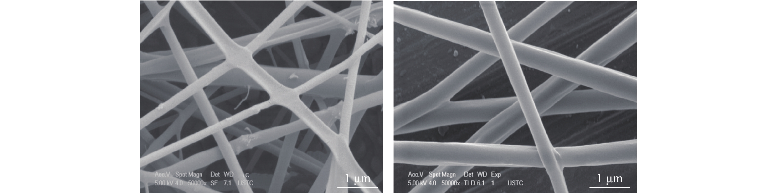
图 2 PSH纤维膜电镜形貌(×50000)
Figure 2. Electron microscopic morphology of PSH fiber membrane (×50000)

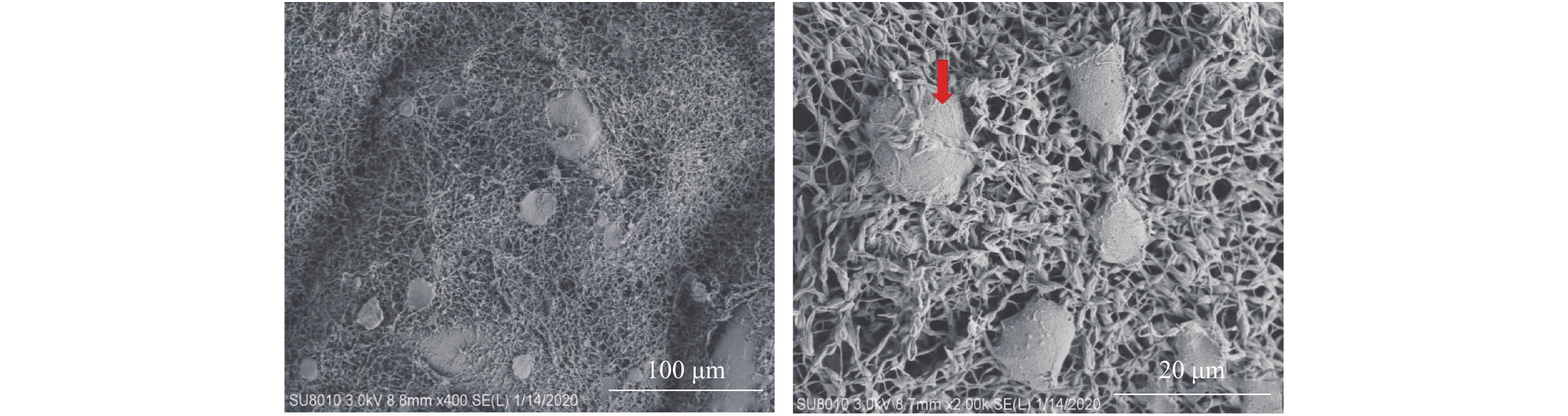
图 4 BMP-2-PSH膜及复合在其上的hPDLSCs细胞(红色箭头所示)(×2 000)
Figure 4. BMP-2-PSH membrane and hPDLSCs cells compounded on it (shown by the red arrow)(×2 000)
-
[1] Nazir M A. Prevalence of periodontal disease, its association with systemic diseases and prevention[J]. Int J Health Sci (Qassim),2017,11(2):72-80. [2] Kinane D F,Stathopoulou P G,Papapanou P N. Periodontal diseases[J]. Nat Rev Dis Primers,2017,3(1):17038. doi: 10.1038/nrdp.2017.38 [3] Han J,Menicanin D,Gronthos S,et al. Stem cells,tissue engineering and periodontal regeneration[J]. Australian Dental Journal,2013,59(s1):117-30. [4] Lin N H,Gronthos S,Bartold P M. Stem cells and future periodontal regeneration[J]. Periodontology,2009,51(1):239-251. doi: 10.1111/j.1600-0757.2009.00303.x [5] Chen F M,Jin Y. Periodontal tissue engineering and regeneration:Current approaches and expanding opportunities[J]. Tissue Eng Part B Rev,2010,16(2):219-255. [6] Zhao L R,Mao J Q,Zhao B J,et al. Isolation and biological characteristics of exosomes derived from periodontal ligament stem cells[J]. Shanghai Kou Qiang Yi Xue,2019,28(4):343-348. [7] Liu J,Chen B,Bao J,et al. Macrophage polarization in periodontal ligament stem cells enhanced periodontal regeneration[J]. Stem Cell Res Ther,2019,10(1):320. doi: 10.1186/s13287-019-1409-4 [8] 黄涛,陈汉. 纳米羟基磷灰石牙体修复材料的生物性能[J]. 中国组织工程研究,2016,20(34):5045-5050. [9] Toth F,Gall JM,Tozser J,et al. Effect of inducible bone morphogenetic protein 2 expression on the osteogenic differentiation of dental pulp stem cells in vitro[J]. Bone,2019,132(12):115214. [10] Park S Y,Kim K H,Gwak E H,et al. Ex vivo bone morphogenetic protein 2 gene delivery using periodontal ligament stem cells for enhanced re‐osseointegration in the regenerative treatment of peri‐implantitis[J]. Journal of Biomedical Materials Research Part A,2015,103(1):38-47. doi: 10.1002/jbm.a.35145 [11] Tan J,Zhang M,Hai Z,et al. Sustained release of two bioactive factors from supramolecular hydrogel promotes periodontal bone regeneration[J]. ACS Nano,2019,13(5):5616-5622. doi: 10.1021/acsnano.9b00788 [12] Zheng DH,Wang XX,Ma D,et al. Erythropoietin enhances osteogenic differentiation of human periodontal ligament stem cells via Wnt/β-catenin signaling pathway[J]. Drug design,Development and Therapy,2019,13(6):2543-2552. [13] Liu H,Zheng J,Zheng T,et al. Exendin-4 regulates Wnt and NF-κB signaling in lipopolysaccharide-induced human periodontal ligament stem cells to promote osteogenic differentiation[J]. International Immunopharmacology,2019,75(9):105801. [14] Yan W,Cao Y,Yang H,et al. CB1 enhanced the osteo/dentinogenic differentiation ability of periodontal ligament stem cells via p38 MAPK and JNK in an inflammatory environment[J]. Cell Proliferation,2019,52(6):e12691. [15] Jia L,Xiong Y,Zhang W,et al. Metformin promotes osteogenic differentiation and protects against oxidative stress-induced damage in periodontal ligament stem cells via activation of the Akt/Nrf2 signaling pathway[J]. Experimental Cell Research,2020,386(2):111717. doi: 10.1016/j.yexcr.2019.111717 [16] Liu Z,Guo L,Li R,et al. Transforming growth factor-β1 and hypoxia inducible factor-1α synergistically inhibit the osteogenesis of periodontal ligament stem cells[J]. International Immunopharmacology,2019,75(8):105834. [17] Xu Q,Liu Z,Guo L,et al. Hypoxia mediates runt-related transcription factor 2 expression via induction of vascular endothelial growth factor in periodontal ligament stem cells[J]. Molecules and Cells,2019,42(11):763-772. -

 下载:
下载:
 点击查看大图

计量
- 文章访问数: 3553
- HTML全文浏览量: 2290
- PDF下载量: 35
- 被引次数: 0
