Clinical Characteristics of Gastric Cancer in Young Patients: A Retrospective Cohort Study
-
摘要:
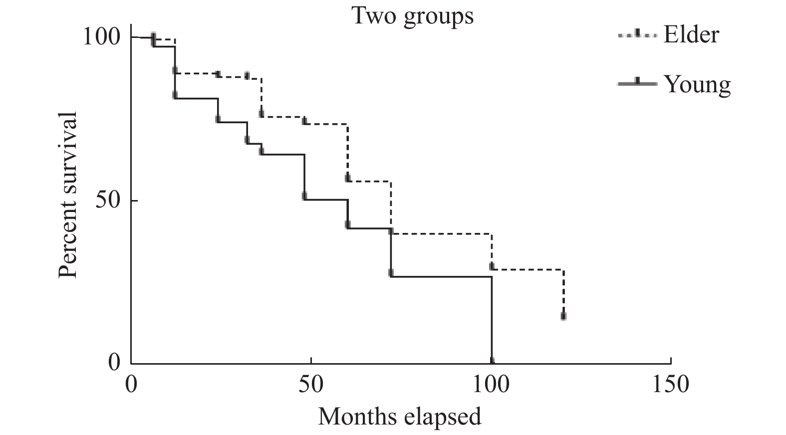
目的 探讨青年胃癌患者的临床特征,分析影响其预后的因素。 方法 回顾性分析上海交通大学医学院附属第九人民医院和复旦大学附属华山医院静安分院2005年1月~2015年12月收治的535例胃癌患者的临床资料,比较分析青年胃癌患者和中老年胃癌患者的临床特征及病理分型特点,探讨可能影响青年患者胃癌的预后因素。 结果 青年组中男女比例高于中老年组,最突出的临床表现为上腹痛,中老年组以消瘦、上消化道出血及上腹部疼痛及不适为主,二者差异有统计学意义(P < 0.0001);在病变部位方面,青年组多以胃窦为主,同时全胃比例高,而中老年患者胃中上1/3占比高于青年组。 H.pylori感染方面:青年组感染率(70.37%,57/81)明显高于中老年组(29.96%,136/454)(P < 0.0001);中老年组较青年组有更高的消化性溃疡史( P < 0.0001);青年组的Lauren分型弥漫型胃癌的比例高于中老年组。青年组5 a总体生存率低于中老年组( P = 0.008)。 结论 青年组中女性比率高,分布以胃窦为主,有更高的H.pylori感染率、组织分型以弥漫型和混合型为多,5 a总体生存率青年组低于中老年组。 Abstract:Objective The rate of gastric cancer in young patients has increased over the past few decades. The aim of this study was to analyze the clinical characteristics of young patients with gastric cancer, and investigate the possible risk factors that may be related to the survival rate of young patients. Methods From January 2005 to December 2015 a series of 535 consecutive patients were admitted to our hospitals because of a gastric cancer. We carried out a retrospective cohort study in 81 patients younger than 40 years old and in 454 patients aged 40 years older. The comparison was involved in the evaluation of patient and tumor characteristics. Results The proportion of the female in the young group was higher than that in the middle-aged and old group. The young patients had significantly more preoperative abdominal pain, while the elder had more weight loss as well as upper gastrointestinal bleeding and abdominal pain. There was no significance in duration of symptoms before the diagnosis between the two groups; In terms of lesion, pathological change occurred at the antrum or full stomach was seen more often in young patients, while elderly patients often had lesions at the upper part of corpus or the fundus. Helicobacter pylori infection and diffuse histological type were significantly associated with younger age. There was also statistically significant difference regarding overall and cancer-related 5-year survival; advanced cancer stage and diffuse histological type were the independent negative prognostic factors influencing cancer-related survival. Conclusion In our study, the proportion of the female in young patiants was higher than that in the middle-aged and old patiants and had more Helicobacter pylori infection, as well as the diffuse histological type of gastric cancer along with poor cancer-related 5-year survival rate, but we do not have sufficient evidence to consider gastric cancer in female patients as a different clinical entity. Further studies are needed to understand carcinogenesis in younger patients, especially females. -
Key words:
- Gastric cancer in youth /
- Clinical characteristics /
- Prognosis
-
表 1 患者一般情况 [n(%)]
Table 1. Patient characteristics [n(%)]
项目 青年组(≤40yr) 中老年组(> 40yr) P (n = 81) (n = 454) 平均年龄(yrs) 33.14 ± 4.52 69.70 ± 11.28 男:女 47∶34 342∶112 0.001* 生化指标 贫血:Hb < 10 g/dL 12(14.81) 114(25.11) 0.044* 低蛋白血症:< 35 g/L 13(16.05) 231(50.88) < 0.000* 合并症 0.645 心血管疾病 0 82(18.06) 高血压 2(2.24) 154(33.92) 脑血管事件 0 68(14.98) 慢性阻塞性肺病 0 36(7.93) 糖尿病 0 45(9.91) 肝硬化 0 9(1.98) 症状 < 0.0001 上腹痛 45(55.56) 91(20.04) 呕吐 1(1.23) 45(9.91) 吞咽困难 0 5(1.10) 乏力 24(29.63) 91(20.04) 厌油、纳差 1(1.23) 45(9.91) 上消化道出血 8(9.88) 114(25.11) 消瘦 16(19.75) 182(40.09) 症状出现时间(月) 10.44 ± 5.48 11.45 ± 8.48 0.304127 中位数 12 9 H.pylori 感染 < 0.000* 阳性:阴性 57∶24 136∶318 消化性溃疡史 8(9.88) 182(40.99) < 0.000* 胃癌家族史 12(14.81) 45(9.91) 0.1877 吸烟 < 0.000* 长期 10(12.35) 200(44.05) 偶尔/从不 71(87.65) 254(55.95) 饮酒 0.3427 酗酒 0(0.00) 5(1.10) 偶尔/从不 81(100.00) 449(98.90) *P < 0.05。 表 2 两组患者肿瘤特征期、组织分型、临床治疗及预后生存比较 [例(%)]
Table 2. Characteristics of gastric cancer [n(%)]
项目 青年组(≤40岁) 中老年组(> 40岁) P (81) (454) 肿瘤部位 < 0.0001* 上1/3 12(14.81) 91(20.04) 中1/3 12(14.81) 136(29.96) 下1/3 41(50.62) 226(49.78) 全胃型 16(19.75) 1(0.22) 组织学分型(Lauren’s 分型 0.014 肠型 36(44.44) 295(64.98) 弥漫型 20(24.69) 68(14.98) 混合型 25(30.86) 91(20.04) TNM分期 0.064 Ⅰ 9(11.11) 23(5.07) Ⅱ 24(29.63) 114(25.11) Ⅲ 28(34.57) 182(40.09) Ⅳ 20(24.70) 135(29.74) 手术 根治性手术 55(67.90) 325(71.59) 0.85 非根治性治疗 26(31.1) 129(28.41) 化疗 新辅助化疗 5(6.17) 0 0.67 化疗 65(80.25) 318(70.04) 生存率(a) 0.008 1 66(81.48) 423(93.17) 3 52(64.20) 342(75.33) 5 34(41.98) 237 **(56.81) 生存时间(月) 4-143(中位数:55) 2-142(中位数:69) 0.81 *P < 0.05;**失访37例。 表 3 全体患者预后因子的Cox回归分析
Table 3. Cox regression analysis of prognostic factors
因素 HR(95%CI) P 肿瘤TNM分期 0.90(0.56-1.23) < 0.01 年龄 0.57(0.46-0.67) < 0.01 性别 0.33(0.21-0.45) > 0.05 组织分型(弥漫型) 1.82(1.05-2.59) < 0.01 HR:危险系数;CI:可信区间;P值:双侧检验。 表 4 H.pylori感染暴露情况比较
Table 4. Odds ratio of H.pylori infection between two groups
项目 青年组 中老年组 OR P H. pylori感染 + 57 136 5.55 < 0.000 − 24 318 OR:比值比。 -
[1] Siegel R L,Miller K D,Jemal A. Cancer statistics[J]. CA Cancer J Clin,2015,65(1):5-29. doi: 10.3322/caac.21254 [2] Allemani C,Weir H K,Carreira H,et al. Global surveillance of cancer survival 1995-2009:Analysis of individual data for 25,676,887 patients from 279 population-based registries in 67 countries(CONCORD-2)[J]. Lancet,2015,385(9972):977-1010. [3] Chen W,Zheng R,Baade P D,et al. Cancer statistics in China,2015[J]. CA Cancer J Clin,2016,66(2):115-132. doi: 10.3322/caac.21338 [4] Takatsu Y,Hiki N,Nunobe S,et al. Clinicopathological features of gastric cancer in young patients[J]. Gastric Cancer,2016,19(2):472-478. doi: 10.1007/s10120-015-0484-1 [5] Fuchs C S,Mayer R J. Medical progress:gastric carcinoma[J]. N Engl J Med,1995,333(1):32-41. doi: 10.1056/NEJM199507063330107 [6] Theuer C P,Virgilio C D,Keese G,et al. Gastric adenocarcinomain patients 40 years of age or younger[J]. Am J Surg,1996,172(5):473-477. doi: 10.1016/S0002-9610(96)00223-1 [7] Zaręba K P,Zińczuk J,Dawidziuk T,et al. Stomach cancer in young people – a diagnostic and therapeutic problem[J]. Gastroenterology Rev,2019,14(4):283-285. doi: 10.5114/pg.2019.90254 [8] Chong V H,Telisinghe P U,Abdullah M S,et al. Gastric cancer in Brunei Darussalam:Epidemiological trend over a 27 year period(1986-2012)[J]. Asian Pac J Cancer Prev,2014,15(17):7281-7285. doi: 10.7314/APJCP.2014.15.17.7281 [9] Park J C,Lee Y C,Kim J H,et al. Clinicopathological aspects and prognostic value with respect to age:An analysis of 3,362 consecutive gastric cancer patients[J]. J Surg Oncol,2009,99(7):395-401. doi: 10.1002/jso.21281 [10] Edge S B,Compton C C. The American Joint Committee on Cancer:The 7th edition of the AJCC cancer staging manual and the future of TNM[J]. Ann Surg Oncol,2010,17(6):1471-1474. doi: 10.1245/s10434-010-0985-4 [11] Saito H,Takaya S,Fukumoto Y,et al. Clinicopathologic characteristics and prognosis of gastric cancer in young patients[J]. Yonago Acta Med,2012,55(3):57-61. [12] Ai-Refaie W B,Hu C Y,Pisters P W,et al. Gastric adenocarcinoma in young patients:A population-based appraisal[J]. Ann Surg Oncol,2011,18(10):2800-2807. doi: 10.1245/s10434-011-1647-x [13] Yao Q,Qi X,Xie S H. Sex diference in the incidence of cardia and non-cardia gastric cancer in the United States,1992~2014[J]. BMC Gastroenterol,2020,20(1):418-425. doi: 10.1186/s12876-020-01551-1 [14] Chen B K,Yang C Y. Mortality from cancers of the digestive system among grand multiparous women in Taiwan[J]. Int J Environ Res Public Health,2014,11(4):4374-4383. doi: 10.3390/ijerph110404374 [15] Noh G Y,Ku H R,Kim Y J,et al. Clinical outcomes of early gastric cancer with lymphovascular invasion or positive vertical resection margin after endoscopic submucosal dissection[J]. Surgical endoscopy,2015,29(9):2583-2589. doi: 10.1007/s00464-014-3973-0 [16] Shin K Y,Jeon S W,Cho K B,et al. Clinical outcomes of the endoscopic submucosal dissection of early gastric cancer are comparable between absolute and new expanded criteria[J]. Gut Liver,2015,9(2):181-187. doi: 10.5009/gnl13417 [17] Delgado Guillena P G, Morales Alvarado V J, Jimeno Ramiro M,et al. Gastric cancer missed at esophagogastroduodenoscopy in a well-defined Spanish population[J]. Dig Liver Dis,2019,51(8):1123-1129. [18] Ren W, Yu J, Zhang ZM, et al. Missed diagnosis of early gastric cancer or high-grade intraepithelial neoplasia[J]. World J Gastroenterol,2013,19(13):2092-2096. [19] Moon H H,Kang H W,Koh S J,et al. Clinic opathological characteristics of asymptomatic young patients with gastric cancer detected during a health checkup[J]. Korean J Gastroenterol,2019,74(5):281-290. doi: 10.4166/kjg.2019.74.5.281 [20] Wu Y L,Yu J X,Xu B. Safe major abdominal operations:hepatectomy,gastrectomy and pancreatoduodenectomy in elder patients[J]. World J Gastroenterol,2004,10(13):1995-1997. doi: 10.3748/wjg.v10.i13.1995 [21] Chadwick G,Groene O,Riley S,et al. Gastric cancers missed during endoscopy in England[J]. Clin Gastroenterol Hepatol,2015,13(7):1264-1270. doi: 10.1016/j.cgh.2015.01.025 [22] Ajdarkosh H,Sohrabi M,Moradniani M,et al. Prevalence of gastric precancerous lesions among chronic dyspeptic patients and related common risk factors[J]. Eur J Cancer Prev,2015,24(5):400-406. doi: 10.1097/CEJ.0000000000000118 [23] Hwang J J,Lee D H,Lee A R,et al. Characteristics of gastric cancer in peptic ulcer patients with Helicobacter pylori infection[J]. World J Gastroenterol,2015,21(16):4954-4960. doi: 10.3748/wjg.v21.i16.4954 [24] Mester M,Morrell A L G,Dias A R,et al. Gastric cancer in young adults:A worse prognosis group?[J]. Rev Col Bras Cir,2019,46(4):e20192256. [25] Kim H J,Kim N,Yoon H,et al. Comparison between resectable Helicobacter pylori-negative and-positive gastric cancers[J]. Gut and liver,2016,10(2):212-219. doi: 10.5009/gnl14416 -


 下载:
下载:


