Assessment of Early Coronary Artery Disease in Patients with Acute Coronary Syndrome
-
摘要:
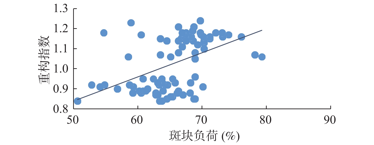
目的 探讨血浆miR-30a与冠状动脉血管内超声(IVUS)在急性冠状动脉综合征(ACS)患者早期冠状动脉病变评估中的作用。 方法 随机入选88例ACS患者,包括稳定性心绞痛组(SA组,42例)和ACS组(46例)。用IVUS检测斑块负荷和重构指数(RI),用Real-time PCR法检测血浆miR-30a的含量。研究ACS组斑块负荷、RI和血浆miR-30a的变化。用Pearson相关性分析斑块负荷、血浆miR-30a和RI的关系。 结果 与SA组比较,ACS组患者斑块负荷增加[(62.82±4.40)%比(68.32±4.59)%];RI增加(0.90±0.03)比(1.15±0.05);血浆miR-30a升高[(8.10±1.09)2-ΔΔct*104比(11.66±1.19)2-ΔΔct*104](P < 0.05)。RI和斑块负荷(r = 0.482,P = 0.000)、血浆miR-30a水平(r = 0.817,P = 0.000)呈正相关。 结论 ACS患者早期已发生冠状动脉正性重构,血浆miR-30a可以作为ACS早期冠状动脉病变的评估指标。 Abstract:Objective To investigate the role of plasma miR-30a and coronary intravascular ultrasound (IVUS) in the early assessment of coronary artery disease in patients with acute coronary syndrome (ACS). Methods 88 ACS patients were randomly selected, including stable angina group (SA group, 42 cases) and ACS group (46 cases). Plaque load and remodeling index (RI) were detected by IVUS, and plasma miR-30a was detected by real-time PCR. The changes of plaque load, RI and plasma miR-30a in ACS group were studied. The relationship between plaque load, plasma miR-30a and RI was analyzed by Pearson correlation. Plaque load and remodeling index (RI) were detected by IVUS, and plasma miR-30a was detected by real-time PCR. The changes of plaque load, RI and plasma miR-30a in ACS group were studied. The relationship between plaque load, plasma miR-30a and RI was analyzed by Pearson correlation. Results Compared with SA group, plaque load in ACS group was increased [ (62.82±4.40)% vs (68.32±4.59)%]; RI increased from (0.90±0.03) to (1.15±0.05); plasma miR-30a[ (8.10±1.09)2-ΔΔct*104 vs (11.66±1.19)2-ΔΔct*104] increased. RI was positively correlated with plaque load (r = 0.482, P = 0.000) and plasma miR-30a level (r = 0.817, P = 0.000). Conclusion Patients with ACS have early coronary artery positive remodeling, and plasma miR-30a can be used as an evaluation indicator of early ACS coronary artery disease. -
Key words:
- Echocardiography /
- microRNAs /
- Acute coronary syndrome /
- Vascular remodeling
-
表 1 2组临床基础资料(
$ \bar x \pm s $ )Table 1. Clinical information of the patients (
$ \bar x \pm s $ )组别 n 年龄
(岁)性别
(男/女)收缩压
(mmHg)舒张压
(mmHg)空腹血糖
(mmol/L)LDL-C
(mmol/L)血肌酐
(μmol/L)SA组 42 62.2 ± 12.4 22/20 126.8 ± 18.1 70.9 ± 11.3 5.2 ± 0.8 2.9 ± 0.5 81.2 ± 16.1 ACS组 46 63.1 ± 9.9* 24/22* 127.6 ± 20.6* 72.6 ± 10.9* 5.2 ± 1.0* 2.7 ± 0.6* 79.8 ± 15.3* 与SA组相比,*P > 0.05。 表 2 2组患者斑块负荷、RI和血浆miR-30a比较(
$ \bar x \pm s $ )Table 2. Comparison of plaque load,RI and plasma miR-30a in 2 groups (
$ \bar x \pm s $ )组别 n 斑块负荷(%) RI 血浆miR-30a(2−ΔΔct*104) SA组 42 62.82 ± 4.40 0.90 ± 0.03 8.10 ± 1.09 ACS组 46 68.32 ± 4.59* 1.15 ± 0.05* 11.66 ± 1.19* 与SA组相比,*P < 0.05。 -
[1] Libby P,Pasterkamp G,Crea F,et al. Reassessing the mechanisms of acute coronary syndromes[J]. Circ Res,2019,124(1):150-160. doi: 10.1161/CIRCRESAHA.118.311098 [2] Kimura S,Sugiyama T,Hishikari K,et al. The clinical significance of echo-attenuated plaque in stable angina pectoris compared with acute coronary syndromes:A combined intravascular ultrasound and optical coherence tomography study[J]. Int J Cardiol,2018,270(1):1-6. [3] Stone G W,Maehara A,Ali Z A,et al. Percutaneous coronary intervention for vulnerable coronary atherosclerotic plaque[J]. J Am Coll Cardiol,2020,76(20):2289-2301. doi: 10.1016/j.jacc.2020.09.547 [4] Yang X,Gai LY,Li P,et al. Diagnostic accuracy of dual-source CT angiography and coronary risk stratification[J]. Vasc Health Risk Manag,2010,6:935-941. [5] Andrews J,Puri R,Kataoka Y,et al. Therapeutic modulation of the natural history of coronary atherosclerosis:Lessons learned from serial imaging studies[J]. Cardiovasc Diagn Ther,2016,6(4):282-303. doi: 10.21037/cdt.2015.10.02 [6] Maehara A,Matsumura M,Ali ZA,et al. IVUS-guided versus OCT-guided coronary stent implantation:A critical appraisal[J]. JACC Cardiovasc Imaging,2017,10(12):1487-1503. doi: 10.1016/j.jcmg.2017.09.008 [7] Ono M,Kawashima H,Hara H,et al. Advances in IVUS/OCT and future clinical perspective of novel hybrid catheter system in coronary imaging[J]. Front Cardiovasc Med,2020,7:119. doi: 10.3389/fcvm.2020.00119 [8] Nguyen P,Seto A. Contemporary practices using intravascular imaging guidance with IVUS or OCT to optimize percutaneous coronary intervention[J]. Expert Rev Cardiovasc Ther,2020,18(2):103-115. doi: 10.1080/14779072.2020.1732207 [9] Reddy S,Kadiyala V,Kashyap J R,et al. Comparison of intravascular ultrasound virtual histology parameters in diabetes versus Non-diabetes with acute coronary syndrome[J]. Cardiology,2020,145(9):570-577. doi: 10.1159/000508886 [10] Marino B C A,Buljubasic N,Akkerhuis M,et al. Adiponectin in relation to coronary plaque characteristics on radiofrequency intravascular ultrasound and cardiovascular outcome[J]. Arq Bras Cardiol,2018,111(3):345-353. [11] Kochergin N A,Kochergina A M,Khorlampenko A A,et al. Vulnerable atherosclerotic plaques of coronary arteries in patients with stable coronary artery disease:12-months follow-up[J]. Kardiologiia,2019,60(2):69-74. [12] Çoner A,Aydınalp A,Müderrisoğlu H. Evaluation of hs-CRP and sLOX-1 levels in moderate-to-high risk acute coronary syndromes[J]. Endocr Metab Immune Disord Drug Targets,2020,20(1):96-103. doi: 10.2174/1871530319666190408145905 [13] Tateishi K,Kitahara H,Saito Y,et al. Impact of clinical presentations on lipid core plaque assessed by near-infrared spectroscopy intravascular ultrasound[J]. Int J Cardiovasc Imaging,2021,37(4):1151-1158. doi: 10.1007/s10554-020-02107-w [14] Merkulova I N,Shariya M A,Mironov V M,et al. Computed tomography coronary angiography possibilities in "High Risk" plaque identification in patients with non-ST-elevation acute coronary syndrome:Comparison with intravascular ultrasound[J]. Kardiologiia,2021,60(12):64-75. doi: 10.18087/cardio.2020.12.n1304 [15] Wei Pan,Yun Zhong,Chuanfang Cheng,et al. miR-30-Regulated autophagy mediates angiotensin II-induced myocardial hypertrophy[J]. PLoS ONE,2013,8(1):53950. doi: 10.1371/journal.pone.0053950 [16] 潘伟,赵娟,罗韶金,等. MiR-30a在血管紧张素Ⅱ诱导的心肌肥厚中的作用[J]. 中国心血管病研究,2016,14(6):567-571. -


 下载:
下载:


