The Currative Effect of Biological Lengthened Handle Total Hip Arthroplasty in the Treatment of Subtrochanteric Fracture Combined with Femoral Head Necrosis
-
摘要:
目的 探讨使用生物型加长柄行全髋关节置换治疗股骨转子下骨折合并股骨头缺血性坏死的可行性及临床疗效。 方法 回顾性分析2016年1月至2020年1月收治的88位患者,其中30例股骨转子下骨折合并股骨头缺血性坏死行I期生物型加长柄关节置换(实验组),20例单纯转子下骨折组行PFNA内固定术(PFNA组),32例单纯股骨头缺血性坏死行人工关节置换术(关节置换组),比较3组的手术时间、围术期出血量、术后住院天数;比较实验组与PFNA组骨折愈合时间;比较实验组与关节置换组术后1,3,6月和末次随访的髋关节Harris评分。 结果 实验组手术时间高于PFNA组及关节置换组,差异有统计学意义(P < 0.05);3组围术期出血量、术后下地活动时间及住院天数,差异有统计学意义(P < 0.05);实验组与PFNA组骨折愈合时间,差异无统计学意义,实验组与关节置换组术后髋关节Harris评分,差异无统计学意义(P < 0.05)。 结论 生物型加长柄I期行全髋关节置换治疗股骨转子下骨折合并股骨头缺血性坏死可取得良好的临床疗效,骨折愈合时间与PFNA术无明显差异。 Abstract:Objective To investigate the feasibility and clinical effect of total hip replacement with the biologically lengthened stem in the treatment of subtrochanteric fractures combined with the necrosis of femoral head. Methods A total of 88 patients admitted from January 2016 to January 2020 were analyzed retrospectively. Among them, 30 cases of subtrochanteric fractures with the avascular necrosis of the femoral head underwent stage I biotype long-stem joint replacement (experimental group), 20 cases of simple subtrochanteric fracture group underwent PFNA internal fixation (PFNA group), and 32 cases of simple avascular necrosis of the femoral head underwent the artificial joint replacement (joint replacement group). The operation time, perioperative blood loss and postoperative hospitalization were compared among the three groupsand the fracture healing time between the experimental group and the PFNA group was compared so as to compare the hip Harris scores of the experimental group and the joint replacement group 1, 3, 6 months after the surgery and the last follow-up. Results The operation time of the experimental group was higher than those of the PFNA group and the joint replacement group, and the difference was statistically significant; The three groups had the statistically significant differences in perioperative blood loss, time of postoperative activities and the length of hospital stay; There was no significant difference in fracture healing time between the experimental group and the PFNA group and there was no statistically significant difference in the Harris score of the hip joint between the experimental group and the joint replacement group. Conclusion The total hip arthroplasty for the treatment of subtrochanteric fractures with the necrosis of femoral head can achieve good clinical results. There is no significant difference between fracture healing time and PFNA surgery. -
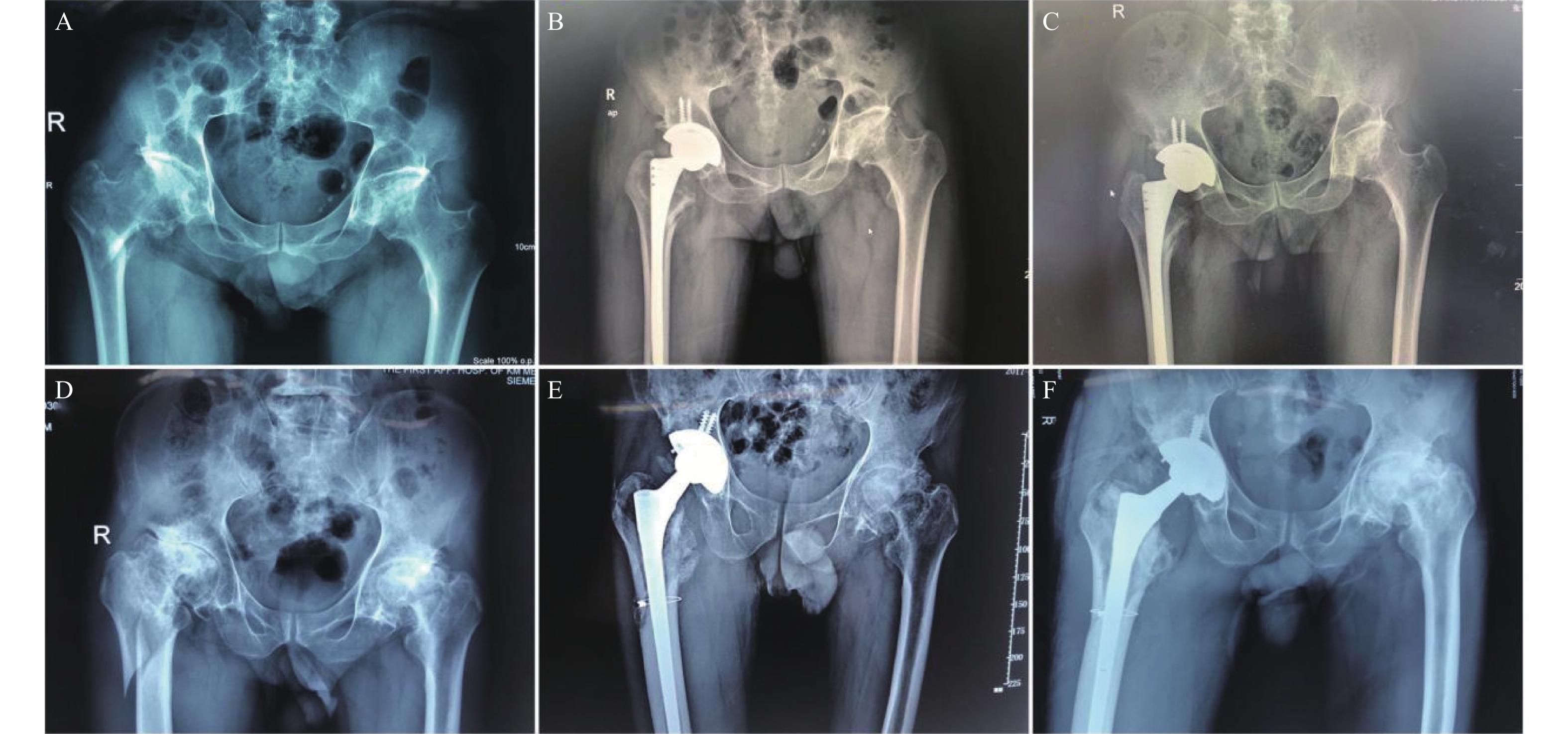
下颌管是下颌骨内重要的解剖结构,起始于下颌孔,其内有下牙槽神经,静脉及动脉穿行,因此下颌管的解剖结构与下颌区多种口腔治疗所致的血管神经损伤有高度的相关性。各类中低位阻生的下颌第3磨牙牙根与下颌管上壁的位置关系密切,即使在使用微创方式进行下颌阻生第3磨牙拔除时,仍然存在不可避免的下颌神经血管束的医源性损伤,其中5%~7%为暂时性损伤,0.5%~1.0%为不可逆神经损伤[1-6]。此外,很多病例报道在进行下颌前磨牙、第1磨牙及第2磨牙根管治疗时可能导致下牙槽神经或颏神经的损伤[7-8],机械性的压迫、化学药品的神经毒性、局部感染以及根管治疗时的直接创伤都可能造成下牙槽神经的损伤[9]。因此,准确地定位下颌管及其走行位置关系对于口腔诊疗有着非常重要的指导意义。
早期有学者运用下颌骨标本对下颌管的局部解剖进行研究,但由于例数太少且方法简单,因而结论的代表性不强[10]。很多影像学的手段也被用来观察下颌管的解剖形态,其中包括X线平片、曲面断层片、计算机X线体层摄影(computed tomography,CT)[11-13]。二维成像的X线平片和曲面断层片不能真实的反应下颌管及其局部解剖结构的三维图像,而 CT的图像重建操作复杂,且放射剂量大,不符合影像学检查“医疗照射正当化”的原则,不适宜在口腔检查中广泛推广。锥形束CT(cone beam computed tomography,CBCT)2001年在美国开始使用并逐步的运用于口腔的颌面部检查[14]。与传统的二维成像技术相比,CBCT图像的主要优点是能清除临近组织的重叠影像,放射剂量低,且三维图像重建快速易操作[15];正因为这些优势,CBCT越来越多的被应用于口腔颌面部的常规诊疗[16]。已有学者证明利用CBCT能够从不同角度和层面清晰地观侧到下颌管及其局部解剖结构[17-18]。
在云南大部分少数民族地区,口腔医疗人员匮乏,口腔医疗技术落后,在颌骨手术、牙种植术、根管治疗及阻生牙拔除术等口腔诊疗中缺乏相应的影像设备进行下颌管三维位置的准确定位及测量,或者仅能凭借二维的根尖片或全景片进行预估,通过本研究可以对云南地区的口腔临床诊疗有一定的指导作用。
1. 资料与方法
1.1 研究对象
收集2018年2月至2019年10月在云南省口腔医院口腔影像科拍摄CBCT的影像资料共339例,根据不同的年龄及性别进行分组,其中青年组(18~39岁)122例,中年组(40~59岁)126例,老年组(60~79岁)91例;男性182例,女性157例。
纳入标准[19]:颌面部对称;下颌骨无外伤及手术史;下颌前磨牙、磨牙牙根发育完成。在CBCT影像资料中,下颌管走行区域能清晰的观察到下颌管;图像无明显严重扭曲变形。排除标准[19]:骨质病理性疏松等代谢性或者免疫系统性疾病;中度或重度牙周病患者;下颌后牙发生脓肿或者根尖炎等导致颌骨或根尖吸收的疾病;下颌后牙区有金属冠、桥修复体及种植体。下颌管及颏孔严重变异,如双下颌管、多颏孔等情况。
1.2 图像采集
使用NEW TOM CBCT设备进行拍摄,拍摄前进行标准化调试,拍摄技师为具有5a以上经验的口腔影像科技师,患者均采用统一标准体位进行拍摄。在扫描过程中嘱患者保持静息状态,预扫描过程中要求患者头部处于放射视野正中位置,放射视野中轴线与患者头颅中轴线一致,如未达到以上要求则重新扫描。将图像数据转换为矩阵512×512的DICOM格式输出,并导入至NNT分析软件,调整三维图像中轴位图像的基准平面与下颌骨下缘平行,在横断面图像界面于颏孔平面标记牙弓曲线,NNT软件自动重建出下颌骨的三维重建图像。
1.3 测量截面选取
在下颌骨三维重建影像上利用冠状面图像对下颌管解剖结构进行观测,将左右两侧每侧测量区域分为五个分区,从远中至近中分别是第3磨牙区、第2磨牙区、第1磨牙区、第2前磨牙区及颏孔区,调整每个分区的基准平面,即调整轴位图像使之与下颌体平行,冠状切线平行于牙体长轴,矢状切线调整至于颊舌侧骨板平行,所有图像三维重建及数据测量均在相同的灰度、对比度及锐化度条件下由同一位医师在同一台电脑上进行。
1.4 测量的指标
在下颌骨的冠状面上测量颏孔区颏孔上缘至牙槽嵴顶的距离,第3磨牙区、第2磨牙区、第1磨牙区及第2前磨牙区测量下颌管上壁内缘至牙槽嵴顶的距离、下颌管下壁内缘至下颌骨下缘骨皮质外侧壁的距离、下颌管颊侧壁内缘至下颌骨颊侧骨板骨皮质外侧缘的距离、下颌管舌侧壁内缘至下颌骨舌侧骨板骨皮质外侧缘的距离及下颌管上壁内缘至牙根根尖的距离。
1.5 统计学处理
用SPSS22.0统计软件包建立数据库并进行数据分析。各指标测量值用(
$ \bar{x} $ ±s)表示;年龄组间的各指标测量值比较采用单因素方差分析,如有差别,用q检验进一步两两比较,性别组间的各指标测量值比较采用两独立样本t检验,P < 0.05为差异有统计学意义。2. 结果
2.1 研究对象基本信息
本实验按照纳入和排除标准共筛选出339例病例作为研究对象,研究对象基本信息,见表1。
表 1 研究对象基本信息统计表(n)Table 1. Demographic data of research objects(n)民族 性别 年龄段 总计 青年组 中年组 老年组 汉族组 男 20 22 18 60 女 19 21 13 53 傣族组 男 24 20 20 64 女 18 18 13 49 哈尼族组 男 19 25 14 58 女 22 20 13 55 总计 122 126 91 339 2.2 颏孔上缘及下颌管上壁内缘到牙槽嵴顶的距离
在年龄组间,颏孔上缘及下颌管上壁内缘至牙槽嵴顶的距离在年龄间的差异具有统计学意义(P < 0.05)。经过两两间比较结果显示,两侧Ⅲ区和右侧Ⅱ区中年组及青年组间无差别(P > 0.05),均高于老年组(P < 0.05),除此之外,其余测量分区均为:中年组 > 青年组 > 老年组(P < 0.05),见表2。
表 2 不同年龄段颏孔上缘及下颌管上壁内缘到牙槽嵴顶的距离[($\bar{x} \pm s$ ),mm]Table 2. The length from the upper edge of the mental foramen and the inner edge of the upper wall of the mandibular canal to the alveolar ridge crest in different age groups[($\bar{x}\pm s$ ),mm]测量截面分区 年龄 F P 青年 中年 老年 左侧 Ⅰ区 15.3 ± 1.3 16.0 ± 1.5 13.9 ± 1.2 61.647 < 0.001 Ⅱ区 17.1 ± 1.4 17.6 ± 1.3 16.0 ± 1.2 21.570 < 0.001 Ⅲ区 16.8 ± 1.5 17.0 ± 1.6 15.2 ± 1.2 43.935 < 0.001 Ⅳ区 15.4 ± 1.3 16.2 ± 1.6 14.4 ± 1.1 43.593 < 0.001 Ⅴ区 14.8 ± 1.4 15.4 ± 1.5 13.9 ± 1.1 30.310 < 0.001 右侧 Ⅰ区 15.4 ± 1.3 16.1 ± 1.5 14.0 ± 1.2 66.188 < 0.001 Ⅱ区 17.3 ± 1.4 17.5 ± 1.4 15.9 ± 1.1 22.273 < 0.001 Ⅲ区 16.8 ± 1.4 16.9 ± 1.7 15.2 ± 1.1 42.923 < 0.001 Ⅳ区 15.5 ± 1.4 16.1 ± 1.7 14.4 ± 1.1 37.096 < 0.001 Ⅴ区 14.8 ± 1.6 15.5 ± 1.5 13.9 ± 1.0 30.905 < 0.001 在性别上,两侧所有检测分区颏孔上缘及下颌管上壁内缘到牙槽嵴顶的距离男性均大于女性,差异有统计学意义(P < 0.05),见表3。
表 3 不同性别颏孔上缘及下颌管上壁内缘到牙槽嵴顶的距离[($ \bar{x}\pm s $ ),mm]Table 3. The length from the upper edge of the mental foramen and the inner edge of the upper wall of the mandibular canal to the alveolar ridge crest in different gender groups[($ \bar{x} \pm s$ ),mm]测量截面分区 性别 t P 男性 女性 左侧 Ⅰ区 15.7 ± 1.6 14.6 ± 1.4 6.848 < 0.001 Ⅱ区 17.4 ± 1.4 16.6 ± 1.4 4.147 < 0.001 Ⅲ区 16.9 ± 1.8 16.0 ± 1.4 5.270 < 0.001 Ⅳ区 15.8 ± 1.6 15.0 ± 1.3 4.591 < 0.001 Ⅴ区 15.1 ± 1.6 14.4 ± 1.2 4.291 < 0.001 右侧 Ⅰ区 15.7 ± 1.6 14.8 ± 1.5 5.369 < 0.001 Ⅱ区 17.4 ± 1.3 16.6 ± 1.4 4.223 < 0.001 Ⅲ区 16.8 ± 1.7 15.9 ± 1.4 5.671 < 0.001 Ⅳ区 15.8 ± 1.7 15.0 ± 1.4 4.690 < 0.001 Ⅴ区 15.1 ± 1.8 14.5 ± 1.1 3.929 < 0.001 2.3 下颌管下壁内缘到下颌骨下缘骨皮质外侧壁的距离
在年龄方面,除右侧Ⅴ区下颌管下壁内缘到下颌骨下缘骨皮质外侧壁的距离在年龄组间差异无统计学意义(P > 0.05),其余检测分区在年龄组间的差异均具有统计学意义(P < 0.05),经过两两间比较结果显示,除左侧Ⅴ区青年组大于中年组及老年组(P < 0.05)外,其余测量分区中年组均大于青年组及老年组(P < 0.05),且其中左侧Ⅲ区及Ⅴ区、右侧Ⅳ区中年组和老年组间无差别(P > 0.05),见表4。
表 4 不同年龄段下颌管下壁内缘到下颌骨下缘骨皮质外侧壁的距离[($ \bar{x} \pm s$ ),mm]Table 4. The length from the inner edge of the inferior wall of the mandibular canal to the lateral wall of the lower edge of the mandible in different age groups[($ \bar{x} \pm s$ ),mm]测量截面分区 年龄 F P 青年 中年 老年 左侧 Ⅱ区 9.2 ± 1.1 9.8 ± 1.0 9.5 ± 1.3 4.087 0.018 Ⅲ区 9.0 ± 1.0 9.6 ± 0.9 9.4 ± 1.1 11.271 < 0.001 Ⅳ区 9.6 ± 1.1 9.8 ± 0.9 10.0 ± 1.1 3.789 0.024 Ⅴ区 11.2 ± 1.4 10.7 ± 1.1 10.5 ± 1.3 9.892 < 0.001 Ⅱ区 9.2 ± 1.1 9.8 ± 1.1 9.6 ± 1.2 5.763 0.004 右侧 Ⅲ区 9.0 ± 1.1 9.7 ± 1.0 9.4 ± 1.1 13.781 < 0.001 Ⅳ区 9.5 ± 1.2 9.9 ± 1.0 10.0 ± 1.1 5.428 < 0.001 Ⅴ区 10.9 ± 1.6 10.7 ± 1.2 10.5 ± 1.3 2.856 0.059 在性别上,两侧所有检测分区下颌管下壁内缘至下颌骨下缘骨皮质外侧壁的距离男女间差异无统计学意义(P > 0.05),见表5。
表 5 不同性别下颌管下壁内缘到下颌骨下缘骨皮质外侧壁的距离[($ \bar{x}\pm s $ ),mm]Table 5. The length from the inner edge of the inferior wall of the mandibular canal to the lateral wall of the lower edge of the mandible in different gender groups[($ \bar{x}\pm s $ ),mm]测量截面分区 性别 t P 男性 女性 左侧 Ⅱ区 9.5 ± 1.2 9.5 ± 1.1 0.042 0.966 Ⅲ区 9.4 ± 1.0 9.2 ± 1.0 1.066 0.287 Ⅳ区 9.8 ± 0.9 9.7 ± 1.2 0.530 0.596 Ⅴ区 10.9 ± 1.5 10.7 ± 1.0 1.264 0.207 右侧 Ⅱ区 9.5 ± 1.2 9.5 ± 1.1 0.060 0.952 Ⅲ区 9.4 ± 1.0 9.3 ± 1.1 0.524 0.600 Ⅳ区 9.8 ± 1.0 9.8 ± 1.2 0.568 0.570 Ⅴ区 10.8 ± 1.4 10.6 ± 1.3 1.028 0.304 2.4 下颌管颊侧壁内缘到下颌骨颊侧骨板骨皮质外侧壁的距离
在年龄方面,两侧Ⅴ区下颌管颊侧壁内缘至颊侧骨板骨皮质外侧壁的距离在年龄组间差异无统计学意义(P > 0.05),其余检测分区在年龄组间的差异均具有统计学意义(P < 0.05)。经过两两间比较结果显示,两侧Ⅱ区老年组和青年组无差别(P > 0.05),均低于中年组(P < 0.05),其余测量分区均为:青年组 > 中年组 > 老年组(P < 0.05),见表6。
表 6 不同年龄段下颌管颊侧壁内缘到下颌骨颊侧骨板骨皮质外侧壁的距离[($ \bar{x} \pm s$ ),mm]Table 6. The length from between the inner edge of the buccal wall of the mandibular canal to the lateral cortical wall of the buccal plate of the mandible in different age groups[($ \bar{x} \pm s$ ),mm]测量截面分区 年龄 F P 青年 中年 老年 左侧 Ⅱ区 4.6 ± 0.7 5.0 ± 0.6 4.7 ± 0.6 5.379 0.005 Ⅲ区 6.3 ± 1.0 5.9 ± 0.7 5.4 ± 0.5 28.512 < 0.001 Ⅳ区 7.4 ± 1.0 6.8 ± 0.6 6.6 ± 0.7 27.267 < 0.001 Ⅴ区 7.0 ± 1.3 6.9 ± 1.0 7.1 ± 0.8 0.608 0.545 Ⅱ区 4.6 ± 0.5 5.1 ± 0.7 4.8 ± 0.7 11.210 < 0.001 右侧 Ⅲ区 6.2 ± 0.9 6.0 ± 0.8 5.4 ± 0.5 30.173 < 0.001 Ⅳ区 7.4 ± 0.9 7.0 ± 0.7 6.6 ± 0.7 26.071 < 0.001 Ⅴ区 7.0 ± 1.2 7.0 ± 0.9 7.1 ± 0.8 0.266 0.767 在性别上,左侧Ⅱ区、Ⅲ区、Ⅳ区和右侧Ⅳ区下颌管颊侧壁内缘至颊侧骨板骨皮质外侧壁的距离男性均大于女性,差异具有统计学意义(P < 0.05),其余测量分区男女间差异无统计学意义(P > 0.05),见表7。
表 7 不同性别下颌管颊侧壁内缘到下颌骨颊侧骨板骨皮质外侧壁的距离[($ \bar{x} \pm s$ ),mm]Table 7. The length from the inner edge of the buccal wall of the mandibular canal to the lateral cortical wall of the buccal plate of the mandible in different gender groups[($ \bar{x} \pm s$ ),mm]测量截面分区 性别 t P 男性 女性 左侧 Ⅱ区 4.9 ± 0.6 4.6 ± 0.6 3.728 < 0.001 Ⅲ区 6.0 ± 0.8 5.8 ± 0.8 1.971 0.049 Ⅳ区 7.1 ± 0.9 6.8 ± 0.7 2.689 0.008 Ⅴ区 7.1 ± 1.1 6.9 ± 1.0 1.624 0.105 Ⅱ区 4.9 ± 0.6 4.8 ± 0.8 1.383 0.168 右侧 Ⅲ区 6.0 ± 0.8 5.9 ± 0.9 0.763 0.446 Ⅳ区 7.1 ± 0.8 6.9 ± 0.8 2.462 0.014 Ⅴ区 7.1 ± 1.0 6.9 ± 1.0 1.773 0.077 2.5 下颌管舌侧壁内缘到下颌骨舌侧骨板骨皮质外侧壁的距离
在年龄方面,两侧Ⅴ区下颌管舌侧壁内缘至下颌骨舌侧骨板骨皮质外侧壁的距离在年龄组间差异无统计学意义(P > 0.05),其余检测分区在年龄组间的差异均具有统计学意义(P < 0.05)。经过两两间比较结果显示,两侧Ⅱ区、Ⅲ区、Ⅳ区老年组大于青年组(P < 0.05),其中左侧Ⅲ区、Ⅳ区中年组和老年组差异无统计学意义(P > 0.05),见表8。
表 8 不同年龄段下颌管舌侧壁内缘到下颌骨舌侧骨板骨皮质外侧壁的距离[($ \bar{x} \pm s$ ),mm]Table 8. The length from the inner edge of the lingual wall of the mandibular canal to the lateral wall of the lingual bone plate of the mandible in different age groups[($ \bar{x} \pm s$ ),mm]测量截面分区 年龄 F P 青年 中年 老年 左侧 Ⅱ区 3.9 ± 0.8 4.1 ± 0.6 4.3 ± 0.5 3.923 0.021 Ⅲ区 3.0 ± 0.6 3.3 ± 0.7 3.4 ± 0.5 12.323 < 0.001 Ⅳ区 2.4 ± 0.5 2.7 ± 0.7 2.8 ± 0.6 12.761 < 0.001 Ⅴ区 2.2 ± 0.5 2.3 ± 0.7 2.3 ± 0.7 1.937 0.146 右侧 Ⅱ区 3.9 ± 0.8 4.0 ± 0.8 4.3 ± 0.4 4.474 0.013 Ⅲ区 3.0 ± 0.7 3.2 ± 0.8 3.4 ± 0.5 9.693 < 0.001 Ⅳ区 2.4 ± 0.5 2.6 ± 0.7 2.9 ± 0.6 11.538 < 0.001 Ⅴ区 2.2 ± 0.6 2.3 ± 0.7 2.4 ± 0.8 2.163 0.117 在性别上,两侧Ⅴ区下颌管舌侧壁内缘至下颌骨舌侧骨板骨皮质外侧壁的距离在性别间差异无统计学意义(P > 0.05),其余检测分区在性别间的差异均具有统计学意义(P < 0.05),见表9。
表 9 不同性别下颌管舌侧壁内缘到下颌骨舌侧骨板骨皮质外侧壁的距离[($ \bar{x} \pm s$ ),mm]Table 9. The length from the inner edge of the lingual wall of the mandibular canal to the lateral wall of the lingual bone plate of the mandible in different gender groups[($ \bar{x} \pm s$ ),mm]测量截面分区 性别 t P 男性 女性 左侧 Ⅱ区 4.2 ± 0.6 4.0 ± 0.7 2.139 0.034 Ⅲ区 3.3 ± 0.6 3.2 ± 0.7 2.152 0.032 Ⅳ区 2.7 ± 0.6 2.6 ± 0.7 2.091 0.037 Ⅴ区 2.3 ± 0.7 2.2 ± 0.6 1.474 0.141 Ⅱ区 4.2 ± 0.7 3.8 ± 0.8 3.240 0.001 右侧 Ⅲ区 3.3 ± 0.6 3.1 ± 0.7 2.892 0.004 Ⅳ区 2.7 ± 0.6 2.5 ± 0.7 2.759 0.006 Ⅴ区 2.4 ± 0.7 2.2 ± 0.6 1.785 0.075 2.6 下颌管上壁内缘到牙根根尖的距离
在年龄方面,所有测量分区下颌管上壁内缘至牙根根尖的距离在年龄组间的差异均具有统计学意义(P < 0.05),经过两两间比较结果显示,两侧Ⅱ区、Ⅲ区、Ⅳ区中年均为:中年组 > 青年组 > 老年组(P < 0.05),两侧Ⅴ区中年组和青年组差异无统计学意义(P > 0.05),均低于老年组(P < 0.05),见表10。
表 10 不同年龄下颌管上壁内缘到牙根根尖的距离[($ \bar{x} \pm s$ ),mm]Table 10. The length from the inner edge of the upper wall of the mandibular canal to the root apex in different age groups[($ \bar{x}\pm s $ ),mm]测量截面分区 年龄 F P 青年 中年 老年 左侧 Ⅱ区 5.1 ± 1.6 6.0 ± 1.3 4.7 ± 1.7 11.885 < 0.001 Ⅲ区 5.9 ± 1.5 6.6 ± 1.1 5.1 ± 1.6 31.522 < 0.001 Ⅳ区 4.7 ± 1.4 5.8 ± 1.4 5.1 ± 1.3 18.929 < 0.001 Ⅴ区 2.3 ± 1.3 2.6 ± 1.9 3.5 ± 1.9 13.081 < 0.001 右侧 Ⅱ区 5.2 ± 1.7 6.1 ± 1.1 4.6 ± 1.5 14.127 < 0.001 Ⅲ区 5.8 ± 1.4 6.2 ± 1.3 5.0 ± 1.3 22.384 < 0.001 Ⅳ区 4.8 ± 1.3 5.5 ± 1.8 5.0 ± 1.3 7.704 0.001 Ⅴ区 2.4 ± 1.4 2.5 ± 1.9 3.2 ± 1.5 6.406 0.002 在性别上,两侧Ⅱ区和Ⅲ区下颌管上壁内缘至牙根根尖的距离女性高于男性,右侧Ⅴ区男性高于女性,差异具有统计学意义(P < 0.05),其余测量分区在性别间的差异无统计学意义(P > 0.05),见表11。
表 11 不同性别下颌管上壁内缘到牙根根尖的距离[($ \bar{x} \pm s$ ),mm]Table 11. The length from between the inner edge of the upper wall of the mandibular canal to the root apex in different gender groups[($ \bar{x} \pm s$ ),mm]测量截面分区 性别 t P 男性 女性 左侧 Ⅱ区 5.1 ± 1.5 5.6 ± 1.7 −2.163 0.032 Ⅲ区 5.7 ± 1.6 6.2 ± 1.4 −3.300 0.001 Ⅳ区 5.2 ± 1.5 5.2 ± 1.4 0.306 0.760 Ⅴ区 2.9 ± 1.9 2.5 ± 1.7 1.808 0.071 Ⅱ区 5.1 ± 1.5 5.7 ± 1.6 −2.577 0.011 右侧 Ⅲ区 5.6 ± 1.5 6.0 ± 1.3 −2.490 0.013 Ⅳ区 5.2 ± 1.5 5.1 ± 1.6 0.403 0.687 Ⅴ区 2.8 ± 1.7 2.4 ± 1.5 2.428 0.016 3. 讨论
3.1 下颌管在垂直方向上的位置关系
本研究中,每个测量分区下颌管至牙槽嵴顶及下颌骨下缘的距离中年组大于其他年龄组,这与戴慧颖等[11]对65例病例的CBCT测量结果一致,以上研究结果符合人体的发育规律即随着年龄的增长,至中年时期颌骨发育完成且位置相对恒定,随着牙槽嵴增龄式的生理性吸收,老年时期牙槽嵴高度有所降低,间接导致下颌管距牙槽嵴顶的距离逐渐变短。每个测量截面下颌管至牙槽嵴顶的距离男性均大于女性,这与Ozturk等[20]的研究结果一致,Ozturk等通过测量100例病例的下颌骨左右两侧下颌管至牙槽嵴顶的距离得出男性平均比女性增加了2.00 mm。
以往采用下颌骨标本进行解剖学研究一直被认为是研究下颌管走行的金标准,但是由于下颌骨标本量较小,且不能进行性别及年龄的比较,在进行解剖时存在人为的标本损害,对于精细结构的测量容易产生误差。本实验在CBCT扫描后得到的影像学资料中直接测量下颌管的解剖结构,可研究的样本量大且容易收集,测量结果精确度高,对于在下颌管走形区域进行口腔诊疗时可以充分参考CBCT的测量结果指导手术设计和方案选择。
3.2 下颌管在颊舌方向上的位置关系
老年人的下颌管在颊舌向的位置更偏向于颊侧,青年人更偏向于舌侧,这与Khorshidi[21]等对242例病例的CBCT影像学测量结果一致。吕婴[22]研究报道,下颌骨在颊舌向及垂直向的生长发育可以从青春后期持续至成人期,其生长周期可以延伸至25岁乃至40岁,关于下颌管在颊舌向的位置变化与年龄的关系还需要从生长发育及增龄性骨吸收等方面进行更深层次的研究。
在性别上,大部分测量分区下颌管至颊舌侧骨板的距离男性均大于女性,Tepper等[23]研究报道,颌骨内矿物质含量在性别上具有差异,男性颌骨内矿物质含量高于女性,女性骨小梁较男性稀疏,且随着年龄的增长,男性颌骨矿物质含量基本不变甚至有所增加,而女性体内激素水平的改变会引起颌骨骨质疏松,从而导致下颌骨的骨皮质变薄,所以下颌管在颊舌方向上的位置还需综合骨矿物质增龄性改变来进行研究。
3.3 下颌管与牙根根尖的位置关系
下颌第2前磨牙、第1磨牙及第2磨牙牙根根尖至下颌管的距离中年组大于其他年龄段,下颌管至牙根根尖距离最远处为下颌第2前磨牙区,最近处为下颌第2磨牙区,这与下颌管在垂直方向上由颏孔区-第2前磨牙区-第2磨牙区呈“V”走行规律相关。
在性别上,两侧第2前磨牙区和第1磨牙区下颌管上壁到牙根根尖的距离女性高于男性,与金柱坤等[24]对68例中国人下颌管至后牙牙根根尖距离的研究结论相反,可能是由于金柱坤等[24]采用的是螺旋CT进行测量,CBCT与螺旋CT相比,分辨率较高,层距较小,因此本研究的结论更具有客观性。
综上所述,在下颌管走形区域进行牙槽外科手术、牙拔除术、牙髓治疗时,明确下颌管与牙根根尖的位置关系,在安全范围内开展诊疗有利于减少术后出血、麻痹等的不良并发症的发生;采用CBCT对下颌管的解剖结构和三维位置进行精确的测量分析,结合计算机设计和计算机辅助技术及三维打印技术,可在术前设计制作出高精度的种植导板及外科手术导板,并指导手术方案的选择,在导板的辅助下可提高手术安全性,结合CBCT检测技术的数字化微创诊疗已经成为未来的研究新兴领域。
-
表 1 一般资料比较(n)
Table 1. Comparsion of general data (n)
一般情况 实验组 PFNA组 关节置换组 年龄(岁) 69.43 ± 5.32 71.05 ± 8.47 68.16 ± 6.25 侧别(左) 18 12 20 (右) 12 8 12 骨折分型(RUSSELL-TAYLOR IA型) 6 6 (RUSSELL-TAYLOR IB型) 24 14 股骨头坏死分期(ARCO III期) 0 8 (ARCO IV期) 30 24 表 2 临床疗效比较
Table 2. Comparsion of clinical outcomes
组别 手术时间(min) 术中出血量(mL) 术后下地活动时间(d) 住院天数(d) 骨折愈合时间(d) 实验组 92.97 ± 11.75 198.13 ± 31.74 4.53 ± 1.41 8.20 ± 1.49 12.23 ± 1.26 PFNA组 67.95 ± 14.00 152.25 ± 13.65 12.80 ± 2.02 8.25 ± 1.83 12.25 ± 1.25 关节置换组 64.63 ± 9.07 100.78 ± 22.83 2.41 ± 0.56 8.19 ± 1.80 H/F 61.098 277.314 67.266 0.063 P < 0.001* < 0.001* < 0.001* 0.969 0.975 *P < 0.05。 表 3 随访资料比较(分)
Table 3. Comparsion of follow-up data (scores)
组别 术后1月Harris评分 术后3月Harris评分 术后6月Harris评分 末次随访Harris评分 实验组 78.53 ± 4.754 80.23 ± 4.739 83.90 ± 4.020 86.83 ± 3.788 关节置换组 77.45 ± 5.549 79.75 ± 6.060 85.30 ± 4.857 88.05 ± 4.524 T 0.715 0.301 −1.068 −0.993 P 0.479 0.766 0.293 0.327 -
[1] Egol K A. Subtrochanteric femur fracture below an arthritic hip:Opinion:Open reduction and internal fixation in conjunction with total hip arthroplasty[J]. J Orthop Trauma,2005,19(1):60-61. doi: 10.1097/00005131-200501000-00012 [2] Gallagher C A,Jones C W,Kimmel L,et al. Osteoarthritis is associated with increased failure of proximal femoral fracture fixation.[J]. Int Orthop,2019,43(5):1223-1230. doi: 10.1007/s00264-018-4014-8 [3] Bohndorf K,Roth A. Imaging and classification of avascular femoral head necrosis[J]. Orthopade,2018,47(9):729-734. [4] Rizkalla J M,Nimmons S J B,Jones A L. Classifications in brief:The russell-taylor classification of subtrochanteric hip fracture[J]. Clin Orthop Relat Res,2019,477(1):257-261. [5] 周宗科,翁习生,曲铁兵,等. 中国髋、膝关节置换术加速康复——围术期管理策略专家共识[J]. 中华骨与关节外科杂志,2016,9(1):1-9. doi: 10.3969/j.issn.2095-9985.2016.01.001 [6] 张盘军,王勇,蒋建农,等. Ⅰ期全髋关节置换术治疗股骨转子下骨折合并同侧股骨头坏死[J]. 实用骨科杂志,2020,26(9):778-781. [7] 邓煜,夏于欣,白新文,等. 生物型加长柄髋关节置换治疗老年不稳定性转子间骨折中股骨近端不同重建方法的疗效对比[J]. 中国组织工程研究,2019,23(12):1805-1811. doi: 10.3969/j.issn.2095-4344.1120 [8] Kim J Y,Kong G M. Subcapital femoral neck fracture after treatment of an intertrochanteric fracture with proximal femoral intramedully nail[J]. Coll Physicians Surg Pak,2016,26(1):78-79. [9] 徐风瑞,何明武,姚忠军,等. PFNA内固定与人工股骨头置换术治疗高龄骨质疏松性股骨粗隆间骨折的比较[J]. 中国骨与关节损伤杂志,2016,31(2):130-133. doi: 10.7531/j.issn.1672-9935.2016.02.006 [10] Cui Q,Liu YS,Li DF,et al. Cemented hip hemiarthroplasty clinical observations on unstable intertrochanteric fracture in elderlies[J]. Eur J Trauma Emerg Surg,2016,42(5):651-656. doi: 10.1007/s00068-015-0566-0 [11] 熊健斌,彭伟秋,孙宏志. 髋部骨折高龄患者术后死亡的相关因素回顾性分析[J]. 中国矫形外科杂志,2014,22(18):1066-1069. [12] 陈楠生,林炳基,张子宏. 交锁髓内钉与锁定钢板治疗股骨干骨折的疗效比较[J]. 实用手外科杂志,2019,33(2):245-246. doi: 10.3969/j.issn.1671-2722.2019.02.039 [13] 曹启斌,舒钧,浦波. 股骨转子下骨折手术治疗及分型进展[J]. 中国矫形外科杂志,2012,20(10):906-908. [14] Hassankhani E G,Omidi-Kashani F,Hajitaghi H,et al. How to treat the complex unstable intertrochanteric fractures in elderly patients? DHS or arthroplasty[J]. Arch Bone Jt Surg,2014,2(3):174-179. [15] 李建刚,李超英. 老年股骨颈骨折半髋置换选择骨水泥型和生物型假体的临床观察[J]. 中国矫形外科杂志,2016,24(14):1258-1263. [16] Talsnes O,Vinje T,Gjertsen J E,et al. Perioperative mortality in hip fracture patients treated with cemented and uncemented hemiprosthesis:A register study of 11,210 patients[J]. Int Orthop,2013,37(6):1135-1140. doi: 10.1007/s00264-013-1851-3 [17] Vermesan D,Prejbeanu R,Poenaru D V,et al. Do intramedullary implants improve survival in elderly patients with trochanteric fractures? A retrospective study[J]. Clin Ter,2015,166(3):e140-e145. [18] Middleton R G,Uzoigwe C E,Young P S,et al. Peri-operative mortality after hemiarthroplasty for fracture of the hip:Does cement make a difference?[J]. Bone Joint J,2014,96-B(9):1185-1191. doi: 10.1302/0301-620X.96B9.33935 [19] 蒋垚,张先龙,沈灏,等. 全髋人工关节置换术治疗股骨转子下骨折伴股骨头缺血性坏死[J]. 中华创伤杂志,2005,21(11):16-19. 期刊类型引用(2)
1. 倪毅. 口腔锥形束CT在下颌牙种植位点线性测量精度中的应用. 现代医用影像学. 2024(03): 405-407 . 百度学术
2. 薛凯洋,胡光,韩浩,吴来迪,杜田丰. 基于CBCT对成年人磨牙后管与下颌骨关系的研究. 河南医学研究. 2023(01): 21-25 . 百度学术
其他类型引用(2)
-