

Platelet-rich Plasma Promotes the Proliferation of Human Endometrial Mesenchymal Stem Cells (EnMSCs) through the PI3K/AKT/mTOR Signaling Pathway
-
摘要:
目的 探讨PRP促进EnMSCs增殖的机制,为EnMSCs的扩增及临床应用提供理论基础。 方法 将EnMSCs随机分为对照组、2%PRP组、2%PRP + LY294002组,CCK-8法检测细胞增殖情况;流式细胞仪监测细胞周期及Western Blot和ELISA检测细胞中p-PI3K、AKT、p-AKT、mTOR及p-mTOR的蛋白表达。 结果 (1) CCK-8结果显示:与对照组相比较,2%PRP组EnMSCs的增殖水平显著较高(P < 0.05);与2%PRP组相比,2%PRP+LY294002组EnMSCs的增殖水平显著较低( P < 0.05);(2)流式细胞仪分析细胞周期结果显示:2%PRP组G2/M期细胞比例显著高于对照组和2%PRP+ LY294002组,G0/G1细胞比例明显低于对照组和2%PRP+ LY294002组,差异均有统计学意义( P < 0.05);(3) 2%PRP组EnMSCs中p-PI3K、AKT、p-AKT、mTOR、p-mTOR的蛋白表达明显高于对照组及2%PRP+LY294002组,差异均有统计学意义( P < 0.05)。 结论 EnMSCs的增殖由PI3K/AKT/ mTOR信号通路调控,PRP通过促进AKT、mTOR的蛋白表达及其磷酸化以激活该通路,促进EnMSCs由G1期向G2期转变,从而促进EnMSCs的增殖。 -
关键词:
- 富血小板血浆 /
- 子宫内膜间充质干细胞 /
- PI3K/AKT/mTOR /
- 细胞增殖 /
- 细胞周期
Abstract:Objective To explore the mechanism of PRP promoting EnMSCs proliferation, and to provide a theoretical basis for the expansion and clinical application of EnMSCs. Methods EnMSCs were randomly divided into the control group, 2%PRP group and 2%PRP + LY294002 group. Cell proliferation was detected by CCK-8 method. Cell cycle was monitored by flow cytometry and protein expressions of p-PI3K, AKT, p-AKT, mTOR and p-mTOR were detected by Western Blot. Results (1) CCK-8 results: Compared with the control group, the proliferation level of EnMSCs in 2%PRP group was significantly higher (P < 0.05). Compared with the 2%PRP group, the proliferation level of EnMSCs in the 2%PRP+LY294002 group was significantly lower ( P < 0.05). (2) The results of cell cycle analysis by flow cytometry showed that the proportion of cells in G2/M phase in the 2% PRP group was significantly higher than that in the control group and 2% PRP+LY294002 group, and the proportion of cells in G0/G1 phase was significantly lower than that in the control group and 2% PRP+ LY294002 group. All of these had statistically significant differences ( P < 0.05). (3) The protein expressions of p-PI3K, AKT, p-AKT, mTOR and p-mTOR in EnMSCs in the 2%PRP group were significantly higher than those in the control group and the 2%PRP+LY294002 group, with statistical significance ( P < 0.05). Conclusion The proliferation of EnMSCs is regulated by the PI3K/Akt/mTOR signaling pathway. PRP activates this pathway by promoting the protein expression and phosphorylation of AKT and mTOR, thereby promoting the proliferation of EnMSCs. -
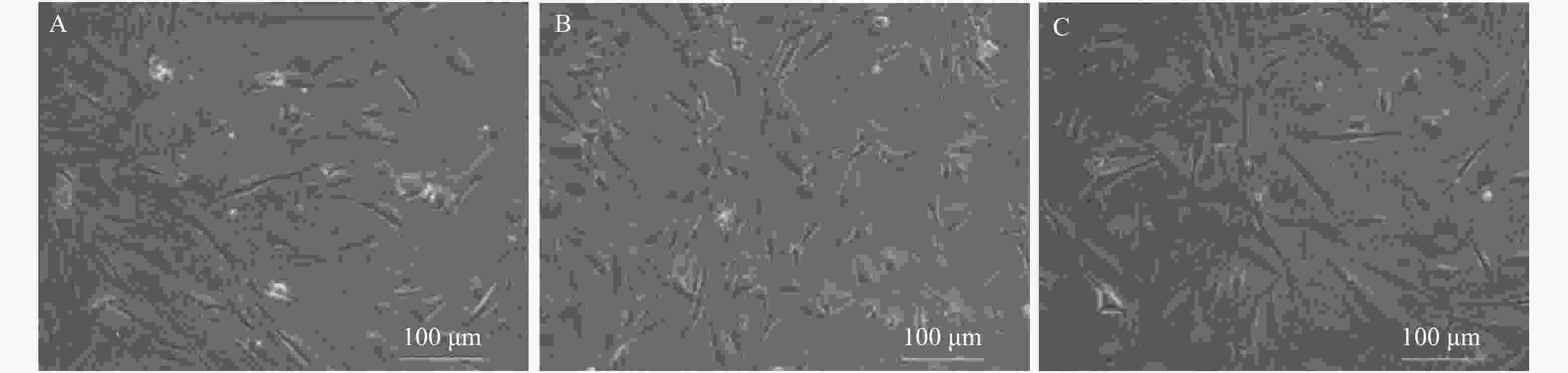
图 1 各组48h EnMSCs生长情况(×200)
A:对照组;B:2%PRP组;C:2%PRP + LY294002组
Figure 1. The 48 h EnMSCs proliferation phenomena for each group (×200)

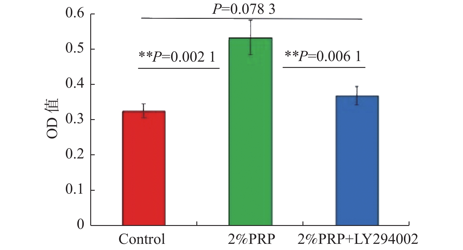
图 2 3组CCK-8增殖结果比较
与对照组比较,**P < 0.01。
Figure 2. The CCK-8 proliferation results comparison between the three groups
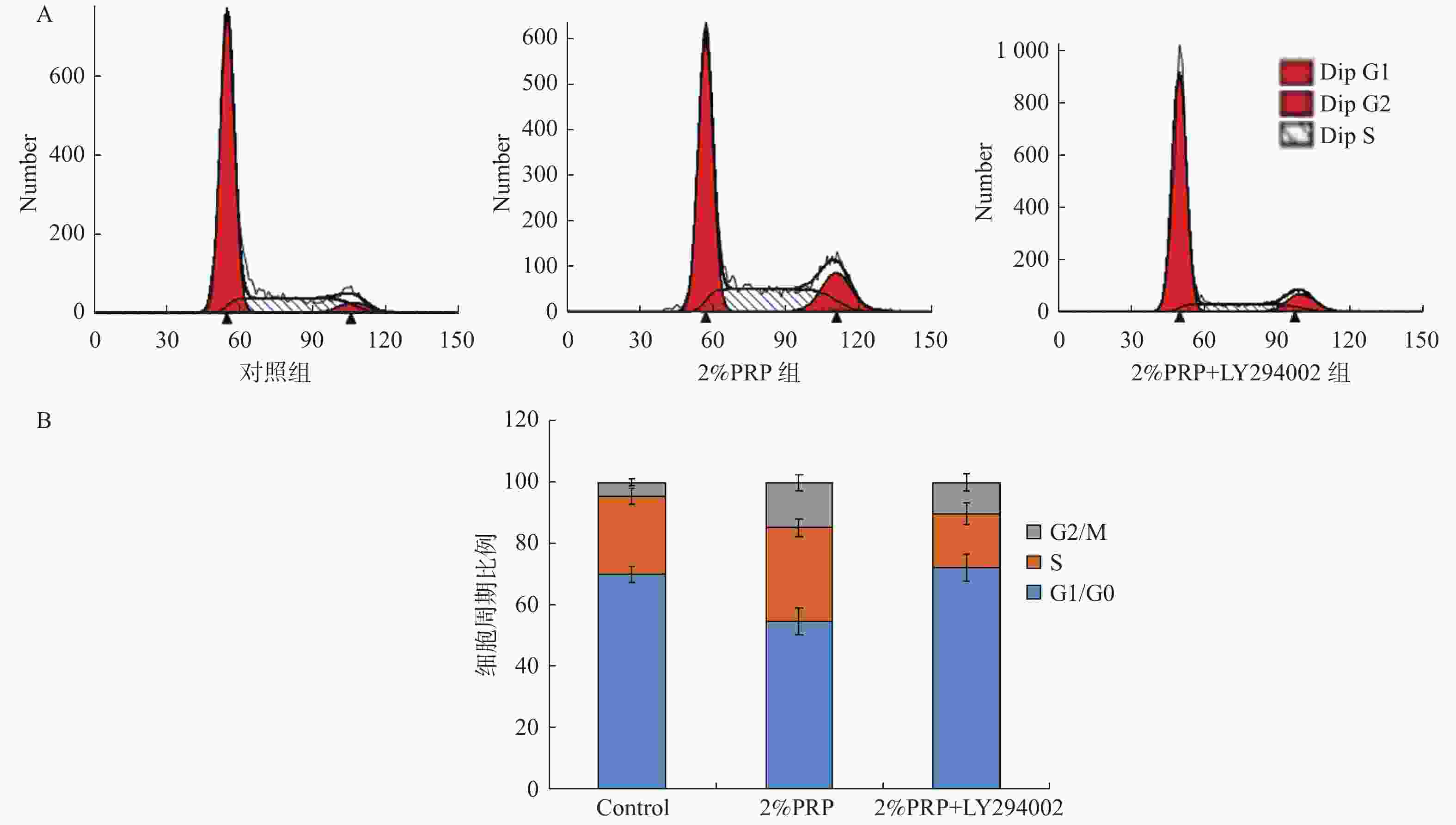
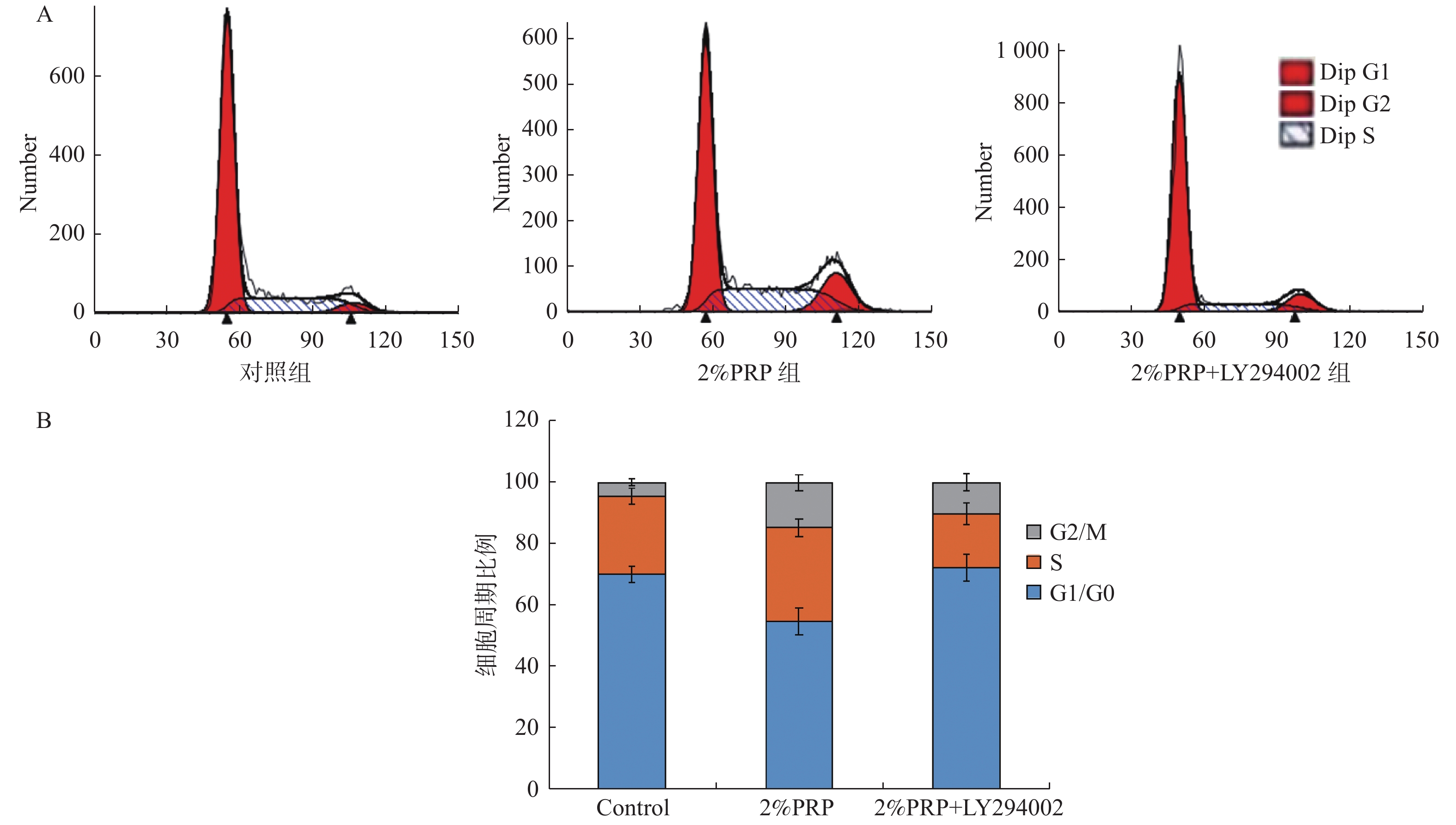
图 3 各组流式细胞周期及细胞周期比例图
A:各组流式细胞周期图;B:细胞周期比例图。
Figure 3. The flow cell cycle and cell cycle proportion gram in three groups
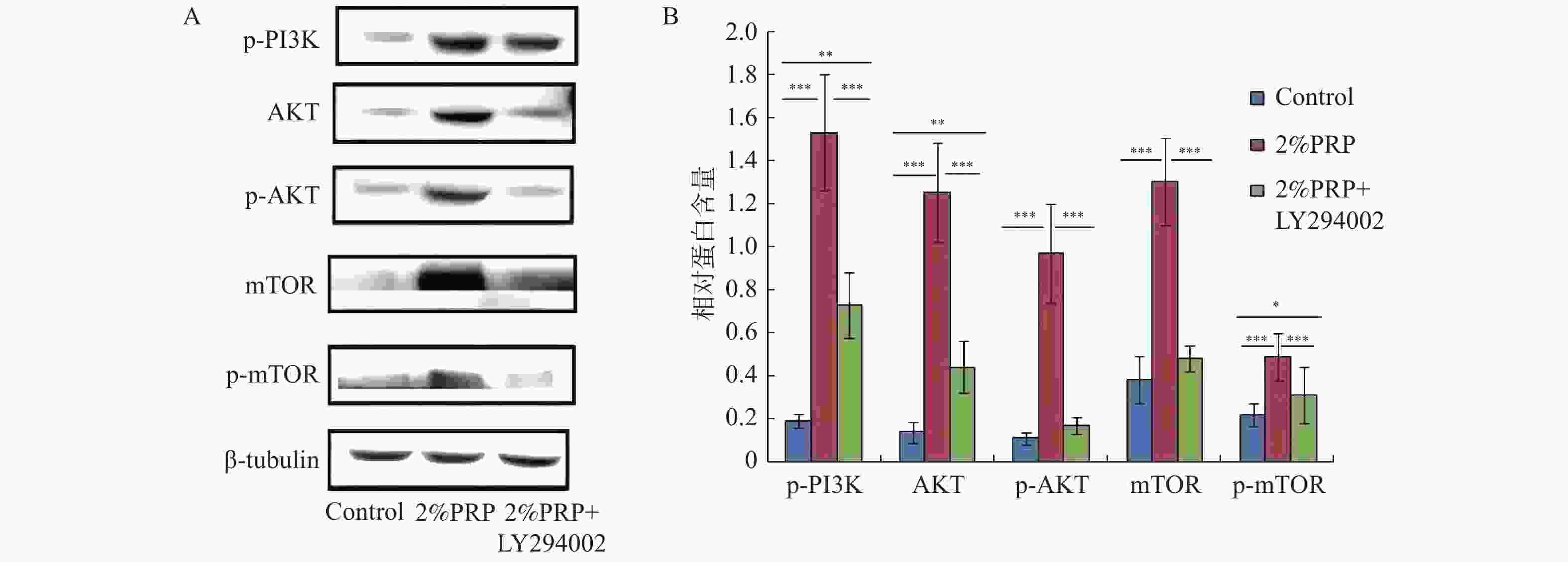
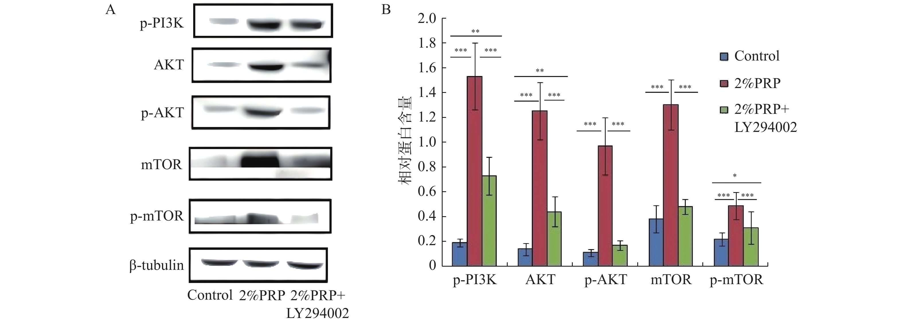
图 4 3组p-PI3K、AKT、p-AKT、mTOR、p-mTOR蛋白表达水平比较
A:Western blot结果;B:ELISA结果。
Figure 4. The p-PI3K, AKT, p-AKT, mTOR, and p-mTOR proteins demonstration levels comparison between the three groups
表 1 3组EnMSCs细胞周期比例(%)比较(n = 3)
Table 1. The EnMSCs cell cycle proportion comparison in the three groups (n = 3)
组别 G1/G0(%) S(%) G2/M(%) 对照组 70.04 ± 2.67 25.49 ± 2.68 4.47 ± 1.04 2%PRP组 54.63 ± 4.23**▲ 30.66 ± 2.85*▲ 14.71 ± 2.72**▲ 2%PRP+ LY294002组 72.16 ± 4.26 17.45 ± 3.49** 10.39 ± 2.84** 与对照组比较,*P < 0.05, **P < 0.01;与2%PRP+ LY294002组比较, ▲P < 0.01。  下载: 导出CSV
-
[1] Wang K,Jiang Z,Webster K A,et al. Enhanced cardioprotection by human endometrium mesenchymal stem cells driven by exosomal microRNA-21[J]. Stem Cells Transl Med,2017,6(1):209-222. doi: 10.5966/sctm.2015-0386 [2] Khadivi F,Koruji M,Akbari M,et al. Application of platelet-rich plasma (PRP) improves self-renewal of human spermatogonial stem cells in two-dimensional and three-dimensional culture systems[J]. Acta Histochem,2020,122(8):151627. doi: 10.1016/j.acthis.2020.151627 [3] Aghajanova L,Houshdaran S,Balayan S,et al. In vitro evidence that platelet-rich plasma stimulates cellular processes involved in endometrial regeneration[J]. J Assist Reprod Genet,2018,35(5):757-770. doi: 10.1007/s10815-018-1130-8 [4] Wang X,Liu L,Mou S,et al. Investigation of platelet-rich plasma in increasing proliferation and migration of endometrial mesenchymal stem cells and improving pregnancy outcome of patients with thin endometrium[J]. J Cell Biochem,2018,120(5):775-782. [5] Lai F,Kakudo N,Morimoto N,et al. Platelet-rich plasma enhances the proliferation of human adipose stem cells through multiple signaling pathways[J]. Stem Cell Res Ther,2018,9(1):107. doi: 10.1186/s13287-018-0851-z [6] Landesberg R,Roy M,Glickman R S. Quantification of growth factor levels using a simplified method of platelet-rich plasma gel preparation[J]. J Oral Maxillofac Surg,2000,58(3):297-301. doi: 10.1016/S0278-2391(00)90058-2 [7] Pötter N,Westbrock F,Grad S,et al. Evaluation of the influence of platelet-rich plasma (PRP),platelet lysate (PL) and mechanical loading on chondrogenesis in vitro[J]. Sci Rep,2021,11(1):20188. doi: 10.1038/s41598-021-99614-0 [8] Oh M,Kim S Y,Park S,et al. Phytochemicals in Chinese chive(allium tuberosum)induce the skeletal muscle cell proliferation via PI3K/Akt/mTOR and smad pathways in C2C12 cells[J]. International Journal of Molecular Sciences,2021,22(5):172-182. [9] Lee H J,Koh S H,Song K M,et al. The Akt/mTOR/p70S6K pathway is involved in the neuroprotective effect of erythropoietin on hypoxic/ischemic brain injury in a neonatal rat model[J]. Neonatology,2016,110(2):93-100. [10] Liu W,Chen B,Zheng Y,et al. Effect of platelet-rich plasma on implant bone defects in rabbits through the FAK/PI3K/AKT signaling pathway[J]. Open Life Sci,2019,14(1):311-317. doi: 10.1515/biol-2019-0034 [11] Sharara F I,Lelea L L,Rahman S,et al. A narrative review of platelet-rich plasma (PRP) in reproductive medicine[J]. J Assist Reprod Genet,2021,38(5):1003-1012. doi: 10.1007/s10815-021-02146-9 [12] Zuo W,Xie B,Li C,et al. The clinical applications of endometrial mesenchymal stem cells[J]. Biopreserv Biobank,2018,16(2):158-164. doi: 10.1089/bio.2017.0057 [13] Yang Y,Iwanaga K,Raso M G,et al. Phosphatidylinositol 3-kinase mediates bronchioalveolar stem cell expansion in mouse models of oncogenic K-ras-induced lung cancer[J]. PLoS One,2008,3(5):e2220. doi: 10.1371/journal.pone.0002220 [14] Sunayama J,Sato A,Matsuda K,et al. Dual blocking of mTor and PI3K elicits a prodifferentiation effect on glioblastoma stem-like cells[J]. Neuro Oncol,2010,12(12):1205-1219. doi: 10.1093/neuonc/noq103 [15] Burnouf T,Strunk D,Koh M B,et al. Human platelet lysate:Replacing fetal bovine serum as a gold standard for human cell propagation?[J]. Biomaterials,2016,76(1):371-387. [16] Chen Q J,Chen L,Wu S K,et al. rhPDGF-BB combined with ADSCs in the treatment of achilles tendinitis via miR-363/PI3 K/Akt pathway[J]. Mol Cell Biochem,2018,438(1-2):175-182. doi: 10.1007/s11010-017-3124-8 [17] Stiles C D,Capone G T,Scher C D,et al. Dual control of cell growth by somatomedins and platelet-derived growth factor[J]. Proc Natl Acad Sci USA,1979,76(3):1279-1283. doi: 10.1073/pnas.76.3.1279 [18] Kakudo N,Morimoto N,Kushida S,et al. Platelet-rich plasma releasate promotes angiogenesis in vitro and in vivo[J]. Med Mol Morphol,2014,47(2):83-89. doi: 10.1007/s00795-013-0045-9 [19] Zhang S,Li P,Yuan Z,et al. Effects of platelet-rich plasma on the activity of human menstrual blood-derived stromal cells in vitro[J]. Stem Cell Res Ther,2018,9(1):48. doi: 10.1186/s13287-018-0795-3 [20] 郑利强,江琼,伍亚民,等. APS通过PI3K/AKT信号通路促进神经干细胞增殖[J]. 基础医学与临床,2016,36(10):1359-1363. [21] Loibl M,Lang S,Hanke A,et al. Leukocyte-reduced platelet-rich plasma alters protein expression of adipose tissue-derived mesenchymal stem cells[J]. Plast Reconstr Surg,2016,138(2):397-408. doi: 10.1097/PRS.0000000000002388 [22] Xie X,Wang Y,Zhao C,et al. Comparative evaluation of MSCs from bone marrow and adipose tissue seeded in PRP-derived scaffold for cartilage regeneration[J]. Biomaterials,2012,33(29):7008-7018. doi: 10.1016/j.biomaterials.2012.06.058 -

 点击查看大图

计量
- 文章访问数: 4680
- HTML全文浏览量: 2484
- PDF下载量: 62
- 被引次数: 0
