

miR-145 Regulates the Expression of OCT4 in Human Endometrial Stromal Cells and Promotes the Development of Endometriosis
-
摘要:
目的 通过体外细胞试验探讨miR-145在子宫内膜异位症(Endometriosis ,EMs)中的调控机制。 方法 蛋白印迹检测OCT4等相关蛋白的表达水平,通过相关分析和双荧光素酶报告试验用于评估miR-145和OCT4之间的关联,CCK8试剂盒检测细胞活力,流式细胞术检测细胞凋亡,通过Transwell法检测hESCs的迁移。 结果 前期临床试验发现异位子宫内膜组织miR-145表达上调,OCT4表达下调。在细胞实验中,miR-145过表达可显著促进hESCs的增殖和迁移(P < 0.01),但抑制hESCs的凋亡(P < 0.05)。miR-145模拟物转染hESCs后,OCT4、Bax和MMP1等蛋白表达水平降低(P < 0.01),Bcl-2蛋白表达水平升高(P < 0.05)。敲除miR-145逆转了上述结果,并通过靶向OCT4显著抑制了hESCs的增殖(P < 0.05)。 结论 研究结果表明,miR-145表达增加通过抑制OCT4蛋白表达,可能通过促进子宫内膜基质细胞上皮细胞间质转化(Epithelial-mesenchymal Transition,EMT)过程,从而在促进EMs的发展中起到一定的作用。 Abstract:Objective To further explore the regulatory mechanism of miR-145 in Endometriosis (EMs) through cell experiments. Methods miR-145 or octamer-binding transcription factor 4 (OCT4)gene and protein expression levels in cells were examined using reverse transcription-quantitative PCR and western blotting, respectively. Correlation analyses and dualluciferase reporter assays were performed to assess the association between miR-145 and OCT4. A Cell Counting Kit-8 assay was performed to test cell viability, while cell apoptosis was measured using flow cytometry. Moreover, the migration of hESCs was measured via Transwell assays. Results In cell assays, overexpression of miR-145 signifcantly promoted proliferation and migration (P < 0.01), but inhibited the apoptosis of hESCs (P < 0.05). Furthermore, the transfection of hESCs with miR-145 mimics decreased the protein expression levels of OCT4, Bax and MMP1 (P < 0.01), as well as increased the protein expression of Bcl-2. However, knockdown of miR-145 reversed these results and significantly inhibited the proliferation of hESCs by targeting OCT4 (P < 0.05). Conclusion The present results suggested that knockdown of miR-145 signifcantly suppressed the development of EMs by targeting OCT4, which may serve as a potential target for the treatment of EMs. -
Key words:
- Endometriosis /
- miRNA-145 /
- OCT4 /
- EMT /
- Human endometrial stromal cells
-

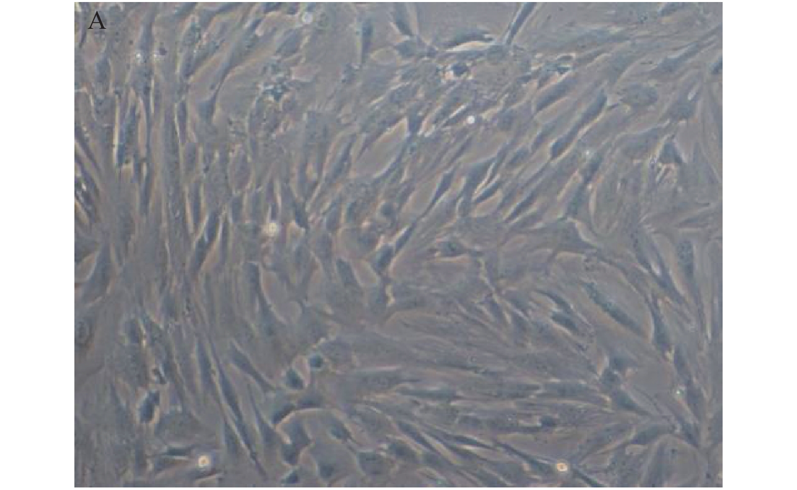
图 1 人体子宫内膜异位病灶组织原代培养子宫内膜基质细胞显微镜下可以看到成功分离的 hESCs (×100)
Figure 1. Primary culture of hESCs from human endometriosis (×100)

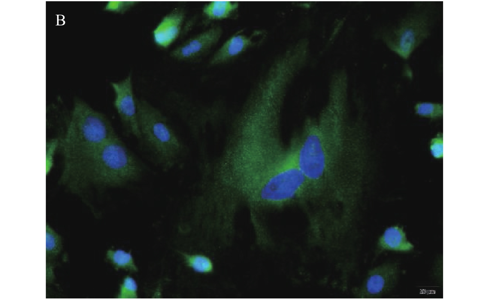
图 2 波形蛋白染色进一步证实了分离出子宫内膜基质细胞在用miR-145抑制剂、NC或miR-145 mimic转染hESCs 48小时后,绿色荧光表示波形蛋白,蓝色荧光显示 DAPI (×40)
Figure 2. Vimentin staining further confirmed the isolation of endometrial stromal cells

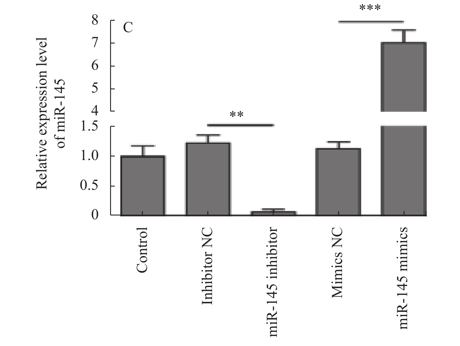
图 3 用 miR-145抑制剂、NC或miR-145模拟物转染hESCs 48 h使用定量逆转录PCR计算miR-145相对表达量
**P < 0.01,***P < 0.001
Figure 3. Transfection efficiency was evaluated using reverse transcription-quantitative PCR

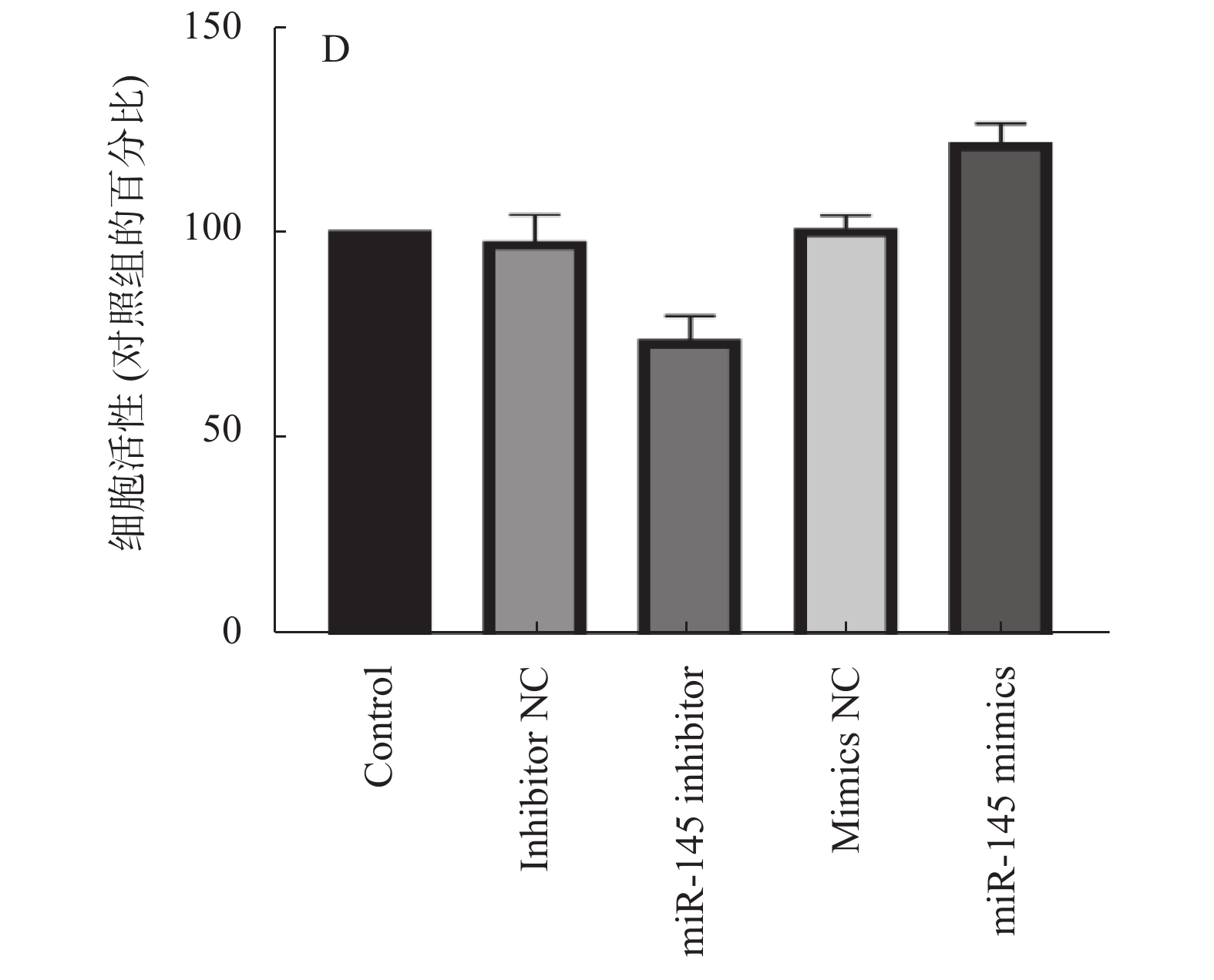
图 4 CCK-8检测hESCs的增殖
Figure 4. Proliferation of hESCs was detected with a Cell Counting Kit-8 assay

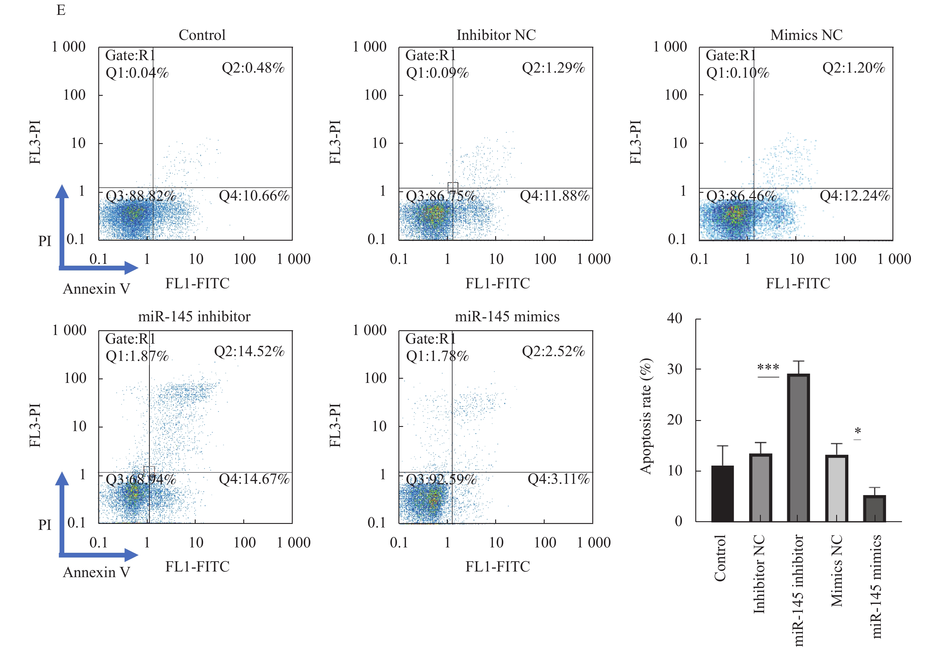
图 5 Annexin V和PI双染色后,流式细胞仪检测hESCs的凋亡率
*P < 0.05,***P < 0.001。
Figure 5. Apoptotic rate of hESCs was investigated via FACS after double staining with Annexin V and PI

图 6 用 Transwell 法测定 hESCs 的迁移
**P < 0.01。
Figure 6. Migration of hESCs was measured using the Transwell assay

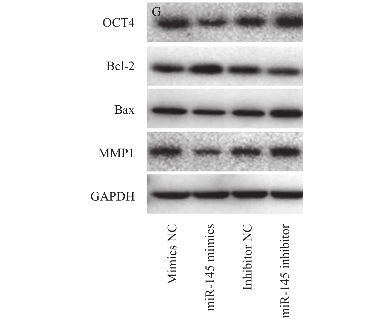
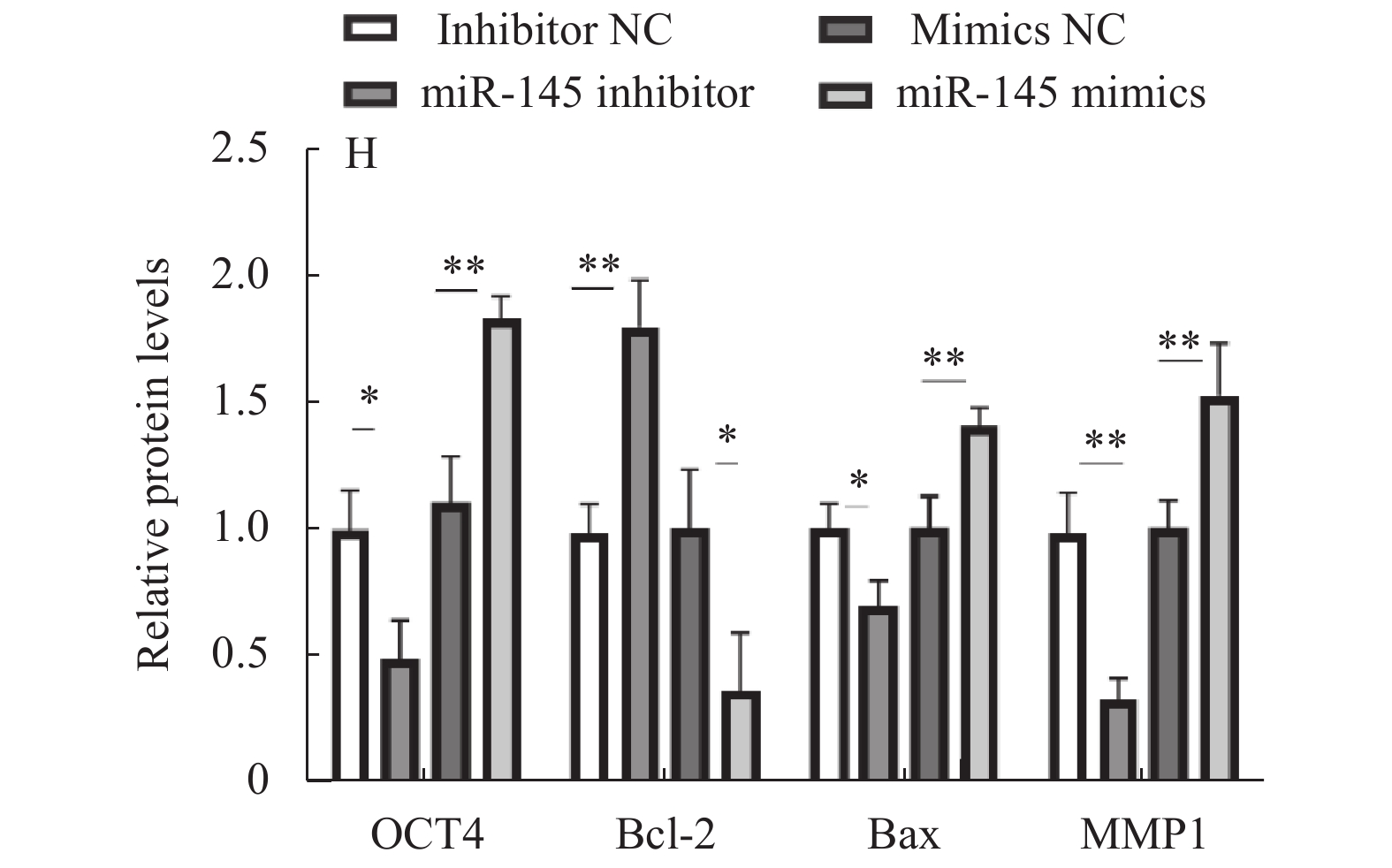
图 7 蛋白印迹检测hESCs中的OCT4、Bax、Bcl-2和MMP1表达
Figure 7. Expression levels of OCT4, Bax, Bcl 2 and MMP1 in hESCs were detected via western blotting

图 8 以 GAPDH 表达量为内参半定量 OCT4、Bax、Bcl- 2和 MMP1 的表达
*P < 0.05,**P < 0.01。
Figure 8. Relative expression was semi-quantified by normalizing to GAPDH expression

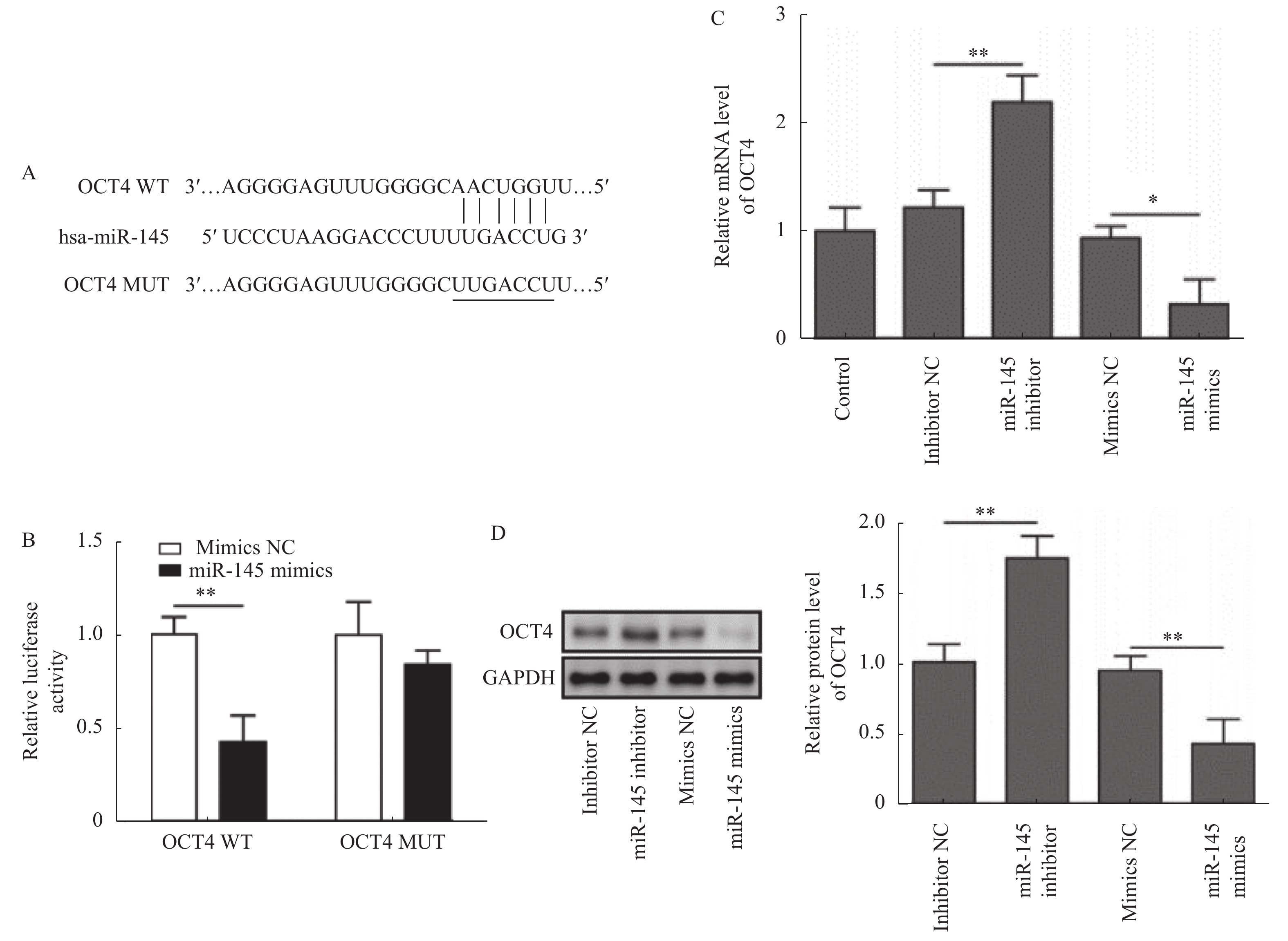
图 9 OCT4 靶向作用于 miR-145
A:OCT4 基因结构分析显示 miR-145 在其 3'-UTR 存在潜在靶点。B:荧光素酶在用 WT 或 MUT OCT4 3'-UTR 质粒和 miR-145 模拟物共转染 hESCs 后,使用双荧光素酶报告基因检测活性(hESCs 未处理或用 NC、miR - 145 模拟物或抑制剂处理后)。C:通过反转录定量 PCR 检测 OCT4 在 hESCs 中的表达。D: 蛋 白印迹检测 OCT4 蛋白在 hESCs 中的表达。**P < 0.01,*P < 0.05。
Figure 9. OCT4 is a direct target of miR-145

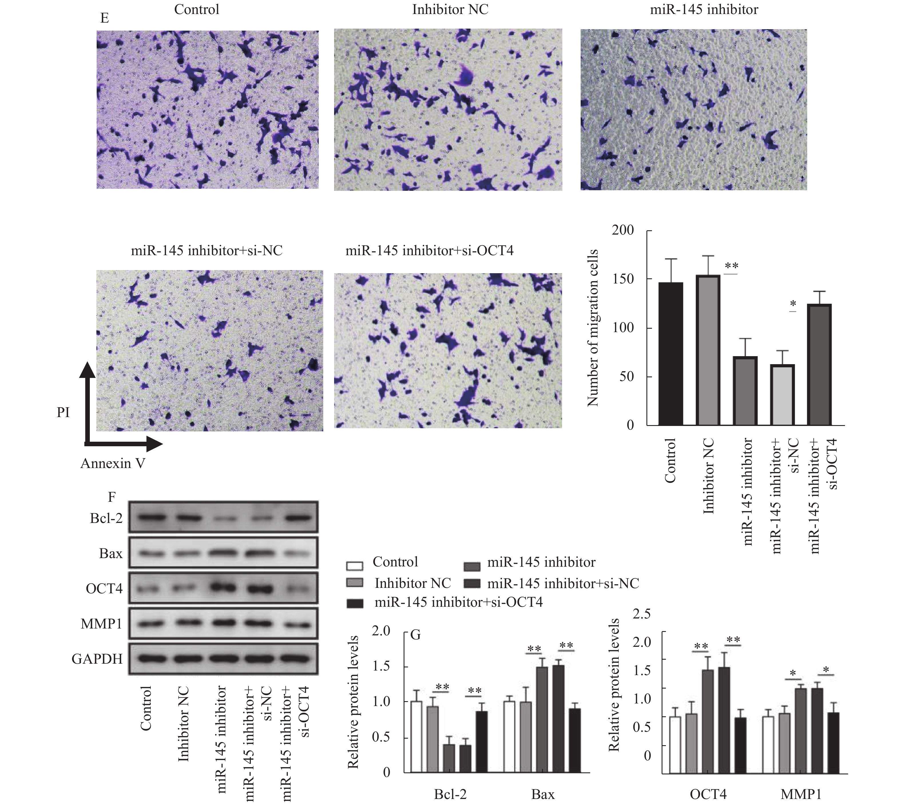
图 10 miR-145 基因敲除可通过靶向 OCT4 抑制 EMs 的发展
A:通过反转录定量 PCR 检测 OCT4 mRNA 表达。B:蛋白印迹检测 OCT4 蛋白在 hESCs 中的表 达。C:扩散在对照的 hESCs 中,使用 CCK - 8 检测抑制剂 NC、miR - 145 抑制剂、miR - 145 抑制 剂+ si - NC 或 miR - 145 抑制剂+ si - OCT4 组。D:Annexin V 和 PI 染色后用流式细胞仪检测 hESCs 的凋亡率。E:使用 Transwell 分析检测 hESCs 的迁移。F:蛋白印迹检测 OCT4、Bax、Bcl-2 和 MMP1 在 hESCs 中的表达水平。G:以 GAPDH 表达为内参 Bcl-2,Bax,OCT4,MMP1 相对表达 的半定量;**P < 0.01,*P < 0.05。
Figure 10. miR-145 knockdown inhibits the development of EMs in vitro by targeting OCT4. hESCs were transfected with si-OCT4
-
[1] Zhou C F,Liu M J,Wang W,et al. miR-205-5p inhibits human endometriosis progression by targeting ANGPT2 in endometrial stromal cells[J]. Stem Cell Research & Therapy,2019,10(1):287. [2] Cipollini M,Luisi S,Piomboni P,et al. Functional polymorphism within NUP210 encoding for nucleoporin GP210 is associated with the risk of endometriosis[J]. Fertility and Sterility,2019,112(2):343. doi: 10.1016/j.fertnstert.2019.04.011 [3] Cheng F,Lu L,Wang H,Cheng H,et al. Expression and signifcance of miR-126 and miR-145 in infertility due to endometriosis[J]. J Coll Physicians Surg Pak,2019,29:585-587. [4] Sanchez A M,Pagliardini L,Cermisoni g C,et al. Does endometriosis influence the embryo quality and/or development? Insights from a large retrospective matched cohort study[J]. Diagnostics (Basel),2020,10:83. doi: 10.3390/diagnostics10020083 [5] Wang D,Luo Y,Wang g,et al. Circular RNA expression profles and bioinformatics analysis in ovarian endometriosis[J]. Mol genet genomic Med,2019,7:e00756. [6] Zhang Y, Yan J and Pan X: miR-141-3p affects apoptosis and migration of endometrial stromal cells by targeting KLF-12[J]. Pflugers Arch, 2019, 471: 1055-1063, [7] Tang W,Chen O,Yao F,et al. miR455 targets FABP4 to protect human endometrial stromal cells from cytotoxicity induced by hydrogen peroxide[J]. Mol Med Rep,2019,20:4781-4790. [8] Marí-Alexandre J,Carcelén A P,Agababyan C,et al. Interplay between microRNAs and oxidative stress in ovarian conditions with a focus on ovarian cancer and endometriosis[J]. Int J Mol Sci,2019,20:5322. doi: 10.3390/ijms20215322 [9] Petracco R,Dias A C O,Taylor H,et al. Evaluation of miR-135a/b expression in endo-metriosis lesions[J]. Biomed Rep,2019,11:181-187. [10] Nagai T,Ishida C,Nakamura T,et al. Focal adhesion kinase-mediated sequences,including cell adhesion,inflammatory response,and fbrosis,as a therapeutic target in Endometriosis[J]. Reprod Sci,2020,27:1400-1410. doi: 10.1007/s43032-019-00044-1 [11] Saare M,Rekker K,Laisk-Podar T,et al. Challenges in endometriosis miRNA studies-from tissue heterogeneity to disease specifc miRNAs[J]. Biochim Biophys Acta Mol Basis Dis,2017,1863:2282-2292. doi: 10.1016/j.bbadis.2017.06.018 [12] Dai Y,Lin X,Xu W,et al. MiR-210-3p protects endometriotic cells from oxidative stress-induced cell cycle arrest by targeting BARD1[J]. Cell Death Dis,2019,10:144. doi: 10.1038/s41419-019-1395-6 [13] Moga M A,Bălan A,Dimienescu O G,et al. Circulating miRNAs as biomarkers for endo-metriosis and endometriosis-related ovarian cancer-an overview[J]. J Clin Med,2019,8:735. doi: 10.3390/jcm8050735 [14] Zhang M,Wang S,Tang L,et al. Downregulated circular RNA hsa_circ_0067301 regulates epithelial-mesenchymal transition in endometriosis via the miR-141/Notch signaling pathway[J]. Biochem Biophys Res Commun,2019,514:71-77. doi: 10.1016/j.bbrc.2019.04.109 [15] Liu Y,Feng W,Liu W,et al. Circulating lncRNA ABHD11-AS1 serves as a biomarker for early pancreatic cancer diagnosis[J]. J Cancer,2019,10:3746-3756. doi: 10.7150/jca.32052 [16] Bashti O, Noruzinia M, garshasbi M, et al. miR-31and miR-145 as potential non-invasive regulatory biomarkers in patients with endometriosis[j]. Cell J, 2018, 20: 293. [17] Cosar E,Mamillapalli R,Ersoy G S,et al. Serum microRNAs as diagnostic markers of endometriosis:A comprehensive array-based analysis[J]. FertilSteril,2016,106:402-;409. [18] Michael A K,grand R S,Isbel L,et al. Mechanisms of OCT4-SOX2 motif readout on nucleosomes[J]. Science,2020,368:1460-1465. doi: 10.1126/science.abb0074 [19] Harpelunde Poulsen K,Nielsen J E,grønkær Toft B,et al. Influence of Nodal signalling on pluripotency factor expression,tumour cell proliferation and cisplatin-sensitivity in testicular germ cell tumours[J]. BMC Cancer,2020,20:349. doi: 10.1186/s12885-020-06820-6 [20] Sadeghi F,Peymaeei F,Falahati M,et al. The effect of Candida cell wall beta-glucan on treatment-resistant LL/2 cancer cell line:In vitro evaluation[J]. Mol Biol Rep,2020,47:3653-3661. doi: 10.1007/s11033-020-05459-7 [21] Chang Y C,Yang Y F,Chiou J,et al. Nonenzymatic function of Aldolase Adownregulates miR-145 to promote the Oct4/DUSP4/TRAF4 axis and the acquisition of lung cancer stemness[J]. Cell Death Dis,2020,11:195. doi: 10.1038/s41419-020-2387-2 [22] Cao H,Wei Y X,Zhou Q,et al. Inhibitory effect of curcumin in human endometriosis endometrial cells via downregulation of vascular endothelial growth factor[J]. Mol Med Rep,2017,16:5611-5617. doi: 10.3892/mmr.2017.7250 [23] Vanhie A,O D,Peterse D,Beckers A,et al. Plasma miRNAs as biomarkers for endometriosis[J]. Hum Reprod,2019,34:1650-1660. doi: 10.1093/humrep/dez116 [24] Wen J,Wang H,Dong T,et al. STAT3-induced upregulation of lncRNA ABHD11-AS1 promotes tumour progression in papillary thyroid carcinoma by regulating miR-1301-3p/STAT3 axis and PI3K/AKT signalling pathway[J]. Cell Prolif,2019,52:e12569. doi: 10.1111/cpr.12569 [25] He D,Yue Z,Liu L,et al. Long noncoding RNA ABHD11-AS1 promote cells proliferation and invasion of colorectal cancer via regulating the miR-1254-WNT11 pathway[J]. J Cell Physiol,2019,234:12070-12079. doi: 10.1002/jcp.27877 [26] Coutinho L M,Ferreira M C,Rocha A L L,et al. New biomarkers in endometriosis[J]. Adv Clin Chem,2019,89:59-77. [27] Zhu H,Cao X X,Liu J,et al. MicroRNA-488 inhibits endometrial glandular epithelial cell proliferation,migration,and invasion in endometriosis mice via Wnt by inhibiting FZD7[J]. J Cell Mol Med,2019,23:2419-2430. doi: 10.1111/jcmm.14078 [28] Bjorkman S and Taylor H S. MicroRNAs in endometriosis:Biological function and emerging biomarker candidatesdagger[J]. Biol Reprod,2019,100:1135-1146. [29] Zhang H,Li g,Sheng X,et al. Upregulation of miR33b promotes endometriosis via inhibition of Wnt/β-catenin signaling and ZEB1 expression[J]. Mol Med Rep,2019,19:2144-2152. [30] Li D,Jiang W,Jiang Y,et al. Preliminary functional inquiryof lncRNA ENST00000433673 in embryo implantation using bioinformatics analysis[J]. Syst Biol Reprod Med,2019,65:164-173. doi: 10.1080/19396368.2018.1563844 [31] Hsu C Y,Hsieh T H,Er T K,et al. MiR-381 regulates cell motility,growth and colony formationthrough PIK3CA in endometriosis-associated clear cell and endometrioid ovarian cancer[J]. Oncol Rep,2018,40:3734-3742. [32] Mashayekhi P,Noruzinia M,Zeinali S,et al. Endometriotic mesenchymal stem cells epigenetic pathogenesis:Deregulation of miR-200b,miR-145,and let7b in a functional imbalanced epigenetic disease[J]. Cell J,2019,21:179-185. [33] GE R and GAO G. Anti-antioxidant impacts of circZNF609 silence in HaCaT cells through regulating miR-145[J]. Artif Cells Nanomed Biotechnol,2020,48:384-392. doi: 10.1080/21691401.2019.1709863 [34] Adammek M,greve B,Kässens N,Schneider C,et al. MicroRNA miR-145 inhibits proliferation,invasiveness,and stem cell phenotype of an in vitro endometriosis model by targeting multiple cytoskeletal elements and pluripotency factors[J]. Fertil Steril,2013,99:1346-1355.e5. doi: 10.1016/j.fertnstert.2012.11.055 [35] Shan W,Chen W,Zhao X,et al. Long noncoding RNA TUg1 contributes to cerebral ischaemia/reperfusion injury by sponging mir-145 to up-regulate AQP4 expression[J]. J Cell Mol Med,2020,24:250-259. doi: 10.1111/jcmm.14712 [36] Zhuang X,Tong H,Ding Y,et al. Long noncoding RNA ABHD11-AS1 functions as a competing endogenous RNA to regulate papillary thyroid cancer progression by miR-199a-5p/SLC1A5 axis[J]. Cell Death Dis,2019,10:620. doi: 10.1038/s41419-019-1850-4 [37] Liu Y,Zhang Z,Yang F,et al. The role of endometrial stem cells in the pathogenesis of endometriosis and their application to its early diagnosis[J]. Biology of Reproduction,2020,102(6):1153-1159. [38] Tiwari D,Ray Das C,Sultana R,et al. Impact of modulation of telomerase and cancer stem-cell marker OCT4 axis incervical cancer pathogenesis with underlying HPV16 infection[J]. J Cell Biochem,2019,121:2782-2791. [39] Proestling K,Birner P,Balendran S,et al. Enhanced expression of the stemness-related factors OCT4,SOX15 and TWIST1 in ectopic endometrium of endometriosis patients[J]. Reprod Biol Endocrinol,2016,14:81. doi: 10.1186/s12958-016-0215-4 [40] Chang J H,Au H K,Lee W C,et al. Expression of the pluripotent transcription factor OCT4 promotes cell migration in endometriosis[J]. Fertil Steril,2013,99:1332-1339.e5. doi: 10.1016/j.fertnstert.2012.11.033 [41] Au H K,Chang J H,Wu Y C,et al. TgF-βI regulates cell migration through pluripotent transcription factor OCT4 in endometriosis[J]. PLoS One,2015,10:e0145256. doi: 10.1371/journal.pone.0145256 [42] Kang K,Niu B,Wu C,et al. The construction and application of lentiviral overexpression vector of goat miR-204 in testis[J]. Res Vet Sci,2020,130:52-58. doi: 10.1016/j.rvsc.2020.02.014 [43] Channakkar A S,Singh T,Pattnaik B,et al. MiRNA-137-mediated modulation of mitochondrial dynamics regulates human neural stem cell fate[J]. Stem Cells,2020,38:683-697. doi: 10.1002/stem.3155 [44] Shariati F,Favaedi R,Ramazanali F,et al. Increased expression of stemness genes REX-1,OCT-4,NANOG,and SOX-2 in women with ovarian endometriosis versus normal endometrium:A case-control study[J]. International Journal of Reproductive Biomedicine,2018,16(12):783-791. [45] Jiang Z,Zhang C,Liu X,et al. Dexamethasone inhibits stemness maintenance and enhances chemosensitivity of hepatocellular carcinoma stem cells by inducing deSUMOylation of HIF-1α and Oct4[J]. Int J Oncol,2020,57(3):780-790. doi: 10.3892/ijo.2020.5097 -

 下载:
下载:

 点击查看大图
计量
- 文章访问数: 3827
- HTML全文浏览量: 2275
- PDF下载量: 28
- 被引次数: 0
