Measurement of Articular Cartilage Thickness of Talus in Normal Adults by MRI
-
摘要:
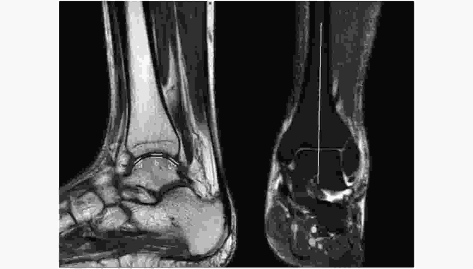
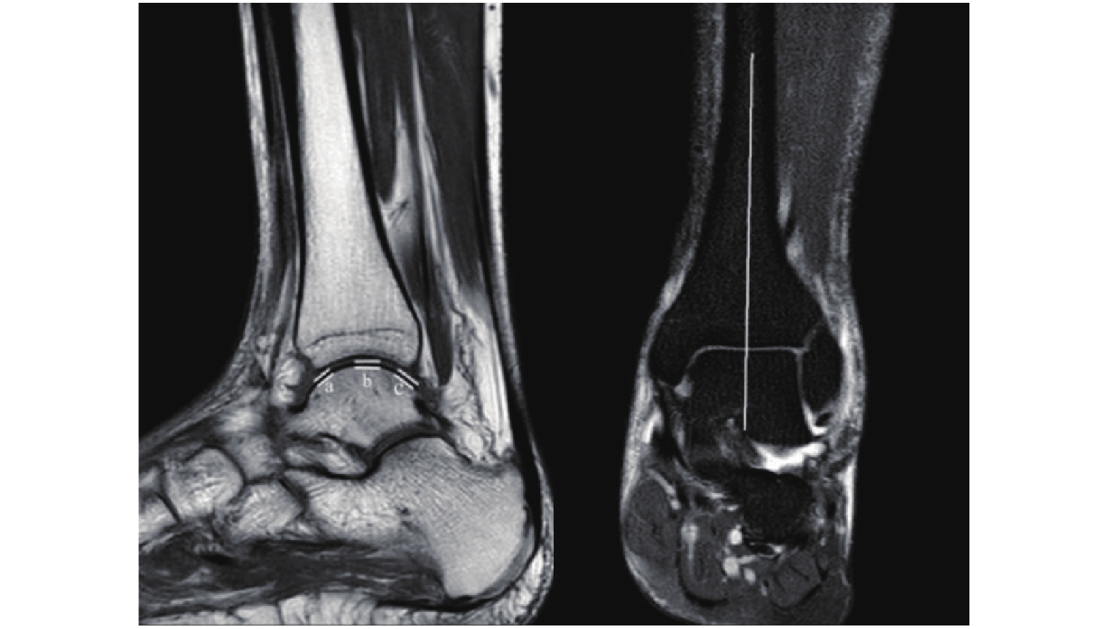
目的 通过MRI测量正常成人距骨表面软骨厚度,得出正常成人距骨软骨厚度的参数以及与性别、年龄、体重、身高的关系。 方法 选择踝关节无关节炎、无外伤史的成人100名,男、女各50名,18~60岁,行MRI检查,通过MRI测量前、中、后距骨关节面软骨厚度,测量结果采用SPSS25.0统计学软件包进行统计学分析,P < 0.05表示差异有统计学意义。 结果 男性前、中、后距骨关节面软骨厚度分别为(1.00±0.18)mm、(1.40±0.21)mm、(0.87±0.18)mm;女性前、中、后距骨关节面软骨厚度分别为(0.96±0.19)mm、(1.29±0.20)mm、(0.86±0.15)mm;男性和女性距骨前、后关节软骨面厚度,差异无统计学意义(P > 0.05),距骨中关节面软骨厚度,差异有统计学意义(P < 0.05),男性与女性年龄,差异无统计学意义(P > 0.05)。 结论 男性距骨中关节面平均软骨厚度大于女性;前后关节面软骨厚度无差异;男、女性身高、体重、年龄与前、中、后关节面软骨厚度均无相关性;在距骨软骨损伤时对需要修复的软骨量提供依据,在距骨解剖假体设计时加入相应软骨厚度。 Abstract:Objective To measure the thickness of cartilage on the surface of the talus of normal adults by MRI and to obtain the parameters of the thickness of the cartilage of the talus of normal adults and the relationship with gender, age, weight and height. Methods 100 adults, 50 males and 50 females, aged 18 ~ 60, without arthritis and trauma history of ankle joint in the outpatient department were selected for MRI examination.The thickness of articular cartilage of anterior, middle and posterior talus was measured by MRI. The measurement results were measured by SPSS 25. 0 statistical software package for statistical analysis. Results The cartilage thicknesses of the anterior, middle, and posterior talus articular surface in men were (1.00±0.18) mm, (1.40±0.21) mm, and (0.87±0.18) mm respectively; the thicknesses of cartilage in the anterior, middle, and posterior talar articular surfaces of women were (0.96±0.18) mm, 0.19) mm, (1.29 ± 0.20) mm, (0.86 ± 0.15) mm respectively; male and female anterior and posterior articular cartilage thicknesses of the talus were not statistically different (P > 0.05), there was significant difference in the thickness of articular cartilage in talus (P < 0.05), and there was no statistically significant difference in age between men and women (P > 0.05). Conclusion The average cartilage thickness of the articular surface of the talus in men is greater than that in women and there is no difference in the thickness of cartilage on the front and rear articular surfaces; There is no correlation between the height, weight, and age of men’s and women’s articular cartilage thickness. The amount of cartilage provides the basis, and the corresponding cartilage thickness is added to the design of the talar anatomical prosthesis. -
Key words:
- MRI /
- Talus /
- Cartilage thickness /
- Method of measurement
-
表 1 男、女性各指标比较
Table 1. Comparison of male and female indicators
指标 男性 女性 t P a(mm) 1.00 ± 0.18 0.96 ± 0.19 1.342 0.183 b(mm) 1.40 ± 0.21 1.29 ± 0.20 2.715 0.008* c(mm) 0.87 ± 0.18 0.86 ± 0.15 0.089 0.930 年龄(岁) 37.00 ± 11.96 41.42 ± 14.10 −1.680 0.096 身高(m) 1.70 ± 0.06 1.64 ± 0.06 5.194 < 0.001* 体重(Kg) 67.61 ± 6.29 58.04 ± 4.47 8.707 < 0.001* a:前关节面软骨厚度;b:中关节面软骨厚度;c:后关节面软骨厚度。*P < 0.05。 表 2 身高与各指标相关性分析
Table 2. Correlation analysis between height and various indexes
指标 r P a(mm) 0.11 0.297 b(mm) 0.05 0.600 c(mm) −0.03 0.764 a:前关节面软骨厚度;b:中关节面软骨厚度;c:后关节面软骨厚度。 表 3 体重与各指标相关性分析
Table 3. Correlation analysis between body weight and various indexes
指标 r P a(mm) 0.08 0.429 b(mm) 0.17 0.099 c(mm) 0.06 0.588 a:前关节面软骨厚度;b:中关节面软骨厚度;c:后关节面软骨厚度。 表 4 年龄与各指标相关性分析
Table 4. Correlation analysis between age and various indicators
指标 r P a(mm) −0.14 0.182 b(mm) −0.09 0.389 c(mm) −0.15 0.131 a:前关节面软骨厚度;b:中关节面软骨厚度;c:后关节面软骨厚度。 -
[1] Steppacher S D,Hanke M S,Zurmühle C A,et al. Ultrasonic cartilage thickness measurement is accurate,reproducible,and reliable-validation study using contrast-enhanced micro-CT[J]. J Orthop Surg Res,2019,14(1):67. doi: 10.1186/s13018-019-1099-8 [2] Wang N,Badar F,Xia Y. Experimental Influences in the accurate measurement of cartilage thickness in MRI[J]. Cartilage,2019,10(3):278-287. doi: 10.1177/1947603517749917 [3] Song K,Pietrosimone B G,Nissman D B,et al. Ultrasonographic measures of talar cartilage thickness associate with magnetic resonance-based measures of talar cartilage volume[J]. Ultrasound Med Biol,2020,46(3):575-581. [4] 俞佳凤,孙岩,张静,等. 喙突下撞击综合征的磁共振成像测量研究[J]. 实用放射学杂志,2020,36(8):1281-1285. doi: 10.3969/j.issn.1002-1671.2020.08.026 [5] Maier J,Black M,Bonaretti S,et al. Comparison of different approaches for measuring tibial cartilage thickness[J]. J Integr Bioinform,2017,14(2):20170015. [6] Giannicola G,Spinello P,Scacchi M,et al. Cartilage thickness of distal humerus and its relationships with bone dimensions:Magnetic resonance imaging bilateral study in healthy elbows[J]. J Shoulder Elbow Surg,2017,26(5):e128-e136. doi: 10.1016/j.jse.2016.10.012 [7] Giannicola G,Scacchi M,Sedati P,et al. Anatomical variations of the trochlear notch angle:MRI analysis of 78 elbows[J]. Musculoskelet Surg,2016,100(Suppl 1):89-95. [8] 吕嘉玲,林伟强,张隐笛,等. 基质诱导的自体软骨细胞移植术的磁共振成像评估及其与软骨下骨的相关性[J]. 实用放射学杂志,2020,36(5):776-779. doi: 10.3969/j.issn.1002-1671.2020.05.022 [9] Giannicola G,Sedati P,Polimanti D,et al. Contribution of cartilage to size and shape of radial head circumference:Magnetic resonance imaging analysis of 78 elbows[J]. J Shoulder Elbow Surg,2016,25(1):120-126. doi: 10.1016/j.jse.2015.07.003 [10] 袁文昭,邓德茂,段高雄,等. MRI量化评分在腕关节类风湿关节炎治疗中的应用价值[J]. 实用放射学杂志,2018,34(9):1410-1413,1457. doi: 10.3969/j.issn.1002-1671.2018.09.025 [11] Cheng Y,Guo C,Wang Y,et al. Accuracy limits for the thickness measurement of the hip joint cartilage in 3-D MR images:Simulation and validation[J]. IEEE Trans Biomed Eng,2013,60(2):517-533. doi: 10.1109/TBME.2012.2230002 [12] Schmitz R J,Wang H M,Polprasert D R,et al. Evaluation of knee cartilage thickness:A comparison between ultrasound and magnetic resonance imaging methods[J]. Knee.,2017,24(2):217-223. doi: 10.1016/j.knee.2016.10.004 [13] 刘三彪,张文涛,唐陆波,等. 距骨骨软骨损伤修复材料研究进展[J]. 足踝外科电子杂志,2021,8(2):56-62. [14] 陈言智,张洪涛,程宇,等. 距骨骨软骨损伤的诊疗现状[J]. 足踝外科电子杂志,2021,8(2):52-55,62. -


 下载:
下载:


