Value of Different Sections of Echocardiography in the Diagnosis of Fetal Congenital Vascular Ring
-
摘要:
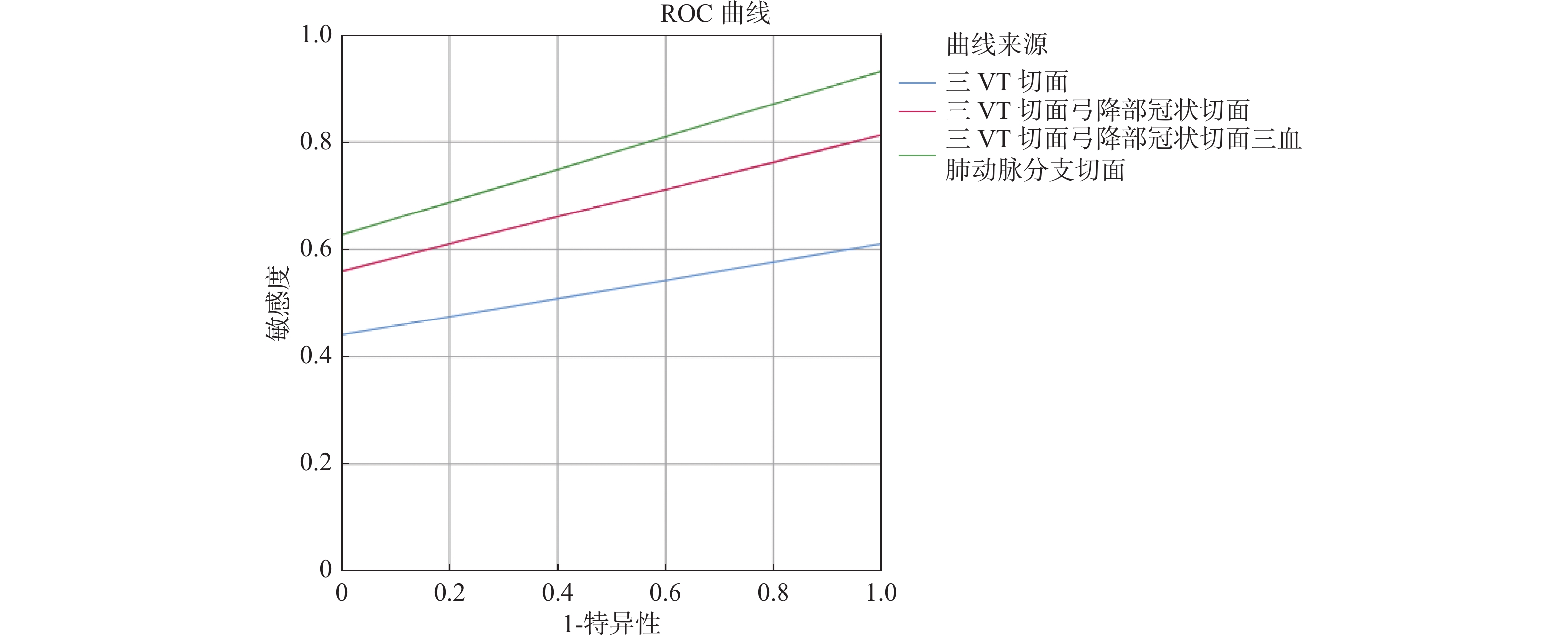
目的 分析超声心动图不同切面诊断胎儿先天性血管环(CVR)的价值。 方法 回顾性分析2018年1月至2021年1月于云南大学附属医院产前诊断的60例CVR超声心动图资料和随访结果,重点观察三血管气管切面(3VT)、弓降部冠状切面、三血管肺动脉分支切面,总结CVR在3VT、弓降部冠状切面、三血管肺动脉分支切面的声像图特点。 对比CVR在3VT、3VT +弓降部冠状切面、3VT +弓降部冠状切面+三血管肺动脉分支切面诊断情况。 结果 60例超声心动图诊断的CVR中,经过引产后病理解剖、产后超声心动图、CT或MRI随访结果证实:共59例确诊为CVR(准确率98.3%),1例误诊(误诊率1.7%)。 3VT +弓降部冠状切面+三血管肺动脉分支切面诊断率高于3VT及3VT +弓降部冠状切面(P < 0.05),3VT +弓降部冠状切面诊断率高于3VT( P < 0.05)。 结论 3VT +弓降部冠状切面+三血管肺动脉分支切面是诊断胎儿CVR的重要切面,诊断符合率高,具有重要的临床应用价值。 -
关键词:
- 超声心动图 /
- 胎儿先天性血管环 /
- 三血管气管切面 /
- 弓降部冠状切面 /
- 三血管肺动脉分支切面
Abstract:Objective To analyze the diagnostic value of different sections of echocardiography in fetal congenital vascular ring (CVR). Methods The echocardiography data and follow-up results of 60 cases of CVR diagnosed in Yunnan University Hospital from January 2018 to January 2021 were retrospectively analyzed. The three vessel trachea section (3VT), the descending arch coronal section and the three vessel pulmonary artery branch section were observed. The ultrasonographic characteristics of CVR in 3VT, coronal section of descending arch and three-vessel pulmonary artery branch were summarized. The diagnosis of CVR in 3VT, 3VT + descending arch coronal section, 3VT + descending arch coronal section + three-vessel pulmonary artery branch section was compared. Results Among 60 cases of CVR diagnosed by echocardiography, 59 cases were confirmed to be CVR (accuracy rate 98.3%), and 1 case was misdiagnosed (misdiagnosis rate 1.7%), which was confirmed by pathological autopsy after labor induction, postpartum echocardiography, CT or MRI follow-up. The diagnostic rate of 3VT + descending arch coronal section + three-vessel pulmonary artery branch section was higher than that of 3VT and 3VT + descending arch coronal section (P < 0.05), and the diagnostic rate of 3VT + descending arch coronal section was higher than that of 3VT ( P < 0.05). Conclusion 3VT + coronal section of descending arch + three-vessel section of pulmonary artery branch is an important section for the diagnosis of fetal CVR, with high diagnostic accuracy and important clinical application value. -
表 1 不同切面超声心动图诊断胎儿CVR的确诊、漏误诊情况[n(%)]
Table 1. Diagnosis, omission and misdiagnosis of fetal CVR by echocardiography at different sections [n (%)]
诊断切面 确诊 漏误诊 3VT切面 39(65.0) 21(35.0) 3VT切面+弓降部冠状切面 51(85.0)* 9(15.0) 3VT切面+弓降部冠状切面+
三血管肺动脉分支切面59(98.3)*# 1(1.7) χ2 23.693 P 0.000 跟3VT切面比较,*P < 0.05;与3VT切面+弓降部冠状切面比较, #P < 0.05。 表 2 不同切面超声心动图诊断不同类型CVR的准确率[n(%)]
Table 2. Diagnostic accuracy of different types of CVR by echocardiography at different sections [n(%)]
类型 3VT切面 3VT切面+弓降部冠状切面 3VT切面+弓降部冠状切面+三血管肺动脉分支切面 DAA 2(100.0) 2(100) 2(100) RAA-ALSA-LDA 10(66.7) 13(86.6) 15(100) LAA-ARSA-LDA 27(65.9) 36(87.8) 41(100) PAS 0(0) 0(0) 1(50) 表 3 胎儿先天性血管环的超声表现
Table 3. Ultrasonic findings of fetal congenital vascular rings
类型 n 3VT切面 弓降部冠状切面 三血管肺动脉分支切面 DAA 2 “O” 形环包绕气管 双弓上均发出颈总动脉和锁骨下动脉 未见异常 RAA-ALSA-LDA 15 “U” 形环包绕气管、1例肺动脉左侧可见左上腔静脉, 左锁骨下动脉与左侧动脉导管相连,起源于降主动脉 1例肺动脉左侧可见左上腔静脉,余病例正常 LAA-ARSA-LDA 41 “C” 形环包绕气管、2例肺动脉细窄 左、右锁骨下动脉均发自降主动脉起始部,降主动脉先发出左锁骨下动脉,再发出右锁骨下动脉 2例肺动脉及分支细窄、余病例正常 PAS 2 未见明显异常 未见明显异常 1例左肺动脉起源于右肺动脉;另1例左肺动脉细窄 表 4 胎儿CVR合并畸形及妊娠结局(n)
Table 4. Fetal CVR with malformations and pregnancy outcomes (n)
类型 孤立性 合并心内畸形 合并心外畸形 染色体异常 继续妊娠 终止妊娠 RAA-ALSA-LDA 11 3 2 2 12 3 LAA-ARSA-LDA 39 2 0 2 39 2 PAS 0 0 2 0 0 2 DAA 2 0 0 0 0 2 -
[1] 宋宴鹏,胡晓阳,方萍,等. 超声诊断胎儿先天性血管环并Roger氏病1例[J]. 中国临床医学影像杂志,2013,24(12):885. [2] 接连利. 胎儿心脏畸形解剖与超声对比诊断[M]. 北京: 人民卫生出版社, 2016: 358-374. [3] 马佳宁,孙雪,雷文嘉,等. 胎儿先天性血管环的产前超声诊断现状[J]. 临床超声医学杂志,2019,21(8):610-612. doi: 10.3969/j.issn.1008-6978.2019.08.016 [4] 颜华英,张春国,王泓力,等. 产前超声诊断胎儿先天性血管环图像特征及意义[J]. 中国计划生育和妇产科,2020,12(8):45-48. doi: 10.3969/j.issn.1674-4020.2020.08.12 [5] 何怡华. 胎儿超声心动图学[M]. 北京: 人民卫生出版社, 2013: 273-281. [6] 许燕,接连利,姜志荣,等. 三血管观多切面扫查对胎儿先天性血管环的超声诊断价值[J]. 中国超声医学杂志,2015,31(9):807-809. [7] 李胜利. 胎儿畸形产前超声与病理解剖图谱 胸腔、心脏和腹部分卷[M]. 北京: 人民军医出版社, 2015: 389-429. [8] Tamayo-Espinosa T,Erdmenger-Orellana J,Becerra-Becerra R,et al. Right-side aortic arch with aberrant left subclavian artery and Kommerrll's diverticulum. Acause of vascular ring[J]. Arch Cardiol Mex,2017,87(4):345-348. [9] 张大娟,梁喜. 迷走右锁骨下动脉的产前超声诊断及临床结局[J]. 中国优生与遗传学杂志,2015,23(12):88-89. [10] Svirsky R,Reches A,Brabbing-Goldstein D,et al. Association of aberrant right subclavian artery with abnormal karyotype and microarray results[J]. Prenat Diagn,2017,37(8):808-811. [11] 汪荣华,林建寨、陈景钗,等. 迷走右侧锁骨下动脉的产前超声诊断技巧及意义[J]. 现代医学影像学,2018,27(3):727-730. [12] 顾小宁,杨敏,刘勇. 右位主动脉弓合并迷走左锁骨下动脉的产前超声诊断及染色体核型分析[J]. 中国超声医学杂志,2016,32(8):684-687. doi: 10.3969/j.issn.1002-0101.2016.08.004 [13] 张烨, 何怡华, 孙琳, 等. 胎儿孤立完全性血管环常见类型产前超声诊断研究[J/CD]. 中华医学超声杂志(电子版), 2016, 13(8): 577-581. [14] 苏晓婷,王志斌,陈涛涛,等. 二维超声结合时空关联成像诊断先天性血管环[J]. 中国医学影像技术杂志,2015,31(7):1071-1074. [15] 朱艳芳,梁翠英,张建辉,等. 三血管气管切面联合HDFI在胎儿迷走锁骨下动脉诊断中的应用价值[J]. 广东医科大学学报,2019,37(4):454-457. doi: 10.3969/j.issn.1005-4057.2019.04.030 [16] 彭卉,陈曦,肖咸英,等. 弓降部冠状切面对完全性血管环的产前诊断应用价值[J]. 实用医学影像杂志,2019,20(3):280-281. [17] 王新霞,栗河舟,王铭,等. 肺动脉吊带的产前超声特征及三血管气管-肺动脉分叉的应用(附三例报告并文献复习)[J]. 中国临床超声医学影像杂志,2016,27(9):670-672. [18] Bravo C,Gámez F,Pérez R,etal. Fetal aortic arch anomalies:key sonographic views for their differential diagnosis and clinical implications using the cardiovascular system sonographic evaluation protocol[J]. J Ultrasound Med,2016,35(2):237-251. doi: 10.7863/ultra.15.02063 [19] 陈琳,周柳英,杨林华,等. STIC-HDlive flow技术诊断胎儿完全性血管环的价值讨论[J]. 中国产前诊断杂志(电子版),2018,1(10):38-43. -


 下载:
下载:


