

Expressions and Clinical Significance of LEA and CDw75 in Colorectal Adenocarcinoma
-
摘要:
目的 探讨LEA、CDw75在大肠腺癌组织中的表达及其临床意义。 方法 免疫组化方法检测11例正常大肠黏膜、43例大肠腺瘤、51例大肠腺癌不伴有淋巴结转移、21例大肠腺癌伴有淋巴结转移组织中LEA和CDw75的表达,并随访到45例大肠腺癌患者,对检测结果进行统计学分析和预后评估。 结果 (1)LEA和CDw75在正常大肠黏膜、大肠腺瘤、大肠腺癌组织中的表达呈逐步升高态势(P < 0.05);并与大肠腺癌分化程度有一定关系(P < 0.05),但与患者是否伴有淋巴结转移无关(P > 0.05);(2)LEA和CDw75在绒毛状腺瘤中的表达高于管状腺瘤(P < 0.05);(3)Cox回归生存分析结果显示患者性别、年龄、临床分期、LEA和CDw75表达是影响大肠腺癌患者预后的因素。 结论 (1)LEA和CDw75在大肠腺癌发生发展中有一定作用;(2)患者的性别、年龄、临床分期、LEA和CDw75表达强弱可作为大肠腺癌预后判断的参考指标。 Abstract:Objective To investigate the expression and clinical significance of LEA and CDw75 in human colorectal adenocarcinoma. Methods The expressions of LEA and CDw75 were detected by immunohistochemistry staining in 11 cases of normal colorectal normal mucosa, 43 cases of colorectal adenomas, 51 cases of colorectal adenocarcinoma without lymph node metastasis, and 21 cases of colorectal adenocarcinoma with lymph node metastasis. Forty-five patients with colorectal adenocarcinoma were followed up, and the results of immunohistochemistry and follow-up were used for statistical analysis and prognosis analysis. Results 1. The expression of LEA and CDw75 in normal colon mucosa, colorectal adenoma and colorectal adenocarcinoma was gradually increased (P < 0.05). It is related to the differentiation degree of colorectal adenocarcinoma (P < 0.05). It was not related with lymph node metastasis (P > 0.05). 2. The expression of LEA and CDw75 was higher in villous adenomas than in tubular adenomas (P < 0.05). 3. Cox regression survival analysis showed that patient gender, age, clinical stage, LEA and CDw75 expression were the prognostic factors of colorectal adenocarcinoma. Conclusions 1. LEA and CDw75 may play a role during the development of colorectal adenocarcinoma. 2. Patient gender, age, clinical stage, LEA and CDw75 expression intensity can be used as prognostic indicators of colorectal adenocarcinoma. -
Key words:
- LEA /
- CDw75 /
- Colorectal adenocarcinoma /
- Prognosis /
- Immunohistochemistry
-

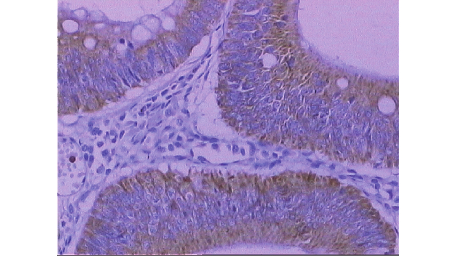
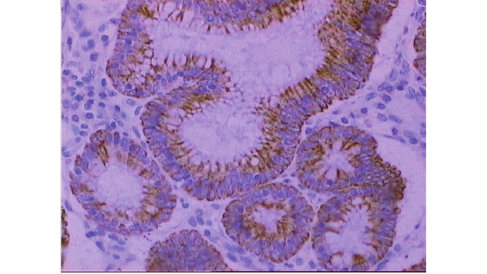
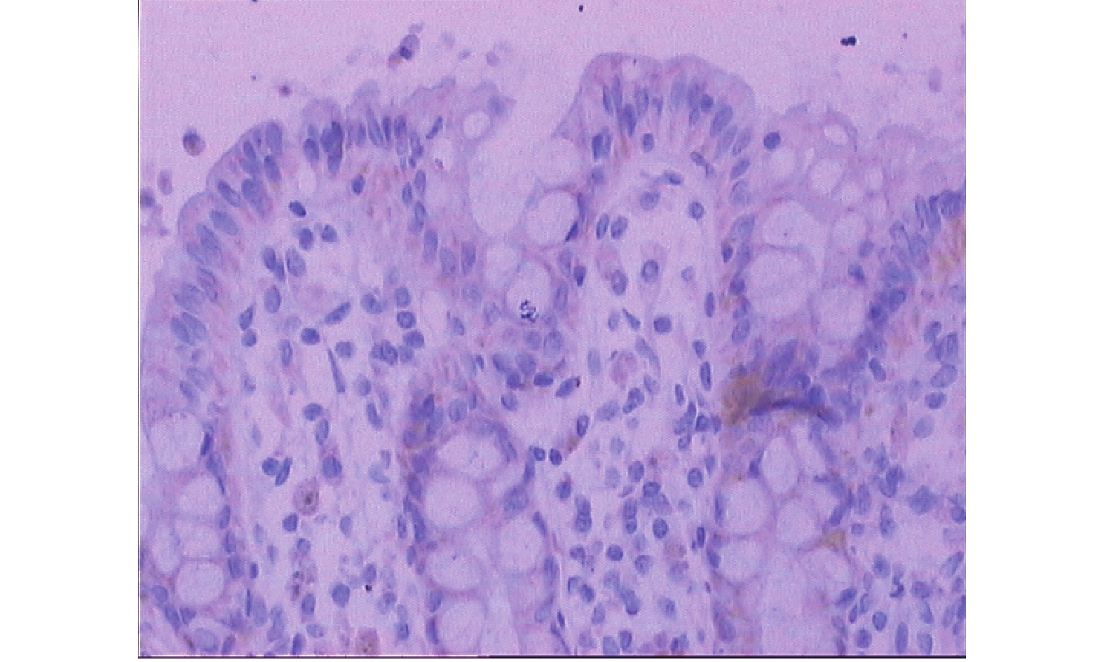
图 1 正常大肠黏膜LEA呈弱阳性表达(SP×200)
Figure 1. The weakly positive expression of LEA in normal colorectal mucosa (SP×200)

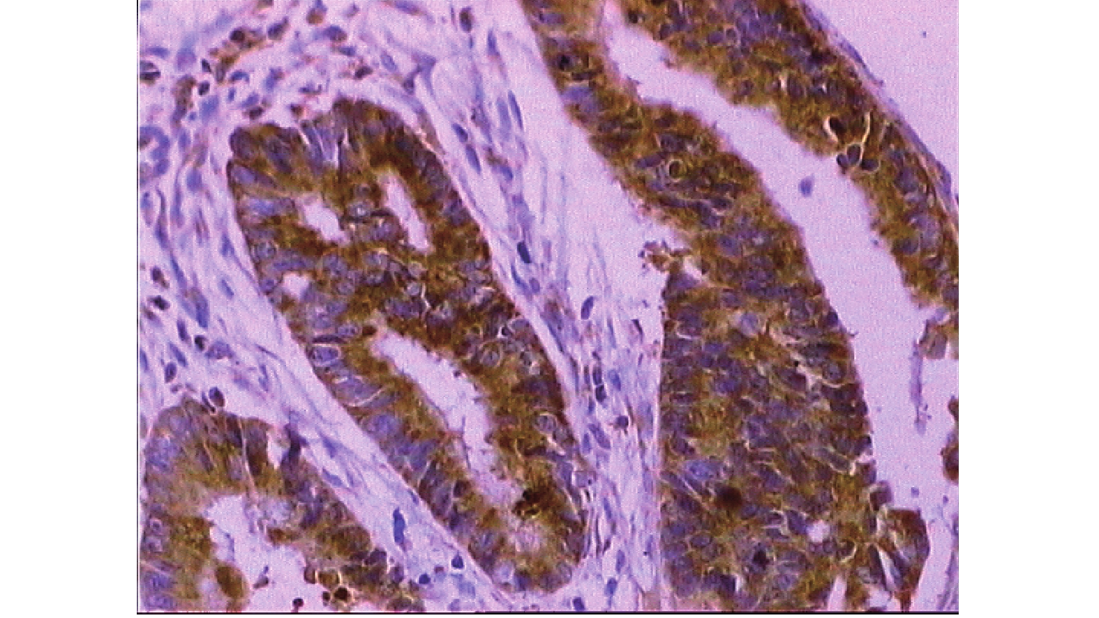
图 2 大肠腺瘤LEA呈阳性表达(SP×200)
Figure 2. The positive expression of LEA in colorectal adenoma (SP×200)

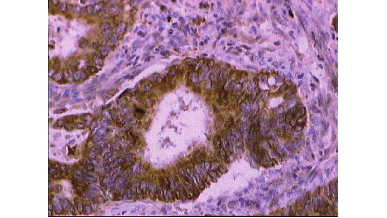
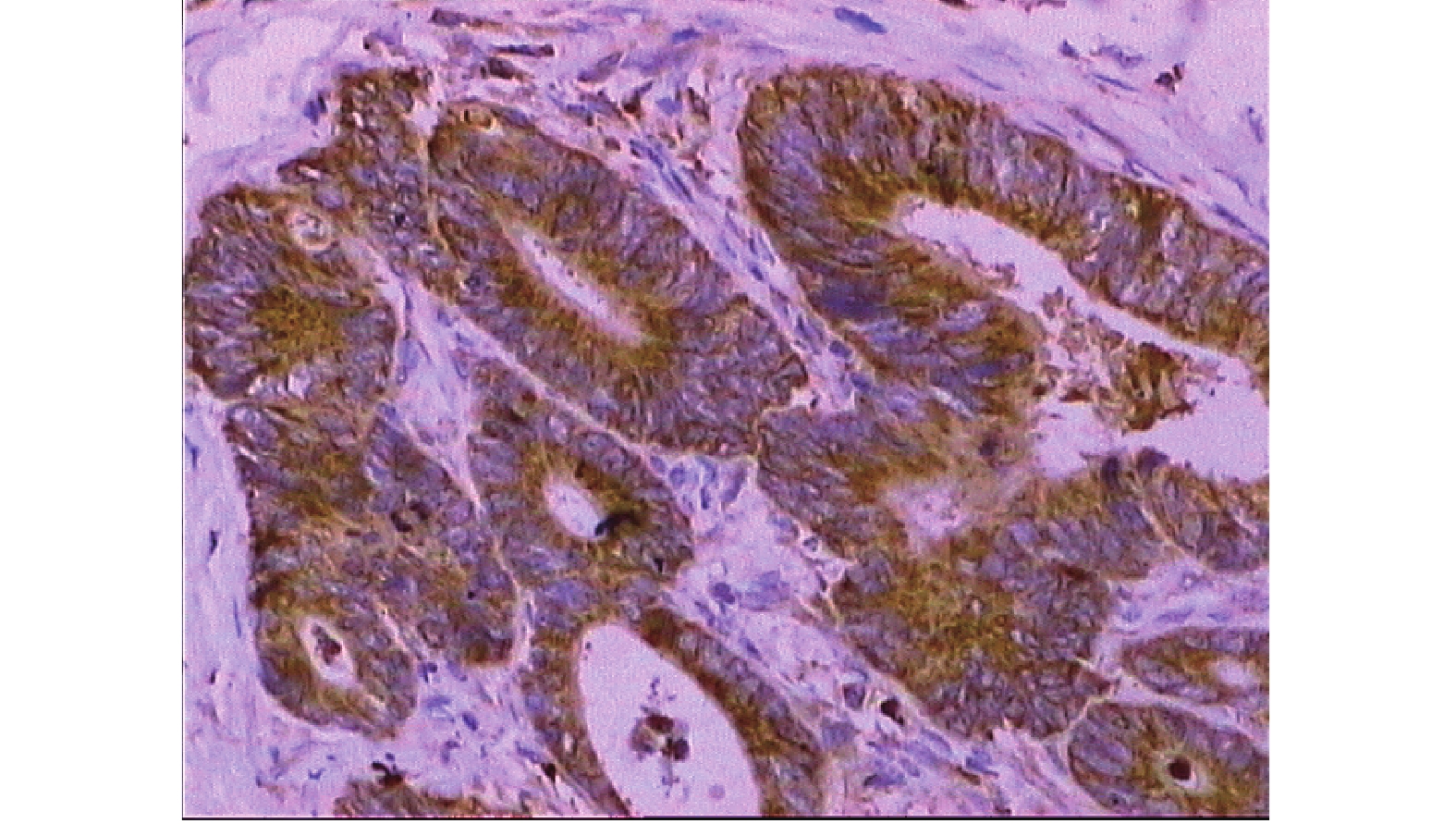
图 3 淋巴结无转移大肠腺癌LEA呈强阳性表达(SP×200)
Figure 3. The strong positive expression of LEA in colorectal adenocarcinoma without regional lymph node metastasis (SP×200)

图 4 淋巴结有转移大肠腺癌LEA呈强阳性表达(SP×200)
Figure 4. The strong positive expression of LEA in colorectal adenocarcinoma with regional lymph node metastasis (SP×200)

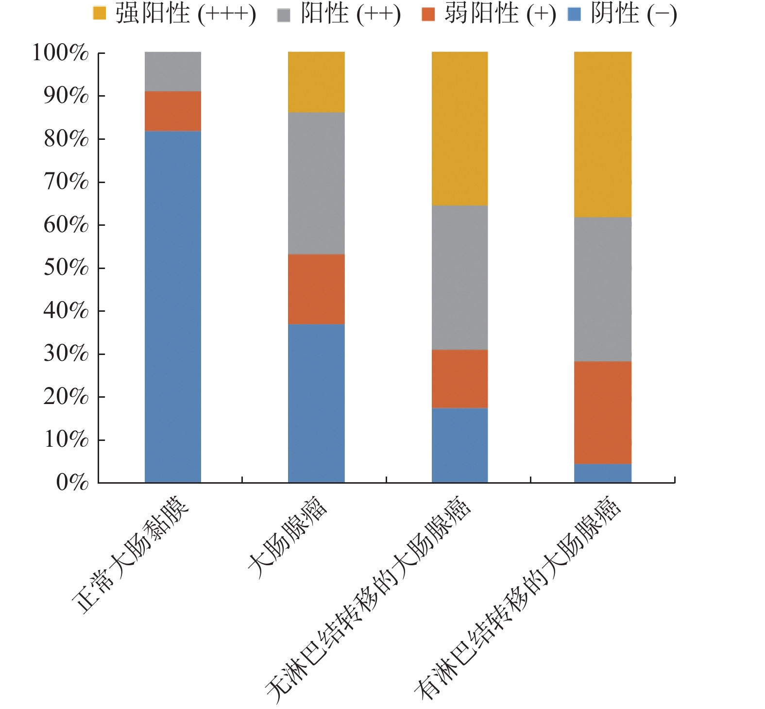
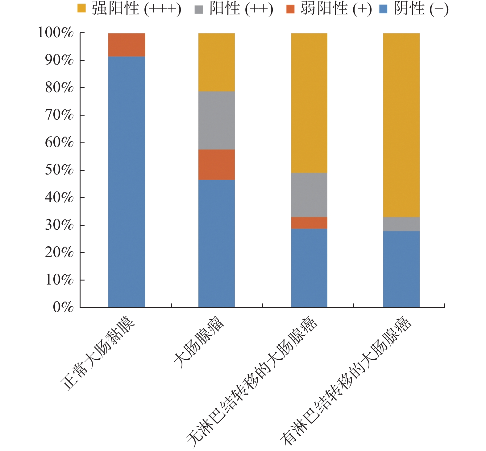
图 5 LEA在正常大肠黏膜、大肠腺瘤和大肠腺癌组织中的表达分布
Figure 5. Distribution of LEA expression in normal colorectal mucosa, colorectal adenoma and colorectal adenocarcinoma

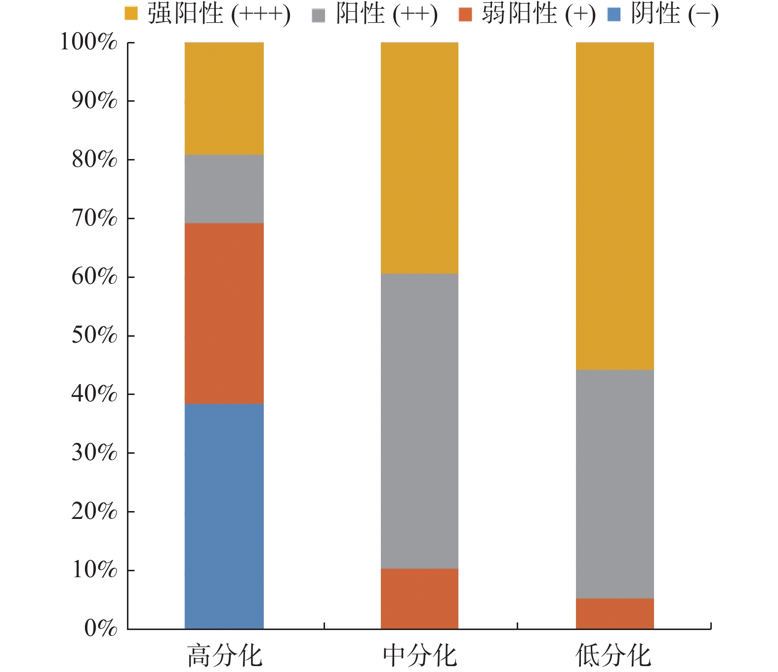
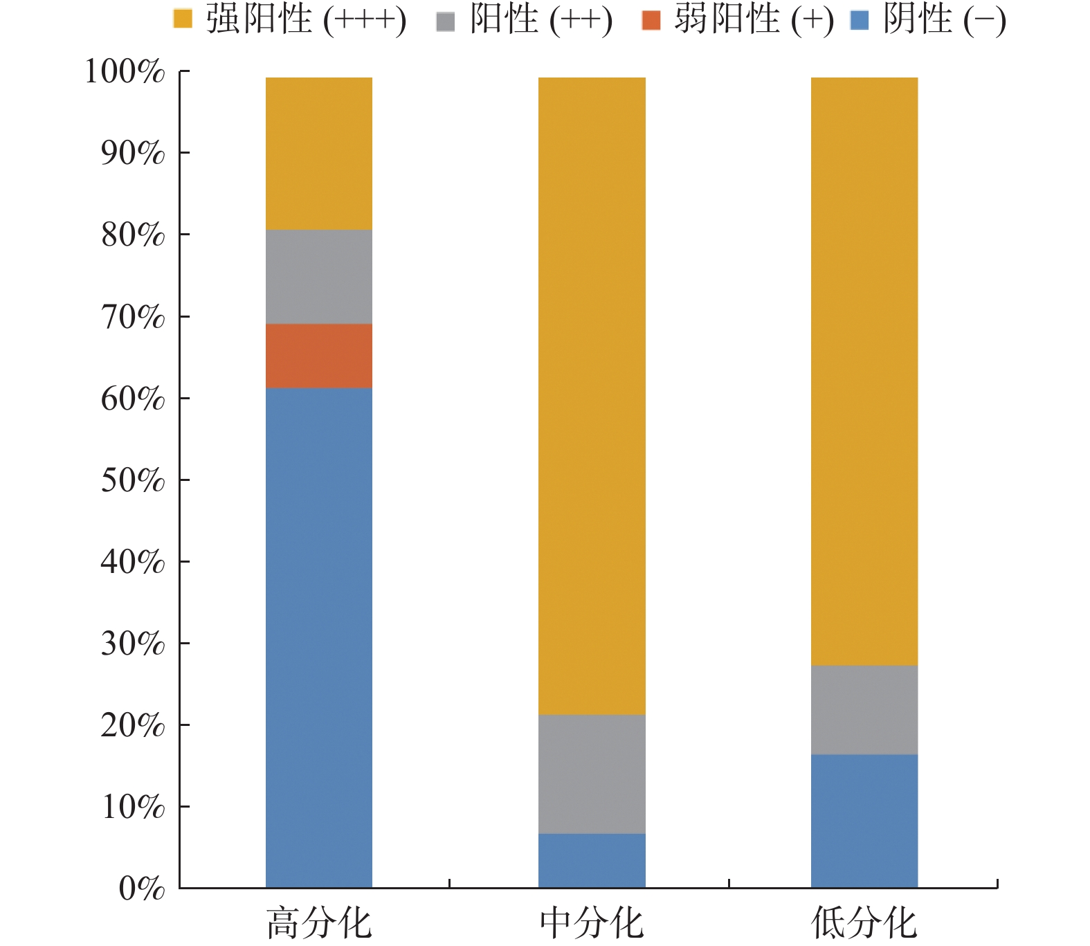
图 6 LEA在不同分化程度大肠腺癌中的表达分布
Figure 6. Distribution of LEA expression in colorectal adenocarcinoma with different degrees of differentiation

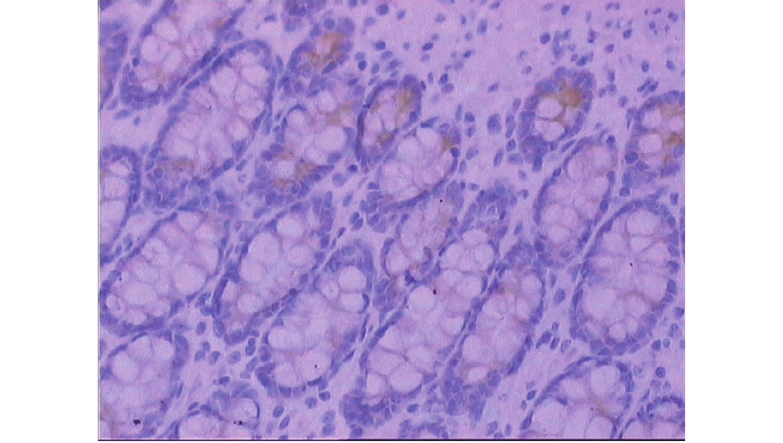
图 8 正常大肠黏膜CDw75呈弱阳性表达(SP×200)
Figure 8. The weakly positive expression of CDw75 in normal colorectal mucosa (SP×200)

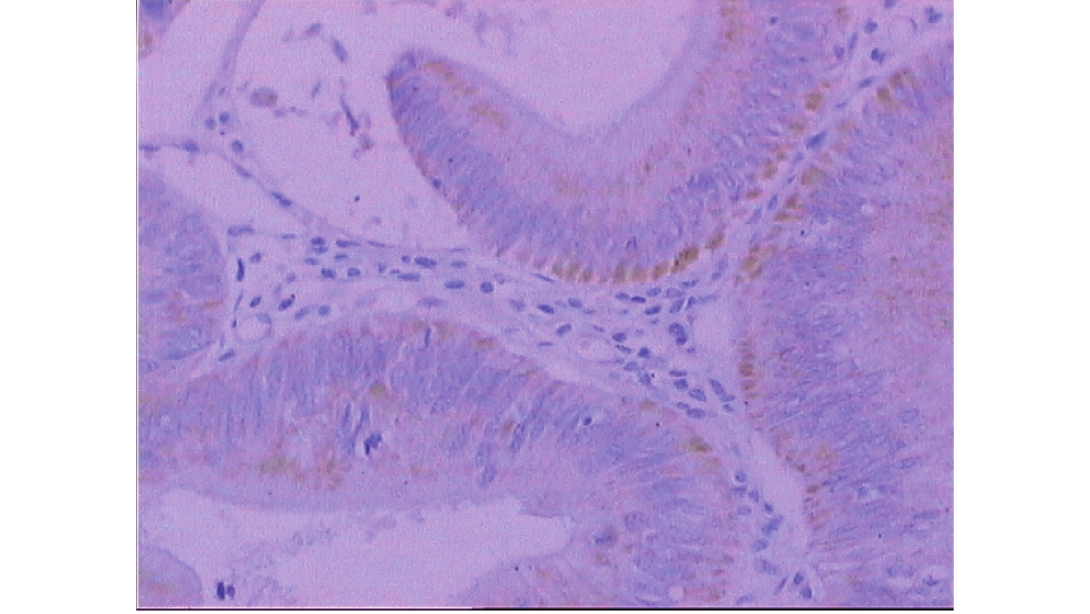
图 9 大肠腺瘤组织CDw75呈阳性表达(SP×200)
Figure 9. The positive expression of CDw75 in colorectal adenoma (SP×200)

图 10 淋巴结无转移的大肠腺癌组织CDw75呈强阳性表达(SP×200)
Figure 10. The strong positive expression of CDw75 in colorectal adenocarcinoma without regional lymph node metastasis (SP×200)

图 11 淋巴结有转移的大肠腺癌组织CDw75呈强阳性表达(SP×200)
Figure 11. The strong positive expression of CDw75 in colorectal adenocarcinoma with regional lymph node metastasis (SP×200)

图 12 CDw75在正常大肠黏膜、大肠腺瘤和大肠腺癌组织中的表达分布
Figure 12. Distribution of CDw75 expression in normal colorectal mucosa, colorectal adenoma and colorectal adenocarcinoma

图 13 CDw75在不同分化程度的大肠腺癌中的表达分布
Figure 13. Distribution of CDw75 expression in colorectal adenocarcinoma with different degrees of differentiation

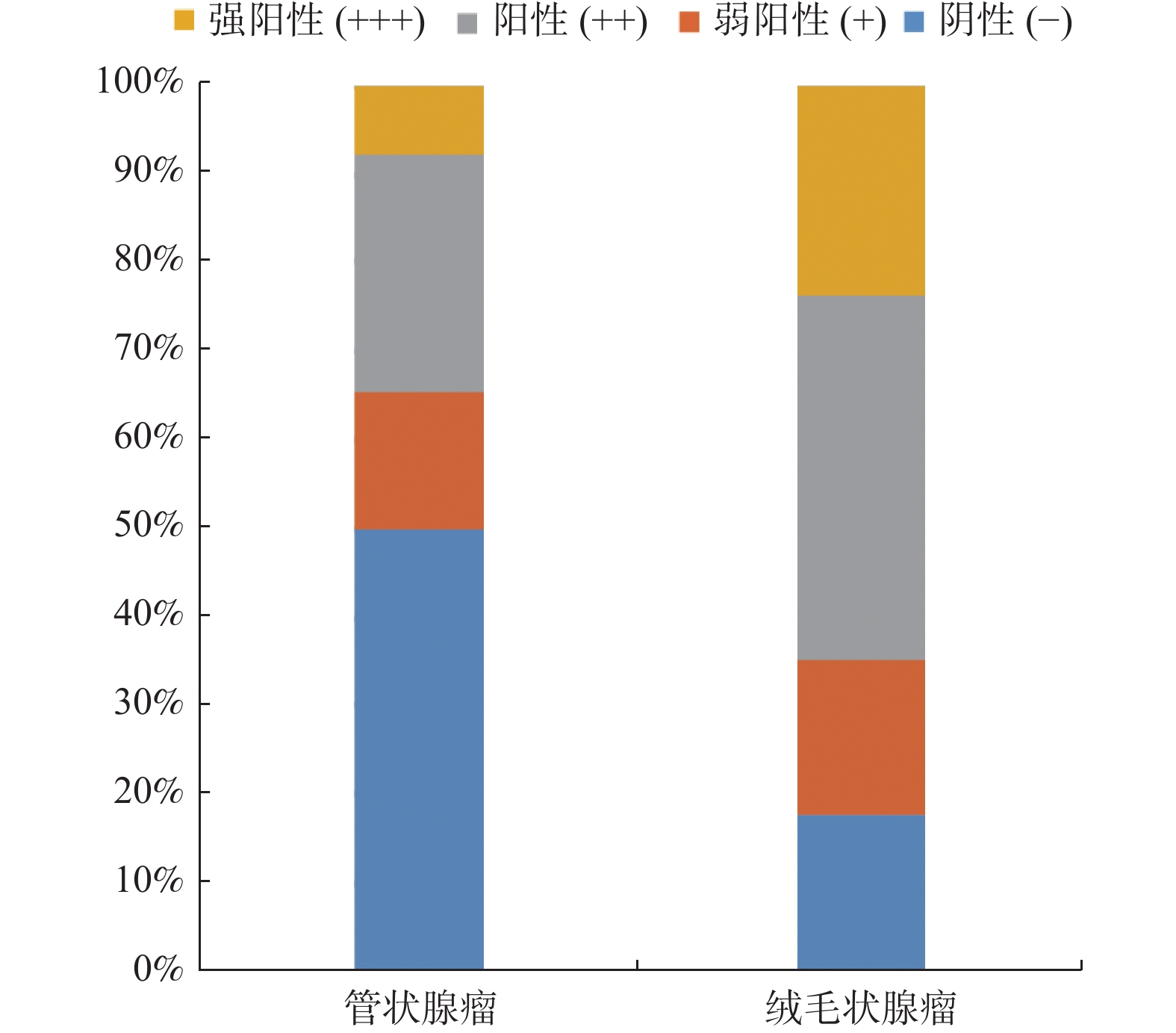
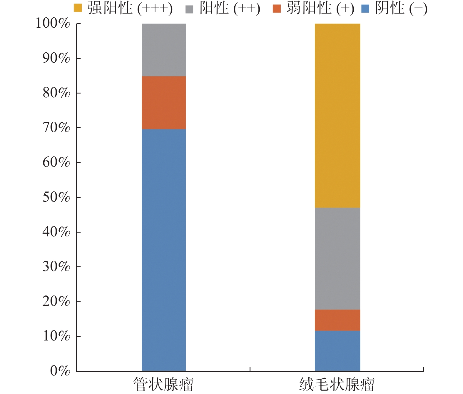
图 14 CDw75在大肠腺瘤中的表达分布
Figure 14. Distribution of CDw75 expression in colorectal adenoma
表 1 LEA在正常大肠黏膜、大肠腺瘤、大肠腺癌中的表达(n)
Table 1. The expression of LEA in normal colorectal mucosa,colorectal adenoma and colorectal carcinoma (n)
组别 n LEA表达 P − + ++ +++ 正常大肠黏膜 11 9 1 1 0 0.000▲,0.000▲▲ 大肠腺瘤 43 16 7 14 6 0.045▲,0.048▲▲ 无淋巴结转移的大肠腺癌 51 9 7 17 18 有淋巴结转移的大肠腺癌 21 1 5 7 8 检验水准α = 0.083。▲与无淋巴结转移大肠腺癌比较,▲▲与有淋巴结转移大肠腺癌比较。  下载: 导出CSV
表 2 LEA在不同分化程度大肠腺癌中的表达(n)
Table 2. The expression of LEA in colorectal adenocarcinoma with different degrees of differentiation (n)
组别 n LEA表达 P − + ++ +++ 高分化大肠腺癌 26 10 8 3 5 0.001*,0.000** 中分化大肠腺癌 28 0 3 14 11 低分化大肠腺癌 18 0 1 7 10 检验水准α = 0.017。*与中分化大肠腺癌比较,**与低分化大肠腺癌比较。
下载: 导出CSV
表 3 LEA在大肠腺瘤中的表达(n)
Table 3. The expression of LEA in colorectal adenoma (n)
组别 n LEA表达 P − + ++ +++ 管状腺瘤 26 13 4 7 2 0.023* 绒毛状腺瘤 17 3 3 7 4 检验水准α = 0.05。*P < 0.05。
下载: 导出CSV
表 4 CDw75在正常大肠黏膜、大肠腺瘤、大肠腺癌中的表达(n)
Table 4. The expression of CDw75 in normal colorectal mucosa,colorectal adenoma and colorectal carcinoma (n)
组别 n CDw75表达 P − + ++ +++ 正常大肠黏膜 11 10 1 0 0 0.001▲,0.000▲▲ 大肠腺瘤 43 20 5 9 9 0.042▲▲ 无淋巴结转移的大肠腺癌 51 15 2 8 26 有淋巴结转移的大肠腺癌 21 6 0 1 14 检验水准α = 0.083。▲与无淋巴结转移大肠腺癌比较,▲▲与有淋巴结转移大肠腺癌比较。
下载: 导出CSV
表 5 CDw75在不同分化程度大肠腺癌中的表达(n)
Table 5. The expression of CDw75 in colorectal adenocarcinoma with different degrees of differentiation (n)
组别 n CDw75表达 P − + ++ +++ 高分化大肠腺癌 26 16 2 3 5 0.000*,0.001** 中分化大肠腺癌 28 2 0 4 22 低分化大肠腺癌 18 3 0 2 13 检验水准α = 0.017。*与中分化比较,**与低分化比较。
下载: 导出CSV
表 6 CDw75在大肠腺瘤中的表达(n)
Table 6. The expression of CDw75 in colorectal adenoma (n)
组别 n CDw75表达 P − + ++ +++ 管状腺瘤 26 18 4 4 0 0.000* 绒毛状腺瘤 17 2 1 5 9 检验水准α = 0.05。*P < 0.05。
下载: 导出CSV
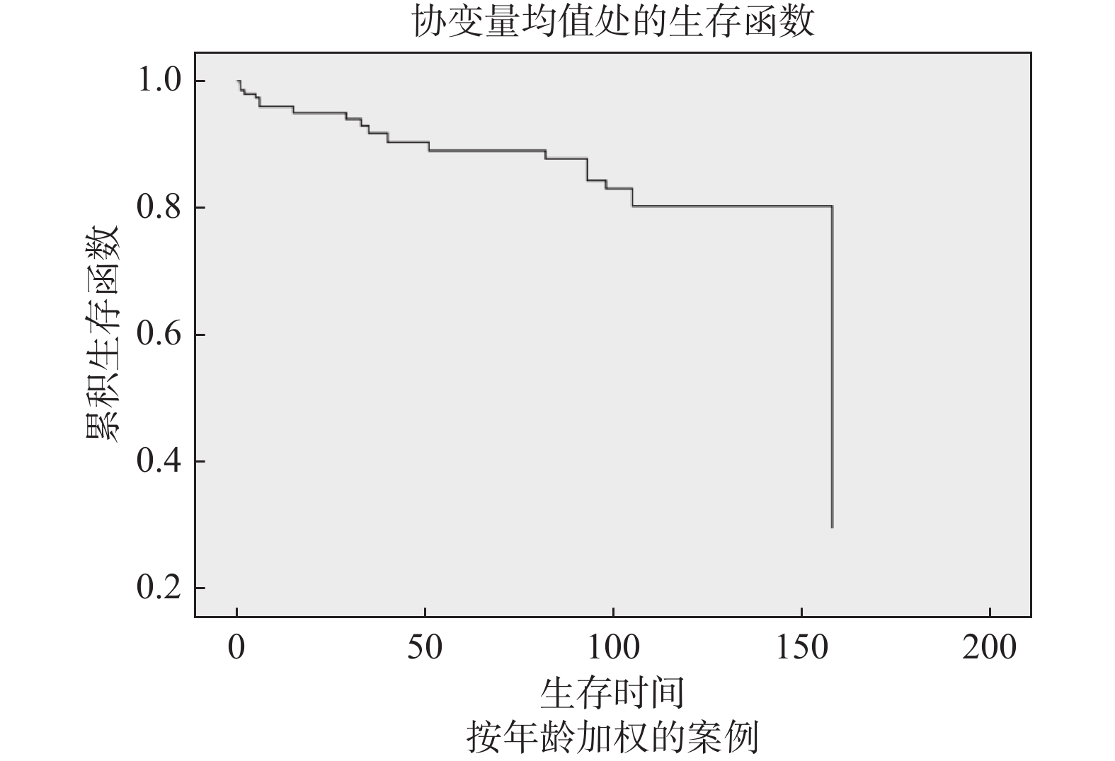
表 7 多因素 COX比例风险回归模型
Table 7. Multivariate COX proportional hazards regression model
影响因素 β SE Wald P RR 95%CI 性别 −0.656 0.073 80.428 0.000 0.519 0.450~0.599 年龄 0.038 0.003 169.684 0.000 1.038 1.032~1.044 临床分期 0.808 0.039 427.070 0.000 2.242 2.077~2.421 淋巴结转移 −0.416 0.079 27.819 0.000 0.66 0.565~0.770 LEA表达 1.010 0.067 226.549 0.000 2.747 2.408~3.133 CDw75表达 1.363 0.095 207.480 0.000 3.908 3.246~4.704
下载: 导出CSV
-
[1] Pourhoseingholi M A,Zali M R. Colorectal cancer screening:time for action in Iran[J]. World J Gastrointest Oncol,2012,4(4):82-83. doi: 10.4251/wjgo.v4.i4.82 [2] Center M M,Jemal A,Smith R A,et al. Worldwide variations in colorectal cancer[J]. CA Cancer J Clin,2009,59(6):366-378. doi: 10.3322/caac.20038 [3] Brenner D E,Rennert G. Fecal dna biomarkers for the detection of colorectal neoplasia:attractive,but is it feasible[J]. J Natl Cancer Inst,2005,97(15):1107-1109. doi: 10.1093/jnci/dji244 [4] Bleday R,Song J,Walker E S,et al. Characterization of a new monoclonal antibody to a cell surface antigen on colorectal cancer and fetal gut tissues[J]. Cancer,1986,57(3):433-440. doi: 10.1002/1097-0142(19860201)57:3<433::AID-CNCR2820570305>3.0.CO;2-Q [5] 王明武,宋今丹. 人侵袭性结肠癌细胞内质网的超微结构[J]. 中国医科大学学报,2000,29(6):401-403. doi: 10.3969/j.issn.0258-4646.2000.06.001 [6] Dezheng Yuan,Hang Chen,Shuo Wang,et al. Identification of LEA,a podocalyxin-like glycoprotein,as a predictor for the progression of colorectal cancer[J]. Cancer Med,2018,7(10):5155-5166. doi: 10.1002/cam4.1765 [7] WHO Classification of Tumours Editorial Board. World Health Organization classification of tumours [M]//Naglegaal I D, Arends M J, Odze R D, et al. Digestive System Tumours-Tumours of the Colon and Rectum. 5th Edition. Switzerland: World Health Organization, 2019: 157-192. [8] Bayascas J R. PDK1:the major transducer of PI 3-kinase actions[J]. Curr Top Microbiol Immunol,2010,346(1):9-29. [9] 万德森,陈功. 结直肠癌的流行病学及其危险因素研究近况[J]. 实用癌症杂志,2000,15(2):220-222. [10] 万德森. 结直肠癌流行趋势及对策[J]. 癌症,2009,28(9):897-902. doi: 10.3321/j.issn:1000-467X.2009.09.001 [11] Rajaratnam S G,Dennett E R. Development of metachronous neoplasms after colorectal cancer resection:absence of synchronous neoplasms predicts a lower risk[J]. N Z Med J,2009,122(1294):61-66. [12] Shi C,Gao F,Gao X,et al. A novel anti-VEGF165 monoclonal antibody-conjugated liposomal nanocarrier system:Physical characterization and cellular uptake evaluation in vitro and in vivo[J]. Biomed Pharmacother,2015,69(1):191-200. [13] 万文徽. 血清肿瘤标志的临床应用[J]. 实用癌症杂志,1998,13(4):316-317. doi: 10.3969/j.issn.1001-5930.1998.04.039 [14] 王之章,姜卫国,宋今丹,等. 抗结肠癌单克隆抗体ND-1在结肠癌组织的标记[J]. 中国医科大学学报,1991,20(2):98-101. [15] 刘小平,刘恩卿,姜卫国,等. 肿瘤相关抗原LEA在大肠腺瘤组织中的表达[J]. 中国医科大学学报,1995,24(1):38-40. [16] White K N,Hope D B. Characterization of aspirin hydrolase of guinea-pig liver cytoplasm[J]. Biochim Biophys Acta,1984,785(3):132-137. doi: 10.1016/0167-4838(84)90137-7 [17] Hakomori S. Aberrant glycosylation in tumors and tumor-associated carbohydrate antigens[J]. Adv Cancer Res,1989,52(1):257-331. [18] 李连弟,鲁凤珠,张思维,等. 中国恶性肿瘤死亡率20年变化趋势和近期预测分析[J]. 中华肿瘤杂志,1997,19(1):3-9. doi: 10.3760/j.issn:0253-3766.1997.01.001 [19] Gruszewska E,Chrostek L. The alterations of glycosylation in malignant diseases[J]. Pol Merkur Lekarski,2013,34(199):58-61. [20] Óscar Mariño-Crespo,Almudena Fernández-Briera,Emilio Gil-Martín. Identification of proteins with the CDw75 epitope in human colorectal cancer[J]. Oncol Lett,2018,15(1):580-587. [21] Shiozaki K,Yamaguchi K,Takahashi K,et al. Regulation of sialyl lewis antigen expression in colon cancer cells by sialidase neu4[J]. J Biol Chem,2011,286(24):21052-21061. doi: 10.1074/jbc.M111.231191 [22] Yang S H,Jiang J K,Chang S C,et al. Clinical significance of ca19-9 in the follow-up of colorectal cancer patients with elevated preoperative serum ca19-9[J]. Hepatogastroenterology,2013,60(125):1021-1027. [23] 蒋小猛,于亚杰,吴莺,等. hTERT和CDw75在胃癌癌组织中的表达及意义[J]. 山东医药,2009,1(1):86-87. doi: 10.3969/j.issn.1002-266X.2009.01.056 [24] 夏磊,王元书,崔淑香. 唾液酸转移酶与肿瘤进展和转移[J]. 国际肿瘤学杂志,2009,36(2):86-88. doi: 10.3760/cma.j.issn.1673-422X.2009.02.002 [25] Li M,Vemulapalli R,Ullah A,et al. Downregulation of a human colonic sialyltransferase by a secondary bile acid and a phorbol ester[J]. Am J Physiol,1998,274(1):599-606. -

 点击查看大图
计量
- 文章访问数: 3738
- HTML全文浏览量: 2569
- PDF下载量: 28
- 被引次数: 0
