Application of Multimodal Ultrasound in Post-operative Monitoring of Transplanted Livers by Donors after the Brain Death
-
摘要:
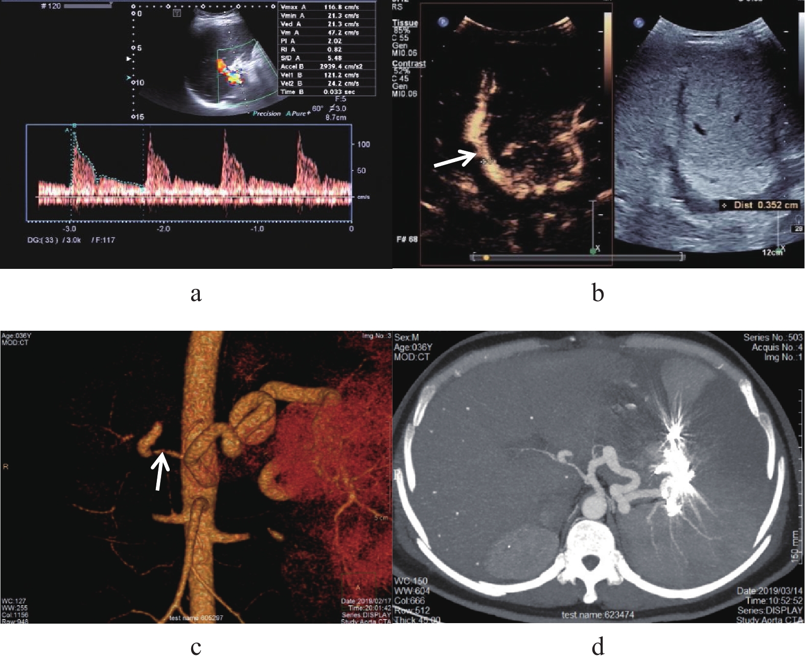
目的 联合应用二维灰阶超声、CDFI(彩色多普勒血流成像)、PW(频谱多普勒)及CEUS(超声造影)对脑死亡器官捐献肝移植术后实现灰阶、血流、微灌注等检查,探讨多模态超声在肝移植术后监测中的应用价值。 方法 选取2018年1月至2021年12月昆明市第一人民医院脑死亡器官捐献(donors after brain death,DBD)移植肝患者57例,所有患者肝移植术后均进行常规超声实时、动态连续监测,对移植肝血管显示困难、血流充盈差、可疑狭窄或血栓、移植肝内或肝周出现不能确定的异常病变进一步行CEUS检查,分析超声检查对移植肝术后各种并发症的诊断,检查结果与其他影像学检查、临床、病理、实验室检查进行对照。 结果 多模态超声提示有血管并发症者7例,其中肝动脉狭窄2例(3.5%),门静脉血栓2例(3.5%),下腔静脉血栓1例(1.2%),下腔静脉狭窄2例(3.5%);胆道并发症4例(7.0 %):其中胆道狭窄3例(5.2%),胆汁瘤1例(1.2%),均经临床证实;一般常见并发症:胸腹腔积液57例(100%),肝内及肝周血肿31例(54.3%)。多模态超声漏诊肝动脉假性动脉瘤1例(1.2%)。 结论 多模态超声检查作为肝移植术后的首选检查方法,能较准确的诊断肝移植术后并发症。 Abstract:Objective To explore the application value of multi-mode ultrasound in post-liver transplantation monitoring by combining two-dimensional gray scale ultrasound, CDFI, Spectral Doppler and CEUS in gray scale, blood flow and microperfusion examination after the liver transplantation. Methods In this study, 57 patients with liver transplants donated by those after brain death (DBD) in our hospital from January 2018 to December 2021 were selected. All the patients underwent the real-time, dynamic and continuous monitoring by conventional ultrasound after the liver transplantation. Results There were 7 cases of vascular complications, including 2 cases of hepatic artery stenosis (3.5%), 2 cases of portal vein thrombosis (3.5%), 1 case of inferior vena cava thrombosis (1.2%), and 2 cases of inferior vena cava stenosis (3.5%). There were 4 biliary complications (7.0%), including 3 biliary stenosis (5.2%) and 1 choleoma (1.2%), which were clinically confirmed. The common complications were pleural and abdominal effusion in 57 cases (100%), intrahepatic and perihepatic hematoma in 31 cases (54.3%). False aneurysm of hepatic artery was missed by multimodal ultrasound in 1 case (1.2%). Conclusion As the first choice after the liver transplantation, multimodal ultrasound can accurately diagnose the complications after the liver transplantation. -
Key words:
- Liver transplantation /
- DBD /
- Multimodal ultrasound /
- CEUS
-
表 1 超声监测肝移植术后各种并发症的发生率(%)
Table 1. The incidence of complications after liver transplantation was monitored by ultrasound(%)
并发症 n 发生率 肝动脉狭窄 2 3.5 门静脉血栓 2 3.5 下腔静脉血栓 1 1.2 下腔静脉狭窄 2 3.5 胆道狭窄 3 5.2 胆汁瘤 1 1.2 胸腹腔积液 57 100 肝内及肝周血肿 31 54.3 表 2 超声造影病例的原因和造影特征
Table 2. Causes and features of cases with contrast-enhanced ultrasound
造影原因 n 超声造影特征及结果 可疑肝动脉局部狭窄
肝动脉显示困难1
8肝动脉局部狭窄
1例肝门区肝动脉显影,内径纤细、间断显示,呈“串珠样;肝实质内灌注不良,
提示肝动脉狭窄;7例表现为肝动脉管腔内造影剂显影良好,肝动脉未见明显狭窄下腔静脉可疑血栓 1 下腔静脉内见造影剂充盈缺损,提示附壁血栓形成(部分栓塞) 门静脉内可疑血栓 2 门静脉管腔内均见造影剂充盈缺损,提示附壁血栓形成(部分栓塞) 肝后段下腔静脉可疑狭窄 2 均表现为肝后段下腔静脉吻合口变细狭窄 下腔静脉显示困难,血流充盈差 1 下腔静脉内造影剂显影良好,未见明显狭窄 -
[1] 李仁冬,白磊,何翼彪,等. 扩大标准供肝质量术前评估现状与展望[J]. 中华器官移植杂志,2017,38(3):188-191. doi: 10.3760/cma.j.issn.0254-1785.2017.03.013 [2] 肖春华,王妍,迟昆燕,等. 彩色多普勒超声在诊断肝移植术后并发症中的价值[J]. 中国介入影像与治疗学,2009,6(5):421-424. [3] 郑荣琴,任杰,许尔蛟. 肝移植超声检查[J]. 中华医学超声杂志(电子版),2010,1(5):558-569. [4] 宋洁,肖春华,周凯,等. 超声造影观察肝移植术后罕见血管并发症2例[J]. 临床超声医学杂志,2014,16(11):728-732. [5] 郑树森. 我国肝脏移植技术展望[J]. 中国医药指南,2003,1(5):46-47. doi: 10.3969/j.issn.1671-8194.2003.05.022 [6] 李秋萍,华扬,刘佳宾,等. 不同Mori分型椎动脉颅内段重度狭窄的彩色多普勒血流成像特点和血流动力学参数分析[J]. 中国脑血管病杂志,2019,16(6):281-287. doi: 10.3969/j.issn.1672-5921.2019.06.001 [7] 宋洁,肖春华. 超声观察肝移植术后胆汁瘤[J]. 中国医学影像技术,2008,24(10):1504. doi: 10.3321/j.issn:1003-3289.2008.10.053 [8] 张芬,肖春华,宋洁. 彩色多普勒超声在肝移植术后血管并发症中的临床应用[J]. 中国卫生产业,2014,11(6):116-117. [9] 李飞,王东平,何晓顺,等. 心脏死亡器官捐赠供受者术前及术后早期临床指标对肝移植预后的影响[J]. 中华器官移植杂志,2013,34(8):473-476. doi: 10.3760/cma.j.issn.0254-1785.2013.08.008 [10] 孟宪春. 原发性肝癌的螺旋CT诊断分析[D]. 延边: 延边大学硕士学位论文, 2007. [11] 杨扬,陈规划,蔡常洁,等. 肝移植术后血管并发症的诊断与治疗[J]. 中国实用外科杂志,2003,5(23):280-283. [12] 丁世兰,肖春华,等. 超声在肝移植术后肝动脉并发症中的应用[J]. 影像研究与医学应用,2018,2(18):80-81. doi: 10.3969/j.issn.2096-3807.2018.18.048 -


 下载:
下载:


