A Retrospective Study of 64-row 128-slice Spiral CT Digital Imaging Data of Normal Adult Coracoid Process
-
摘要:
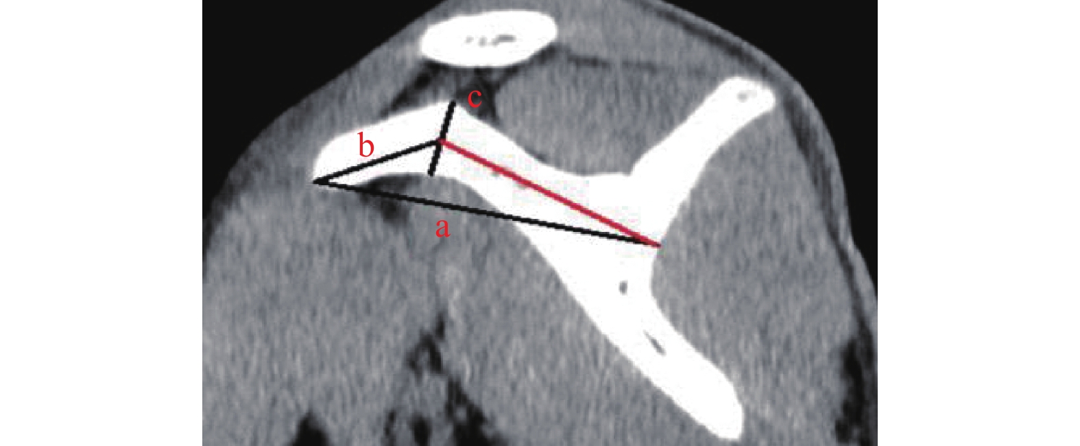
目的 探讨正常成人喙突的64排128层螺旋CT影像学特征,为临床喙突安全手术提供参考。 方法 随机抽取于2019年 01月至 2022年05月在云南省中医医院就诊的成年患者行肩关节64排128层螺旋CT扫描的影像 CT片121例资料作为样本,以肩关节矢状面为测量对象,测量喙突、喙突膝部的长度,测量喙突尖至喙突膝部中点的距离及喙突基底部、喙突尖的高度,再以肩关节矢状面为测量对象,测量喙突尖基底部、喙突膝部、喙突基底部的宽度;测量结果采用SPSS27.0统计学软件包进行统计学分析,基本描述正态分布计量资料采用平均值、中位数、标准差、最大值、最小值、 $\bar x \pm s $ 表示,正态分布计量资料2组间比较采用两独立样本t检验,相关性分析采用Pearson相关性分析。结果 喙突长度为(59.92±6.11)mm;喙突膝部长度为(14.05±2.26)mm;喙突尖至喙突膝部中点的距离为(22.59±3.65)mm;喙突尖基底部的高度为(9.86±1.71)mm;喙突尖高度为(7.22±1.79)mm;喙突尖的宽度为(9.14±2.08)mm;喙突膝部的宽度为(13.79±2.70)mm;喙突基底部的宽度为(21.10±4.18)mm;经两独立样本t检验,除了喙突尖的高度,男性与女性患者组间的喙突影像测量距离均有统计学意义(P < 0.05);左侧与右侧患者组间的喙突长度、喙突尖至喙突膝部中点的距离及喙突膝部的宽度均有统计学意义(P < 0.05);经Pearson相关性分析男性和女性喙突长度与喙突尖至喙突膝部中点的距离、喙突尖的宽度与喙突膝部的宽度均有相关性(P < 0.05)。 结论 喙突长度、宽度均与男女性别、左右侧有差异,其中无论是男性还是女性喙突长度与喙突尖至喙突膝部中点的距离、喙突尖的宽度与喙突膝部的宽度均有一定的关联;因此在临床喙突手术过程中或者相关器械耗材设计的时候应注意性别、左右侧以及它们之间的关联性。 -
关键词:
- 喙突 /
- 成年人 /
- 64排128层螺旋CT /
- 影像学 /
- 回顾性研究
Abstract:Objective To explore the imaging features of 64-row, 128-slice spiral CT of the normal adult coracoid process to provide a reference for clinical coracoid process safety surgery. Methods The data of 121 adult patients who underwent 64-row 128 slice spiral CT scanning of shoulder joints from January 2019 to May 2022 in Yunnan Provincial Hospital of traditional Chinese medicine were randomly selected as samples. The sagittal plane of the shoulder joint was taken as the measurement object to measure the length of the coracoid process and coracoid process knee, the distance from the coracoid process tip to the midpoint of coracoid process knee, and the height of the coracoid process base and coracoid process tip, and then the sagittal plane of the shoulder joint was taken as the measurement object to measure the base of coracoid process tip Width of coracoid process knee and coracoid process base; The measurement results were statistically analyzed by spss27.0 statistical software package. The basic description of normal distribution measurement data was expressed by mean, median, standard deviation, maximum value, minimum value and $\bar x \pm s $ . the normal distribution measurement data were compared by two independent sample t-test. Pearson correlation analysis was used for correlation analysis.Results The length of the coracoid process is 59.92±6.11 mm; the length of the coracoid knee is 14.05±2.26 mm; the distance from the coracoid tip to the midpoint of the coracoid knee is 22.59±3.65 mm; the height of the base of the coracoid tip is 9.86±1.71 mm; the height of the coracoid tip is 7.22±1.79 mm; the width of the coracoid tip is 9.14±2.08 mm; the width of the coracoid knee is 13.79±2.70 mm; the width of the coracoid base is 21.10±4.18 mm; Sample t-test, in addition to the height of the coracoid process tip, the coracoid image measurement distance between male and female patient groups was statistically significant (P < 0.05); the coracoid process length, coracoid process tip between left and right patient groups The distance to the midpoint of the coracoid knee and the width of the coracoid knee were statistically significant (P < 0.05); the length of the coracoid process in males and females and the length of the coracoid process to the midpoint of the coracoid knee were statistically significant (P < 0.05). The distance, the width of the coracoid tip and the width of the coracoid knee were all correlated (P < 0.05). Conclusion The length and width of the coracoid process are different from male and, gender and left and right, among which, the length of the coracoid process and the distance from the tip of the coracoid process to the midpoint of the knee of the coracoid process, the width of the tip of the coracoid process and the width of the knee of the coracoid process are different in both male and female. There are certain correlations; therefore, attention should be paid to gender, left and right, and the correlation between them during clinical coracoid surgery or the design of related equipment and consumables. -
Key words:
- Coracoid process /
- Adult /
- 64-slice 128-slice spiral CT /
- Imaging /
- Retrospective study
-
结核病是由结核分枝杆菌感染引起的慢性传染病,2021年全球结核病报告显示[1]:2020年全球新发结核病患者987万,发病率127/10万。我国估计新发患者84.2万(2019年83.3万),结核病发病率约59/10万(2019年58/10万),我国发病数在全球结核病高负担国家中上升至第2位。结核病属于慢性消耗性疾病,营养不良也是其发病的重要危险因素之一,患者长期的慢性消耗中机体常处于负氮平衡状态,因此易出现不同程度的营养不良[2]。营养不良也是常见的可致免疫功能低下的原因之一,营养不良与发病率和病死率的升高、疾病恢复、获得性耐药及患者生存率和免疫功能等有关[3]。结核病与营养之间着联系复杂且发生发展相互影响,营养支持在结核病的治疗及预后起重要作用[4]。营养与免疫的关系主要有,营养状况是免疫系统和免疫功能的基础,机体的免疫反应和临床疾病可以改变其营养代谢和营养需求量[5]。营养素的缺乏会增加疾病发生风险,影响食物的消化吸收、代谢和机体的生长发育,从而间接影响免疫系统[6-7]。因此,良好的营养状况是免疫系统正常运作和发挥功能的重要保障,也是抵御病原体入侵和促进患者康复的重要因素。目前关于结核病患者营养及免疫方面的实验室指标研究较少,本研究通过对结核病患者实验室相关指标与营养状况及免疫功能的关系及特点分析,可为肺结核病患者开展营养评估和管理,营养支持治疗提供参考。
1. 资料与方法
1.1 研究对象
收集2021年3月至2022年3月在昆明市第三人民医院(昆明结核病防治院)收治的1041例肺结核病患者为研究组,病例多分布在昆明周边地区且覆盖云南全境;其中男性537例,女性504例;年龄8~87岁,中位年龄42岁。另选同期在昆明市第三人民医院健康体检者854例作为对照组,其中男性646例,女性208例;年龄6~85岁,中位年龄41岁。2组在性别构成及年龄分布经卡方检验及曼惠特尼U检验,差异均无统计学意义(χ2 = 0.005,U = 504538;P > 0.05)。
纳入标准:(1)所有诊断均按照《肺结核诊断标准》(WS288-2017)[8];(2)经结核菌痰涂片及痰培养等实验室检查和组织病理学检查确诊为肺结核。排除标准:(1)非结核分枝杆菌病;(2)其它感染性疾病;(3)自身免疫性疾病;(4)合并心、肝、肾等重要器官疾病及严重高血压和精神状况欠佳者;(5)患者为妊娠或哺乳期女性。本研究为回顾性分析,不涉及伦理审查。
1.2 研究方法
将研究对象的外周血进行总蛋白(TP)、白蛋白(ALB)、球蛋白(Glob)、白/球比(A/G)、血红蛋白(Hb)、前白蛋白(PA)、胆固醇(TC)、甘油三酯(TG)、低密度脂蛋白(LDL)、高密度脂蛋白(HDL-C)、成熟T淋巴细胞(CD3)、辅助性T淋巴细胞(CD4+)、细胞毒性T淋巴细胞(CD8+)、CD4+/CD8+、白细胞介素家族(IL)、肿瘤坏死因子(TNF)、干扰素(IFN)等指标检测,检测严格按照实验说明书在相应仪器上完成。
1.3 仪器与试剂
主要仪器∶日立全自动生化分析仪(008AS,日本日立生物科技有限公司),流式细胞仪(Bircyte E6,深圳迈瑞生物医疗电子股份有限公司),希森美康全自动血液分析仪(XT-1800,康佳康科技有跟公司)。主要试剂:细胞因子检测试剂盒(批号:211001,青岛瑞斯凯尔生物科技有限公司),流式细胞检测试剂盒(批号:2021050100,迪亚莱博生物科技有限公司),生化体外诊断试剂(批号:110620,富士胶片和光纯耀化学有限公司)。
1.4 统计学处理
采用 SPSS24.0及GraphPad Prism 8.0统计软件分析及作图。正态分布的计量资料采用t检验或方差分析;非正态分布的计量资料以M (P25,P75)表示,采用Mann-Whitney U 检验;多组比较采用Brown-Forsythe或Kruskal-Wallis检验;细胞因子整体水平间差异采用GraphPad Prism 8.0 嵌套t检验分析;计数资料以例/百分数(n/%)表示,采用χ2检验;相关性分析采用Spearman进行;以P < 0.05为差异有统计学意义。
2. 结果
2.1 对照组与研究组营养及免疫相关指标比较
与对照组比较,研究组营养及免疫相关实验室指标Hb、TP、Alb、Glob、A/G、PA、TC、TG、HDL、LDL、CD3+T、CD4+T、CD8+T、CD4+/CD8+、CD19+B、NK、NKT 表达显著降低,差异均有统计学意义(P < 0.05);细胞因子IL-1β、IL-8、IL-17水平显著升高,IL-4、IL-10、IL-12p70、IFN-γ水平显著降低,差异均有统计学意义(P < 0.05),见表1。
表 1 对照组和研究组相关检测指标水平比较 [$ \bar x \pm s$ /M(P25,P75)]Table 1. Comparison of relevant detection index levels in controland study group [$ \bar x \pm s$ /M(P25,P75)]相关指标 对照组(n = 854) 研究组(n = 1041) t'/U P Hb(g/L) 155.8 ± 16.74 120.4 ± 21.88 27.75 < 0.001* TP(g/L) 74.40 ± 5.864 66.9 ± 7.674 28.64 < 0.001* Alb(g/L) 42.97 ± 4.457 37.42 ± 5.516 28.52 < 0.001* Glob(g/L) 31.43 ± 5.953 28.35 ± 6.061 12.67 < 0.001* A/G 1.426 ± 0.356 1.369 ± 0.328 4.035 < 0.001* PA(mmol/L) 300.5 ± 87.29 222.6 ± 70.51 23.02 < 0.001* TC(mmol/L) 4.634 ± 1.042 4.193 ± 0.982 10.79 < 0.001* TG(mmol/L) 1.22(0.830,1.960) 1.06(0.790,1.465) 637362 < 0.001* HDL(mmol/L) 1.31(1.100,1.540) 1.02(0.830,1.220) 367655 < 0.001* LDL(mmol/L) 3.00(2.470,3.560) 2.64(2.140,3.160) 553012 < 0.001* CD3+T(个/μL) 1202 ± 499.1 1086 ± 456.4 963035 < 0.001* CD4+T(个/μL) 704.5 ± 308.4 628.3 ± 279.4 951581 < 0.001* CD8+T(个/μL) 460.6 ± 234.5 429.1 ± 217.8 1033269 < 0.001* CD4+/CD8+ 1.720 ± 0.763 1.644 ± 0.746 1049776 0.003* CD19+B (个/μL) 246.9 ± 163.4 218.3 ± 158.9 285140 < 0.001* NK (个/μL) 241.7 ± 169.1 207.1 ± 142.8 277918 < 0.001* NKT (个/μL) 194.0(135.3,299.0) 74.0(44.0,118.0) 110096 < 0.001* IL-1β (pg/mL) 3.30(1.710,6.120) 4.03(1.540,7.420) 1017339 0.005* IL-2 (pg/mL) 2.01(1.040,3.363) 1.72(1.080,3.095) 1114912 0.226 IL-4 (pg/mL) 1.65(1.190,2.520) 1.26(1.020,1.790) 829465 < 0.001* IL-5 (pg/mL) 2.13(1.030,3.260) 1.76(1.060,3.038) 1112062 0.106 IL-6 (pg/mL) 3.33(2.118,5.880) 3.18(1.850,6.505) 979359 0.188 IL-8 (pg/mL) 1.84(1.080,3.790) 1.85(1.233,4.740) 1068098 0.029* IL-10 (pg/mL) 1.33(0.890,2.090) 1.23(0.870,1.720) 1055485 < 0.001* IL-12p70 (pg/mL) 1.58(1.220,2.330) 1.48(1.130,2.003) 1031405 < 0.001* IL-17 (pg/mL) 1.86(1.280,3.400) 2.26(1.520,4.510) 917365 < 0.001* TNF-a (pg/mL) 1.70(1.170,2.650) 1.71(1.040,2.820) 931857 0.224 IFN-γ (pg/mL) 4.61(2.310,9.843) 3.65(2.350,8.188) 1069329 0.003* 经校正t检验和曼惠特尼U检验,*P < 0.05。 2.2 研究组营养相关指标及免疫状况分析
2.2.1 营养相关指标程度分析
为了评价研究组与对照组差异有统计学意义的营养及免疫相关指标的表达程度,分析了相关指标低血红蛋白、低白蛋白、高胆固醇、高甘油三酯、低高密度脂蛋白发生率分别为16.98%、59.84%、19.20%、23.42%、62.76%,其程度以轻度和中度为主,重度占比较少,见表2。
表 2 研究组相关指标程度分析 [n(%)]Table 2. Analysis of the degree of relevant indicators in the study group [n(%)]组别 低血红蛋白 低白蛋白 高胆固醇 高甘油三酯 低高密度脂蛋白 轻度 60 (41.38) 412 (80.63) 99 (60.36) 126 (63.00) 163 (30.41) 中度 58 (40.00) 92 (18.00) 56 (34.15) 71 (35.50) 241 (44.96) 重度 27 (18.62) 7 (1.37) 9 (5.49) 3 (1.50) 132 (24.63) 合计 145 511 164 200 536 2.2.2 免疫指标特征分析
根据CD4+T细胞分泌的细胞因子特点,可分为I型(Th1型细胞因子:IL-1β、IL-2、TNF-a、IFN-γ)和II型(Th2型细胞因子:IL-4、IL-5、IL-6、IL-10 )免疫应答,在本研究中肺结核病患者细胞因子水平经嵌套t检验分析,Th1型细胞因子水平表达较Th2型高,I型免疫反应占主导地位(t = 2.506,P < 0.05),见图1。
2.2.3 营养相关指标与免疫状况相关性分析
结核病患者营养与免疫相关指标经Spearman相关性分析显示,营养相关指标CHOL、TG、HGB、HDL-C、LDL-C、TP、ALB、GLOB、PA、A/G与淋巴细胞数、细胞因子水平及免疫相关指标CD3+T、CD4+T、CD8+T、CD4+/CD8+、CD19+B、NK、NKT,IL-1β、IL-2、IL-4、IL-5、IL-6、IL-8、IL-10、IL-12p70、IL-17、TNF-a、IFN-a、IFN-γ都呈正相关(r = 0.461、0.262、0.491;P均 < 0.05),见图2。
2.2.4 不同性别和年龄段营养及免疫相关指标比较
相关指标在不同性别和年龄段水平分析结果显示,在不同性别中男性TP、TG、NKT水平显著高于女性,TC、CD3+T、CD4+T、CD8+T、IL-1β、IL-4、IL-10、IL-12p70表达显著低于女性,差异均有统计学意义(P < 0.05);各年龄段相关指标经单因素方差分析,Hb、TC、TG、CD3+T、CD4+T、CD8+T、CD4+/CD8+、IL-1β表达差异均有统计学意义(P < 0.05),其中41-60岁组和≥61岁组患者CD3+T、CD4+T、CD8+T计数较≤20岁组降低,差异有统计学意义(P < 0.05),见表3、表4。
表 3 不同性别相关检测指标比较 [$ \bar x \pm s$ /M(P25,P75)]Table 3. Comparison of relevant test indicators by gender [$ \bar x \pm s$ /M(P25,P75)]相关指标 性别 t /U P 男性 (n = 581) 女性 (n = 523) Hb(g/L) — TP(g/L) 66.76 ± 7.457 65.80 ± 7.646 2.173 0.030* Alb(g/L) 37.10 ± 5.410 37.11 ± 5.313 0.014 0.989 Glob(g/L) 28.22 ± 5.779 28.04 ± 5.345 0.569 0.569 PA(mmol/L) 223.1 ± 68.67 222.5 ± 71.89 0.138 0.890 TC(mmol/L) 4.054 ± 0.9732 4.332 ± 0.9711 4.884 < 0.001* TG(mmol/L) 1.09(0.830,1.520) 1.04(0.770,1.430) 157520 0.035* HDL(mmol/L) 1.02(0.830,1.210) 1.02(0.840,1.230) 152054 0.999 LDL(mmol/L) 2.60(2.120,3.173) 2.65(2.160,3.160) 150805 0.812 CD3+T (个/μL) 1049 ± 458.4 1124 ± 451.4 555100 < 0.001* CD4+ T(个/μL) 602.2 ± 280.1 655.5 ± 276.2 544894 < 0.001* CD8+T (个/μL) 422.7 ± 230.2 435.8 ± 204.1 577473 0.005* CD4+/CD8+ 1.638 ± 0.790 1.65 ± 0.698 599054 0.163 CD19+B (个/μL) 226.5 ± 171.8 210.1 ± 144.4 138840 0.428 NK (个/μL) 210.1 ± 149.9 204.1 ± 135.2 140958 0.709 NKT (个/μL) 78.00(45.00,129.0) 70.0(43.00,112.0) 134182 0.046* IL-1β (pg/mL) 3.64(1.460,7.150) 4.17(1.623,7.835) 351233 0.026* IL-4 (pg/mL) 1.24(1.010,1.715) 1.31(1.035,1.870) 374319 0.011* IL-8 (pg/mL) 1.76(1.230,4.615) 1.92(1.243,5.015) 377760 0.381 IL-10 (pg/mL) 1.18(0.840,1.680) 1.28(0.900,1.780) 368229 0.003* IL-12p70(pg/mL) 1.42(1.080,1.880) 1.56(1.190,2.098) 354128 < 0.001* IL-17 (pg/mL) 2.30(1.470,4.510) 2.18(1.520,4.520) 138157 0.593 IFN-γ (pg/mL) 3.45(2.200,8.660) 3.84(2.380,9.090) 382754 0.225 经t检验和曼惠特尼U检验,*P < 0.05。 表 4 不同年龄段相关检测指标比较 [$ \bar x \pm s$ /M(P25,P75)]Table 4. Comparison of relevant test indicators in different age groups [$ \bar x \pm s$ /M(P25,P75)]相关指标 年龄组 (岁) ≤20 (n = 129) 21~40 (n = 426) 41~60 (n = 311) ≥61 (n = 175) F/H P Hb(g/L) 118.9 ± 19.56 117.9 ± 21.31 125.3 ± 21.15# 118.5 ± 24.28 8.010 0.046* TP(g/L) 67.17 ± 8.429 65.77 ± 7.325 66.44 ± 7.741 66.68 ± 7.300 1.034 0.376 Alb(g/L) 37.18 ± 4.870 37.07 ± 5.344 37.39 ± 5.423 36.60 ± 5.495 0.556 0.644 Glob(g/L) 28.15 ± 5.276 28.03 ± 5.873 28.06 ± 5.271 28.47 ± 5.541 0.848 0.838 PA(mmol/L) 221.9 ± 78.24 223.7 ± 71.80 223.2 ± 68.94 220.3 ± 65.48 1.743 0.156 TC(mmol/L) 3.70 ± 0.7442 4.13 ± 0.840■ 4.36 ± 1.068■ # 4.23 ± 1.113■ 45.50 < 0.001* TG(mmol/L) 0.89 ± 0.328 1.11 ± 0.650■ 1.41 ± 0.741■ # 1.33 ± 0.661■ # 99.14 < 0.001* HDL(mmol/L) 1.06 ± 0.3176 1.05 ± 0.306 1.04 ± 0.297 1.03 ± 0.298 1.270 0.736 LDL(mmol/L) 2.74 ± 0.900 2.72 ± 0.775 2.66 ± 0.817 2.69 ± 0.817 2.558 0.465 CD3+T (个/μL) 1291 ± 540.6 1166 ± 445.6 1021 ± 420.6■ # 880.8 ± 389.5■ # Δ 161.6 < 0.001* CD4+ T(个/μL) 712.9 ± 314.5 665.3 ± 281.6 606.6 ± 264.8■ # 520.2 ± 240.4■ # Δ 93.99 < 0.001* CD8+T (个/μL) 512.0 ± 238.2 458.4 ± 204.4■ 400.1 ± 215.1■ # 359.8 ± 213.1■ # Δ 135.2 < 0.001* CD4+/CD8+ 1.478 ± 0.470 1.57 ± 0.612 1.74 ± 0.821■ # 1.73 ± 0.9733 24.27 < 0.001* CD19+B (个/μL) 215.4 ± 198.1 209.5 ± 149.5 233.5 ± 159.0 210.1 ± 151.9 5.430 0.143 NK (个/μL) 196.1 ± 143.7 201.7 ± 133.2 218.0 ± 148.2 204.7 ± 154.2 6.459 0.091 NKT (个/μL) 79.50(47.0,108.3) 70.0(42.0,113.0) 78.0(46.0,125.3) 73.0(41.75,137.3) 3.794 0.285 IL-1β (pg/mL) 4.43(1.533,8.098) 4.41(1.790,7.925) 3.76(1.400,7.400) 2.94(1.350,6.185)# 15.97 0.001* IL-4 (pg/mL) 1.28(1.00,1.710) 1.26(1.030,1.793) 1.29(1.020,1.860) 1.25(1.018,1.683) 1.422 0.701 IL-8 (pg/mL) 2.03(1.320,5.253) 1.83(1.228,4.450) 1.84(1.228,4.733) 1.82(1.230,5.330) 2.545 0.467 IL-10 (pg/mL) 1.19(0.860,1.710) 1.23(0.860,1.720) 1.25(0.890,1.690) 1.21(0.870,1.780) 0.180 0.981 IL-12p70(pg/mL) 1.48(1.140,2.020) 1.51(1.155,2.100) 1.48(1.120,1.950) 1.44(1.120,1.850) 3.771 0.287 IL-17 (pg/mL) 2.16(1.520,3.940) 2.13(1.428,4.290) 2.21(1.525,4.388) 2.65(1.540,5.355) 5.747 0.125 IFN-γ (pg/mL) 4.44(2.325,9.41) 3.65(2.318,9.04) 3.11(2.190,8.830) 3.81(2.343,9.29) 4.522 0.210 与≤20岁比较,■P < 0.05;与21~40岁组比较,#P < 0.05;与41~60岁组比较,ΔP < 0.05。*P < 0.05。 3. 讨论
结核病的发生虽然与结核分枝杆菌感染密不可分,但也与机体的营养状况及免疫功能有关,当机体免疫力低下时感染结核分枝杆菌的概率增高,同时营养风险也伴随增高,最终导致其感染率和发病率的进一步提高[9-10]。营养不良一般可分为蛋白质营养不良、蛋白质与能量营养不良及混合性营养不良。本研究分析了肺结核病患者血红蛋白、总蛋白、白蛋白、球蛋白、前白蛋白等水平均明显低于正常对照组,可能的原因有:结核菌毒性反应及抗结核药物会引起患者肝功损伤、刺激胃肠道等不良反应从而降低患者食欲,影响到营养素的摄入及合成;结核病患者病程较长,且机体处于慢性消耗的状态,分解代谢增加,使营养不良发生的几率也随之增加。血清白蛋白水平作为反映营养状况的一个重要指标,也有将白蛋白用于结核病的免疫疗法[11],而低蛋白质等营养不良还可降低免疫细胞的功能,使机体免疫力低下,导致病情反复不易控制,增加患者治疗次数最终形成恶性循环[12]。因此在本研究肺结核病患者易发生蛋白质-能量营养不良,这与柯学等[13]的研究一致,且程度相对较轻。同时在中老年患者中胆固醇和甘油三酯偏高,要警惕该类患者合并心血管疾病的风险。
肺结核患者的免疫以细胞免疫为主,其中CD4+T淋巴细胞起主导作用,其不仅可分泌多种细胞因子促进免疫应答,还可以通过诱导和促进T淋巴细胞的增值分化、B淋巴细胞分泌抗体及巨噬细胞活化等[14],且分泌的细胞因子在结核病的发病及免疫机制中也起着至关重要的作用[15]。本研究中,研究组CD3+T、CD4+T、CD8+T、CD19+B、NK、NKT等细胞绝对值均低于对照组,说明机体营养状况较差时其免疫力下降会导致淋巴细胞表达降低,这与翟云霞等[16]研究一致,细胞因子水平中,促炎因子IL-1β、IL-8、IL-17水平较正常组高,抗炎因子IL-4、IL-10水平较低,提示结核病患者处于较高的炎症状态。同时笔者分析了外周血Th1及Th2型细胞因子水平整体较对照组低,但Th1型降低水平不如Th2型明显,这也提示在本研究中肺结核患者以Th1型免疫反应为主。当然外周血Th1和Th2型免疫反应在结核病的病程中维持动态平衡,通过不同的作用机制影响其发生发展[17],使结核分枝杆菌既能实现免疫逃避,又不对机体造成严重损害,因此有效控制Th1和Th2型免疫反应的平衡,也是防治结核病的重要措施。
通过对不同性别和年龄段的肺结核病患者免疫状况分析,结果显示CD3+T、CD4+T、CD8+T细胞绝对值及IL-1、IL-4、IL-10、IL-12p70水平女性均较男性高,可能的原因是女性的免疫应答比男性强,且相对于男性,女性在一些抵抗传染病方面具有发病率低、病程弱的特点[18]。同时CD3+T、CD4+T、CD8+T细胞绝对数在41~60岁组和≥61岁组较青壮年低,且与年龄成反比,可能因随着年龄的增长,机体免疫系统的功能降低,使其不能正常发挥保护作用,因此要注意老年结核病患者免疫状况。另外,笔者对结核病患者营养状况及免疫功能相关性进行了分析,发现营养相关指标与淋巴细胞数、细胞因子水平及免疫相关指标都呈正相关,这也提示营养素的缺乏会间接影响免疫系统[19]。因此评估肺结核患者的营养状态与免疫功能之间的关系,有助于结核病的早期干预,在临床治疗的同时应重视其营养状况。当然,本研究也存在一些不足之处,作为回顾性研究,缺乏患者的临床资料,在今后的研究中将大样本实验室资料与临床资料紧密结合,使研究更加系统和全面。
综上所述,血清蛋白水平、CD4+T、CD8+T等淋巴细胞数及细胞因子是肺结核病患者营养状况和免疫功能的重要参考指标,这些指标特别是联合指标可简单、方便地为临床用来评估肺结核患者的营养和免疫状态,可对肺结核患者进行评估和干预及结核病的预防和治疗起到积极作用。
-
表 1 基本资料描述
Table 1. Description of basic data
指标 平均值 中位数 SD 最大值 最小值 a 59.92 59.08 6.11 75.76 42.99 b 14.05 14.07 2.26 18.87 8.55 c 22.59 22.52 3.65 31.33 14.74 d 9.86 9.85 1.71 14.36 5.84 e 7.22 7.17 1.79 11.39 3.48 f 9.14 8.66 2.08 17.38 4.56 g 13.79 13.69 2.70 21.29 6.98 h 21.10 21.21 4.18 11.49 30.00 总年龄 49.40 51.00 15.42 80.00 18.00 男性年龄 44.17 41.50 16.14 80.00 18.00 女性年龄 53.35 57.00 13.70 79.00 24.00 表 2 男性和女性情况比较(
$\bar x \pm s $ )Table 2. Comparison of male and female situations (
$\bar x \pm s $ )指标 男性 女性 t P a 63.59 ± 4.92 57.15 ± 5.44 6.714 < 0.001* b 15.52 ± 2.00 12.94 ± 1.77 7.488 < 0.001* c 25.01 ± 3.22 20.76 ± 2.80 7.763 < 0.001* d 10.46 ± 1.83 9.41 ± 1.48 3.502 0.001* e 7.54 ± 1.83 6.97 ± 1.73 1.740 0.085 f 9.69 ± 1.70 8.72 ± 2.26 2.599 0.011* g 14.69 ± 2.31 13.11 ± 2.79 3.319 0.001* h 23.13 ± 3.71 19.57 ± 3.86 5.108 < 0.001* *P < 0.05。 表 3 左侧和右侧情况比较(
$\bar x \pm s $ )Table 3. Comparison of left and right cases (
$\bar x \pm s$ )指标 左侧 右侧 t P a 61.22 ± 5.93 58.76 ± 6.07 2.241 0.027* b 14.39 ± 2.19 13.75 ± 2.29 1.574 0.118 c 23.47 ± 3.85 21.80 ± 3.30 2.562 0.012* d 10.15 ± 1.83 9.60 ± 1.58 1.768 0.080 e 7.49 ± 1.86 6.97 ± 1.69 1.607 0.111 f 9.40 ± 2.20 8.90 ± 1.97 1.320 0.189 g 14.50 ± 2.63 13.16 ± 2.63 2.782 0.006 h 20.68 ± 4.00 21.48 ± 4.33 −1.049 0.296 *P < 0.05。 表 4 a和c相关性分析
Table 4. Correlation analysis of a and c
指标 r P 男性 0.405 0.003* 女性 0.524 < 0.001* *P < 0.05。 表 5 f和g相关性分析
Table 5. Correlation analysis of f and g
指标 r P 男性 0.378 0.006 女性 0.745 < 0.001* *P < 0.05。 -
[1] Murugesan G A,Mohideen S,Kamal K U. An isolated displaced coracoid fracture treated with open reduction internal fixation with 4 mm cannulated cancellous screw[J]. J Orthop Case Rep,2022,12(1):38-40. [2] Li C H,Skalski M R,Matcuk G R Jr,et al. Coracoid process fractures:anatomy,injury patterns,multimodality imaging,and approach to management[J]. Emerg Radiol,2019,26(4):449-458. doi: 10.1007/s10140-019-01683-2 [3] Hsu K L,Yeh M L,Kuan F C,et all. Biomechanical comparison between various screw fixation angles for Latarjet procedure:a cadaveric biomechanical study [published online ahead of print,2022 Apr 6][J]. J Shoulder Elbow Surg,2022,22:S1058-1064. [4] Galvin J W,Kang J,Ma R,Li X. Fractures of the coracoid process:Evaluation,management,and outcomes[J]. J Am Acad Orthop Surg,2020,28(16):e706-e715. doi: 10.5435/JAAOS-D-19-00148 [5] 顾晓清,董芹,沈卫忠,等. 肩关节MRI喙-肱间距与喙突下撞击综合征的相关性[J]. 放射学实践,2021,36(4):520-523. doi: 10.13609/j.cnki.1000-0313.2021.04.019 [6] Chahla J,Marchetti D C,Moatshe G,et al. Quantitative assessment of the coracoacromial and the coracoclavicular ligaments with 3-dimensional mapping of the coracoid process anatomy:A cadaveric study of surgically relevant Structures[J]. Arthroscopy,2018,34(5):1403-1411. doi: 10.1016/j.arthro.2017.11.033 [7] 俞佳凤,孙岩,张静,等. 喙突下撞击综合征的磁共振成像测量研究[J]. 实用放射学杂志,2020,36(8):1281-1285. [8] Ogawa K,Matsumura N,Yoshida A. Nonunion of the coracoid process:a systematic review[J]. Arch Orthop Trauma Surg,2021,141(11):1877-1888. doi: 10.1007/s00402-020-03657-3 [9] 齐欣. 128层螺旋CT三维重建技术在骨盆骨折中的临床价值[J]. 中国医疗器械信息,2022,28(8):93-95. doi: 10.3969/j.issn.1006-6586.2022.08.032 [10] 陈俊. CT三维重建在骨关节创伤疾病诊断中的价值[J]. 影像研究与医学应用,2017,1(4):170-171. doi: 10.3969/j.issn.2096-3807.2017.04.111 [11] 翁泽生,袁丹,林黛英. 64排螺旋CT三维重建成像在髋关节外伤中的应用[J]. 影像研究与医学应用,2021,5(16):195-196. doi: 10.3969/j.issn.2096-3807.2021.16.092 [12] 黄炎,刘川,张振东,等. 基于三维CT重建影像评估Latarjet术后喙突骨块高低的一种方法[J]. 中国骨与关节损伤杂志,2022,37(2):156-158. doi: 10.7531/j.issn.1672-9935.2022.02.011 [13] 任中楷,汪建,于腾波. 喙突解剖学测量及相关性研究[J]. 青岛大学学报(医学版),2020,56(5):513-515. [14] Xin L,Luo J,Chen M,et al. Anatomy and correlation of the coracoid process and coracoclavicular ligament based on three-dimensional computed tomography reconstruction and magnetic resonance imaging[J]. Med Sci Monit,2021,27:e930435. [15] Jia Y,He N,Liu J,et al. Morphometric analysis of the coracoid process and glenoid width:a 3D-CT study[J]. J Orthop Surg Res,2020,15(1):69. doi: 10.1186/s13018-020-01600-1 [16] Nagar M,Tiwari V,Joshi A,et al. A cadaveric morphometric analysis of coracoid process with reference to the Latarjet procedure using the "congruent arc technique"[J]. Arch Orthop Trauma Surg,2020,140(12):1993-2001. doi: 10.1007/s00402-020-03579-0 [17] 保超宇,潘斯学,何光雄,等. MRI测量正常成人距骨关节面软骨的厚度[J]. 昆明医科大学学报,2022,43(5):140-143. 期刊类型引用(1)
1. 何志勇,李竣,王线,陈丹丹,全志文,戴加伟,黄先菊. 大鼠下肢静脉曲张动物模型的建立. 中国实验动物学报. 2024(12): 1556-1564 . 百度学术
其他类型引用(0)
-