Value of Dual-energy CT Spectrum Curve from Primary Tumor in Prediction of Central Occult Lymph Node Metastasis in Papillary Thyroid Cancer
-
摘要:
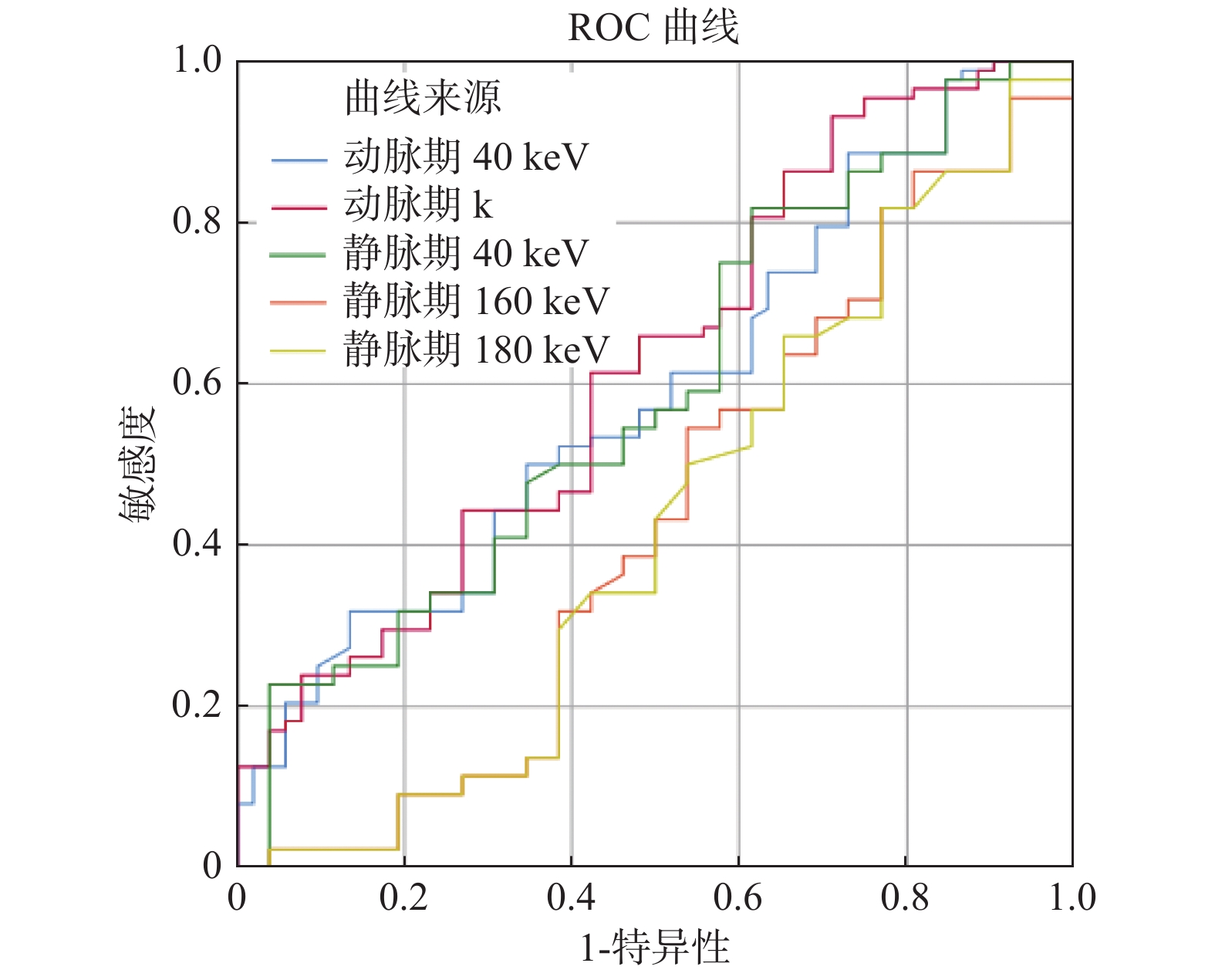
目的 通过分析甲状腺乳头状癌(PTC)癌灶的双能量CT能谱曲线,预测颈部中央区隐匿性淋巴结转移(OLM)的概率,为临床手术方式的制定提供参考。 方法 回顾性收集术前行双能量CT扫描,且术后病理证实为PTC的患者。由放射医师分析CT图像,未检出颈部淋巴结转移者共140例。其中病理证实有中央组淋巴结隐匿性转移者(OLM+)88例、无转移者(OLM−)52例。测量PTC癌灶各单能量图像上的CT值并计算能谱曲线斜率k,分析2组间各参数的差异。 结果 (1)动静脉期OLM+组与OLM−组之间PTC癌灶的karterial、40 keVarterial、40 keVvenous、160 keVvenous、180 keVvenous差异有统计学意义(P均 < 0.050);(2)比较40 keVarterial、karterial、40 keVvenous、160 keVvenous、180 keVvenous的诊断效能,AUC分别为0.590、0.622、0.590、0.429、0.424,当karterial = 2.563时,诊断PTC肿瘤伴中央组OLM的敏感度及准确度分别为93.2%、69.3%;当40 keVvenous = 370.25时,诊断PTC肿瘤伴中央组OLM的特异度为86.5%。 结论 PTC癌灶的能谱曲线预测颈部中央区隐匿性淋巴结转移的有一定的参考价值,其中karterial诊断的敏感度最高、40 keVvenous诊断的特异度最高。 Abstract:Objective To predict the probability of occult lymph node metastasis (OLM) in the central region of the neck by analyzing the dual-energy CT energy spectrum curve of thyroid papillary carcinoma (PTC). Methods Patients with PTC confirmed by pathology before and after operation were retrospectively collected. CT images were analyzed by radiologists. There were 140 cases without cervical lymph node metastasis. Among them, 88 patients with occult lymph node metastasis in the central group (OLM+) and 52 patients without metastasis (OLM−) were confirmed by pathology. The CT value of PTC on each single energy image was measured and the slope (k) of the energy spectrum curve was calculated. The differences in parameters between the two groups were analyzed. Results (1) the karterial, 40 keVarterial, 40 keVvenous, 160 keVvenous and 180 keVvenous of PTC between OLM+ group and OLM− group in arterial and venous phase were significantly different (P < 0.05). (2) The AUC of 40 keVarterial, karterial, 40 keVvenous, 160 keVvenous and 180 keVvenous in PTC were 0.590, 0.622, 0.590, 0.429 and 0.424 respectively. When karterial = 2.563, the sensitivity and accuracy of diagnosing PTC with OLM in the central group were 93.2% and 69.3%. When 40 keVvenous = 370.25, the specificity of PTC with OLM was 86.5%. Conclusion The energy spectrum curve of PTC has a certain reference value in predicting occult lymph node metastasis in the central region of the neck. karterial has the highest sensitivity and 40 keVvenous has the highest specificity. -
图 2 女,35岁,体检发现甲状腺左侧叶结节,病理证实为甲状腺乳头状癌
A、B:动脉期、静脉期40 keV图像,CT值分别为226.4 HU(> 222.15 HU)、229.0 HU(< 370.25 HU);C:能谱曲线,计算能谱曲线斜率为2.747(> 2.563);D:HU染色病理图片,显示有颈部中央区淋巴结转移。
Figure 2. A female patient,35 year old ,nodules in the left lobe of the thyroid gland,pathologically confirmed to be papillary thyroid cancer
表 1 OLM+与OLM−2组间PTC肿瘤各单能量CT值、slope(k)的比较(
$ \bar x \pm s $ )Table 1. Comparison of monoenergy CT values and slope (k) of PTC tumors between OLM+ and OLM− (
$ \bar x \pm s $ )单能量 OLM+ OLM− t P 动脉期 40 keV 335.21 ± 92.81 296.67 ± 80.69 2.489 0.014* 60 keV 175.37 ± 59.11 170.33 ± 46.26 0.527 0.599 80 keV 114.95 ± 30.57 115.23 ± 26.58 0.056 0.956 100 keV 88.87 ± 20.03 90.93 ± 19.89 0.589 0.556 120 keV 75.22 ± 16.24 78.68 ± 18.20 1.162 0.247 140 keV 67.93 ± 14.88 71.91 ± 17.85 1.417 0.159 160 keV 63.66 ± 14.54 67.85 ± 17.84 1.527 0.129 180 keV 60.86 ± 14.48 65.28 ± 17.92 1.595 0.113 k 4.11 ± 1.38 3.43 ± 1.20 2.945 0.004* 静脉期 40 keV 281.12 ± 67.79 253.34 ± 80.71 2.181 0.031* 60 keV 149.80 ± 27.85 143.14 ± 29.51 1.337 0.183 80 keV 99.87 ± 15.28 97.38 ± 18.51 0.859 0.392 100 keV 77.59 ± 12.38 78.88 ± 14.27 0.565 0.573 120 keV 66.31 ± 12.64 69.59 ± 14.16 1.421 0.158 140 keV 60.03 ± 13.31 64.43 ± 14.71 1.819 0.071 160 keV 56.34 ± 13.79 61.33 ± 15.30 1.985 0.049* 180 keV 54.03 ± 13.89 59.38 ± 15.69 2.098 0.038* k 0.42 ± 0.21 0.38 ± 0.17 1.046 0.298 *P < 0.05。 表 2 ROC曲线分析
Table 2. ROC curve analysis
单能量 AUC Youden
指数cut-off
值灵敏度 特异度 准确度 40 keVarterial 0.590 0.220 222.15 81.8% 38.5% 65.7% karterial 0.622 0.203 2.563 93.2% 28.8% 69.3% 40 keVvenous 0.590 0.183 370.25 31.8% 86.5% 52.1% -
[1] Siegel R L,Miller K D,Jemal A. Cancer statistics,2019[J]. CA Cancer J Clin,2019,69(1):7-34. doi: 10.3322/caac.21551 [2] Lee Y K,Hong N,Park S H,et al. The relationship of comorbidities to mortality and cause of death in patients with differentiated thyroid carcinoma[J]. Sci Rep,2019,9(1):11435. doi: 10.1038/s41598-019-47898-8 [3] Lee Y M,Park J H,Cho J W,et al. The definition of lymph node micrometastases in pathologic N1a papillary thyroid carcinoma should be revised[J]. Surgery,2019,165(3):652-656. doi: 10.1016/j.surg.2018.09.015 [4] 陈文达,徐秋贞,王涛,等. 基于非小细胞肺癌原发灶影像组学的隐匿性淋巴结转移预测[J]. 临床放射学杂志,2022,41(4):643-649. doi: 10.13437/j.cnki.jcr.2022.04.008 [5] 王强, 张明星, 金凤山, 等. 基于弹性成像的列线图预测单发临床颈部淋巴结转移阴性甲状腺癌对侧中央淋巴结转移[J], 放射学实践, 2022, 37(5): 632-637. [6] 胡小玲,冉海涛. 超声影像组学评估甲状腺乳头状癌颈部淋巴结转移[J]. 中国超声医学杂志,2022,38(4):367-370. [7] Yeh M W,Bauer A J,Bernet V A,et al. American Thyroid Association statement on preoperative imaging for thyroid cancer surgery[J]. Thyroid,2015,25(1):3-14. doi: 10.1089/thy.2014.0096 [8] 中华医学会放射学分会,中国医师协会放射医师分会,安徽省影像临床医学研究中心. 能量CT临床应用中国专家共识[J]. 中华放射学杂志,2022,56(5):476-487. doi: 10.3760/cma.j.cn112149-20220118-00051 [9] 王学东,刘爱连,田士峰. 单源双能CT能谱成像定量参数评估T2及T3期胃腺癌的价值[J]. 放射学实践,2021,36(11):1408-1413. doi: 10.13609/j.cnki.1000-0313.2021.11.014 [10] 王永丽,杨帆,刘文亚. 能谱CT多参数定量分析预测原发性肺癌病理类型[J]. 中国医学影像技术,2021,37(6):899-903. doi: 10.13929/j.issn.1003-3289.2021.06.025 [11] 朱育婷,陈梦诗,罗敏. 双能量CT小肠造影定量参数评估克罗恩病活动度的可行性研究[J]. 中华炎性肠病杂志,2021,5(4):321-326. doi: 10.3760/cma.j.cn101480-20201230-00142 [12] 宋芹霞,王祥发,刘静,等. 双能量CT成像联合肿瘤指标CEA对晚期肺腺癌EGFR突变的预测价值[J]. 临床放射学杂志,2022,41(5):855-859. doi: 10.13437/j.cnki.jcr.2022.05.012 [13] Wang P,Tang Z,Xiao Z,et al. Dual-energy CT in predicting Ki-67 expression in laryngeal squamous cell carcinoma[J]. Eur J Radiol,2021,140:109774. doi: 10.1016/j.ejrad.2021.109774 [14] Lee J,Song Y,Soh E Y. Central lymph node metastasis is an important prognostic factor in patients with papillary thyroid microcarcinoma[J]. J Korean Med Sci,2014,29(1):48-52. doi: 10.3346/jkms.2014.29.1.48 [15] Kim C, Kim W, Park S J, et al. Application of Dual-Energy Spectral Computed Tomography to Thoracic Oncology Imaging[J]. Korean J Radiol, 2020, 21(7): 838-850. [16] Tian X, Song Q, Xie F, et al. Papillary thyroid carcinoma: an ultrasound-based nomogram improves the prediction of lymph node metastases in the central compartment[J]. Eur J Radiol, 2020, 30(11): 5881-5893. [17] Deniffel D, Sauter A, Dangelmaier J, et al. Differentiating intrapulmonary metastases from different primary tumors via quantitative dual-energy CT based iodine concentration and conventional CT attenuation[J]. Eur J Radiol, 2019, 111: 6-13. [18] 王晨,李绍东,窦沛沛,等. 双能量CT联合肿瘤标记物预测结直肠癌淋巴结转移的价值[J]. 临床放射学杂志,2020,39(12):2481-2485. doi: 10.13437/j.cnki.jcr.2020.12.027 -


 下载:
下载:


