Clinical Application of Ultrasound-guided Peripherally Inserted Central Venous Catheter in Neonates with Vascular Access Difficulties
-
摘要:
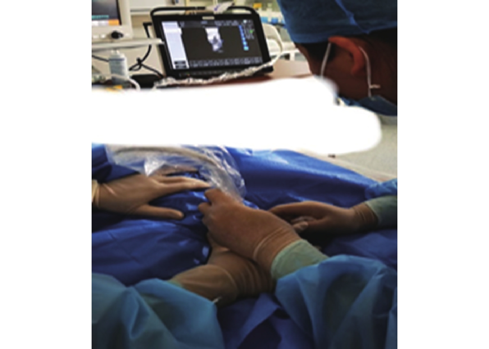
目的 探讨超声引导下经外周置入中心静脉导管(peripherally inserted central catheter,PICC)在新生儿穿刺困难中的临床应用。 方法 选取2019年1月至2021年4月于昆明市儿童医院新生儿科同一组专业的PICC管理团队小组评估为穿刺困难的104例新生儿为研究对象,将其随机分为观察组和对照组,每组各52例。对照组采取传统盲穿置管,观察组采取超声引导下置管,比较2组患儿在穿刺次数、留置时间、一次性穿刺成功率、一次性置管成功率、留置时间等方面的差异。 结果 观察组的平均穿刺次数以及置管时间均少于对照组;观察组一次性穿刺成功率、一次性置管成功率均高于对照组;观察组留置时间长于对照组;差异均有统计学意义 ( P < 0.05)。观察组穿刺时周围组织损伤、经皮氧饱和度下降的发生率均低于对照组;观察组穿刺后维护期并发症的总发生率低于对照组;差异有统计学意义(P < 0.05)。 结论 穿刺困难新生儿行PICC时,经超声引导,在穿刺过程中可有效减少穿刺次数及置管时间,提高一次性穿刺成功率及一次性置管成功率,并且增加了留置时间,值得临床推广。 -
关键词:
- 超声 /
- 新生儿 /
- 经外周置入中心静脉导管 /
- 血管通路 /
- 穿刺困难
Abstract:Objective To investigate the clinical application of peripherally inserted central catheter (PICC) guided by ultrasound in neonates with vascular access difficulties. Methods From January 2019 to April 2021, 104 neonates with vascular access difficulties identified by the PICC management team in the Department of Neonatology of Kunming Children’s Hospital were selected as the research subjects. They were randomly divided into observation group and control group, 52 neonates in each group. The control group received traditional catheterization, while the observation group was treated with ultrasound-guided catheterization. The differences in inserting times, indwelling time, one-time insertion success rate, one-time catheterization success rate, and indwelling time between the two groups were compared. Results The average inserting times and catheterization time of the observation group were less than those of the control group. The success rate of one-time puncture and one-time catheterization in the observation group were higher than those in the control group. The indwelling time in the observation group was longer than that in the control group, and the differences were statistically significant (P < 0.05). The incidence of peripheral tissue injury and percutaneous oxygen saturation decline during puncture in the observation group were lower than those in the control group. The total incidences of postoperative complications in the observation group were lower than that in the control group, the difference was statistically significant (P < 0.05). Conclusion During PICC for neonates with difficult access, ultrasound guidance can effectively reduce the number of puncture and catheterization time, improve the success rate of one-time puncture and one-time catheterization, and increase the indwelling time, which is of clinical application value. -
表 1 2组患儿一般资料比较 [n(%)/(
$ \bar x \pm s$ )]Table 1. Comparison of general data between the two groups [n(%)/(
$ \bar x \pm s$ )]项目 对照组(n = 52) 观察组(n = 52) χ2/t P 性别 0.346 0.556 男性 25(48.1) 28(53.8) 女性 27(51.9) 24(46.2) 穿刺日龄(d) 15.31 ± 9.54 15.21 ± 9.99 0.052 0.958 穿刺体重(kg) 3.30 ± 0.81 3.26 ± 0.79 0.029 0.977 有创呼吸机支持 是 6(11.5) 7(13.5) 0.088 0.767 否 46(88.5) 45(86.5) 无创呼吸机支持 是 11(21.2) 9(17.3) 0.248 0.619 否 41(78.8) 43(82.7) 穿刺部位 0.082 0.941 腋静脉 22(42.3) 17(32.7) 股静脉 16(30.8) 22(42.3) 贵要静脉 2(3.9) 4(7.7) 肘正中静脉 6(11.5) 5(9.6) 颈外静脉 6(11.5) 4(7.7) 表 2 2组患儿穿刺就留置情况比较 (
$ \bar x \pm s$ )Table 2. Comparison of puncture indwelling between the two groups (
$ \bar x \pm s$ )项目 对照组(n = 52) 观察组(n = 52) t/χ2 P 穿刺次数(次) 1.92 ± 0.70 1.21 ± 0.49 5.992 < 0.001* 置管时间(min) 40.77 ± 6.89 38.46 ± 4.45 2.031 0.044* 一次性穿刺成功率 27/52(51.9) 45/52(86.5) 14.625 < 0.001* 一次性置管成功率 24/52(46.2) 44/52(84.6) 16.993 < 0.001* 留置时间(d) 21.13 ± 14.09 27.69 ± 15.78 2.236 0.028* *P < 0.05。 表 3 2组患儿并发症比较 [n(%)]
Table 3. Comparison of complications between the two groups [n(%)]
项目 对照组(n = 52) 观察组(n = 52) χ2 P 穿刺时并发症 周围组织损伤 9(17.3) 2(3.8) 4.981 0.026* 经皮氧饱和度下降 11(21.1) 2(3.8) 7.121 0.008* 穿刺后维护期并发症 静脉血栓 1(1.9) 0(0.0) _# 1.000 导管相关感染 6(11.5) 2(3.8) 2.167 0.141 机械性静脉炎 5(9.6) 2(3.8) 1.378 0.240 *P < 0.05;#Fisher确切概率法。 -
[1] 中国医师协会新生儿科医师分会循证专业委员会. 新生儿经外周置入中心静脉导管操作及管理指南(2021)[J]. 中国当代儿科杂志,2021,23(3):201-212. doi: 10.7499/j.issn.1008-8830.2101087 [2] 于玲,宋淑静,谭彩月,等. B超引导PICC置管中B超探头横轴与纵轴的效果观察[J]. 护理研究,2014,28(36):4545-4546. [3] 蔡志云,李兰,韩秋英. 超声引导下二次改良塞丁格PICC置管术在1~12月龄患儿中的应用[J]. 齐鲁护理杂志,2017,23(16):96-98. doi: 10.3969/j.issn.1006-7256.2017.16.048 [4] 严素芬,谭巧珍,许泽芸. B超定位在早产儿PICC置管中的应用[J]. 齐鲁护理杂志,2020,26(11):128-130. doi: 10.3969/j.issn.1006-7256.2020.11.047 [5] 张丽. B超引导下MST在经外周静脉置入PICC中的应用效果[J]. 国际护理杂杂志,2017,36(19):2726-2728. [6] 陈影洁,陈春贤,简黎,等. B超引导下运用改良塞丁格技术置入PICC的应用[J]. 护理实践与研究,2009,6(10):102-103. doi: 10.3969/j.issn.1672-9676.2009.10.055 [7] 马梦柯,张培莉. PICC相关并发症的影响因素及其护理[J]. 护理研究,2016,30(31):3854-3856. doi: 10.3969/j.issn.1009-6493.2016.31.005 [8] 万永慧,陈芊,邱艳茹. B超引导下经颈内静脉行PICC置管技术在血管通路困难患者中的应用[J]. 护士进修杂志,2016,31(1):68-69. doi: 10.16821/j.cnki.hsjx.2016.01.030 [9] 辛鑫,牛芳芳. 超声引导下PICC置管与传统置管技术成本效果分析研究[J]. 护理管理杂志,2018,18(7):523-526. doi: 10.3969/j.issn.1671-315x.2018.07.016 [10] 许燕萍,商祯茹,Dorazio R M,等. 经外周静脉穿刺中心静脉置管患儿相关性血源感染的危险因素分析[J]. 中国当代儿科杂志,2022,24(2):141-146. [11] 张玮玮,杨峰,王静,等. 超声引导低角度穿刺法在儿科PICC置管中的应用研究[J]. 护士进修杂志,2019,34(23):2178-2181. doi: 10.16821/j.cnki.hsjx.2019.23.015 [12] 王建意. B超引导与传统PICC置管技术的对比研究[J]. 齐鲁护理杂志,2010,16(22):123-124. doi: 10.3969/j.issn.1006-7256.2010.22.089 [13] 王亚静,李杨,孙静,等. 新生儿重症监护病房患儿操作性疼痛现状调查[J]. 护理学杂志,2019,34(11):20-23. doi: 10.3870/j.issn.1001-4152.2019.11.020 [14] 刘玲,周星,朱丽波,等. 静脉内心电图引导经外周中心静脉置管导管尖端定位技术在早产儿的临床应用[J]. 中华新生儿科杂志(中英文),2018,33(6):450-452. doi: 10.3760/cma.j.issn.2096-2932.2018.06.012 [15] 万舸,毛孝容,陈先云. 超声技术在新生儿经外周静脉置入中心静脉导管中的应用研究进展[J]. 实用医院临床杂志,2022,19(1):187-190. doi: 10.3969/j.issn.1672-6170.2022.01.051 [16] 徐云美,姚丽萍,吕晓军,等. 超声引导下贵要静脉穿刺在经外周穿刺中心静脉导管置管术中的应用价值[J]. 中国超声医学杂志,2017,33(6):571-573. doi: 10.3969/j.issn.1002-0101.2017.06.030 [17] 朱丽波,许艳花,胡雪. B超引导联合心房内心电图定位在新生儿PICC中的效果及学习曲线分析[J]. 浙江医学,2021,43(22):2485-2487,2496. doi: 10.12056/j.issn.1006-2785.2021.43.22.2020-3625 [18] 刘玲,朱丽波,奚敏,等. 静脉内心电图引导PICC尖端定位在围术期新生儿的临床应用研究[J]. 中国急救医学,2018,38(z1):299-300. doi: 10.3969/j.issn.1002-1949.2018.z1.287 [19] 任晓玲,陈亚娟,刘敬,等. 超声监测在新生儿经皮外周静脉置入中心静脉导管尖端定位中的应用[J]. 中华实用儿科临床杂志,2019,34(18):1398-1401. doi: 10.3760/cma.j.issn.2095-428X.2019.18.010 [20] 孙利华,朱恩兰,高赟晔,等. 床旁超声多点引导在改良赛丁格技术PICC置管术中的应用[J]. 中国超声医学杂志,2020,36(6):565-567. doi: 10.3969/j.issn.1002-0101.2020.06.025 [21] Xiao W,Yan F,Ji H,et al. A randomized study of a new landmark-guided vs traditional para-carotid approach in internal jugular venous cannulation in infants[J]. Paediatric Anaesthesia,2009,19(5):481-486. doi: 10.1111/j.1460-9592.2009.02989.x [22] Schummer W,Schummer C,Rose N,et a1. Mechanical complications and malpositions of central venous cannulations by experienced operators. Aprospective study of 1794 catheterizations in critically ill patients[J]. Intensive Care Medicine,2007,33(6):1055-1059. doi: 10.1007/s00134-007-0560-z [23] 朱晶文,张雪峰. 早发及晚发型新生儿败血症的临床特征分析[J]. 发育医学电子杂志,2022,10(2):126-131. doi: 10.3969/j.issn.2095-5340.2022.02.008 -


 下载:
下载:


