

Effects of High-frequency Repetitive Transcranial Magnetic Stimulation (rTMS) on Cognitive Function in Patients with Traumatic Brain Injury
-
摘要:
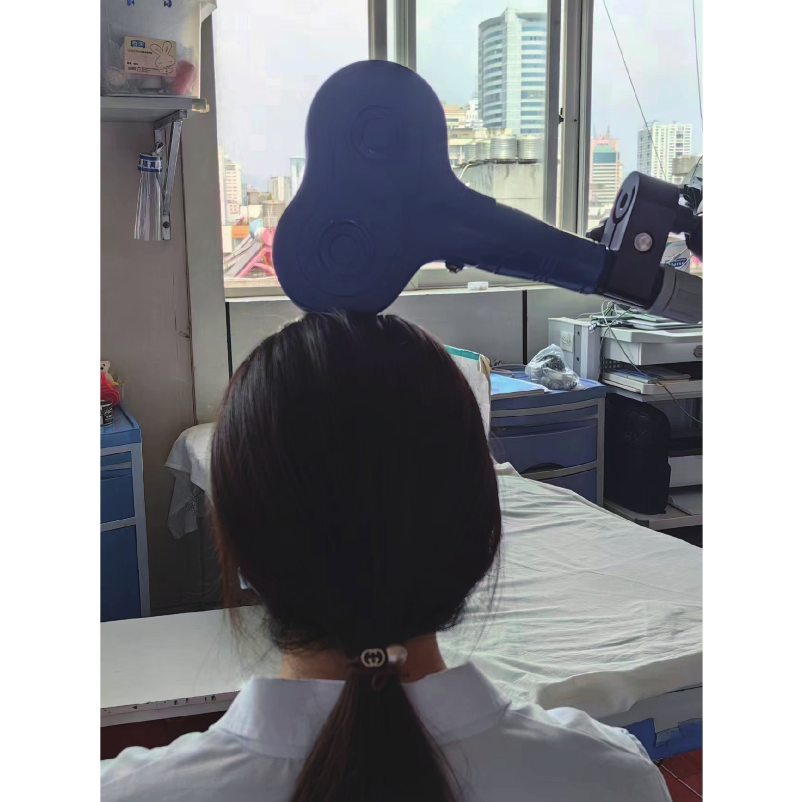
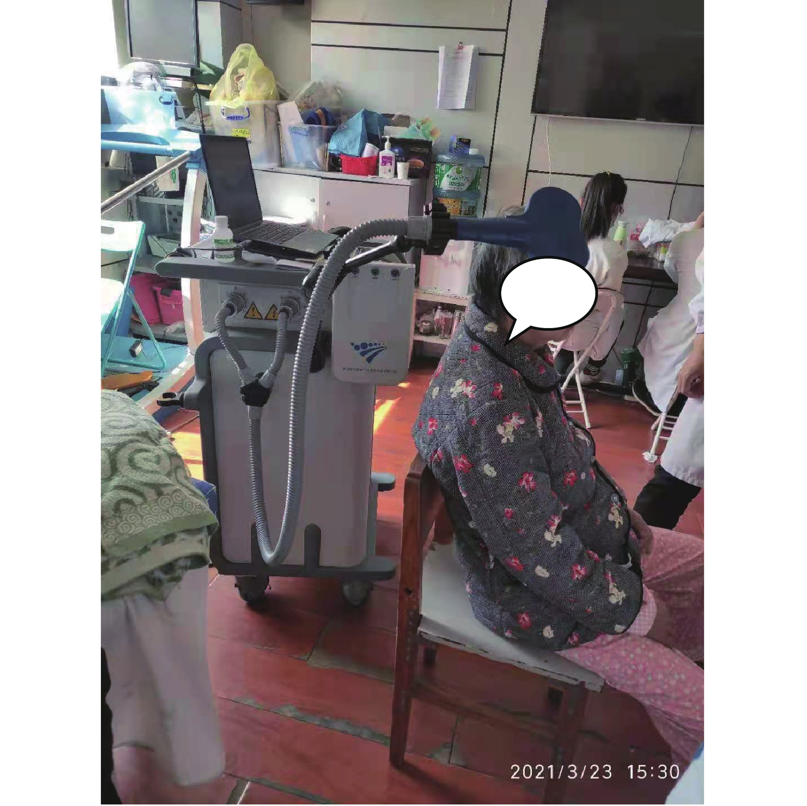
目的 观察高频重复经颅磁刺激(rTMS)对脑外伤患者认知功能的影响。 方法 选取2021年1月至2022年1月昆明市延安医院收治的脑外伤患者160例作为研究对象,按照入院序号采用随机数表法随机分为治疗组、对照组各80例。对照组在常规药物治疗及认知训练的基础上给予rTMS假刺激,治疗组在常规药物治疗及认知训练的基础上给予rTMS真刺激,每日1次,每周治疗6d,共治疗4周。分别于治疗前、治疗4周后采用简易精神状态检查(mini-mental state examination,MMSE)、Rivermead行为记忆测验(rivermead behavioral memory test,RBMT)、蒙特利尔认知评估量表(montreal cognitive assessment,MoCA)对2组患者的认知功能进行评定。 结果 组内比较:治疗组MMSE评分指标(定向能力、记忆能力、回忆能力、计算能力)评分均较治疗前提高(P < 0.05),对照组MMSE评分指标(仅定向能力、即刻记忆能力)评分较治疗前提高(P < 0.05);治疗组RBMT评分指标(记姓、名,记被藏物、记约定、图片再认、即刻路径记忆、定向)评分均较治疗前提高(P < 0.05),对照组RBMT评分指标(仅记姓、名,记被藏物、图片再认、定向)评分较治疗前提高(P < 0.05);治疗组MoCA评分指标(记忆、语言、计算、定向)评分较治疗前提高(P < 0.05),对照组MoCA评分指标仅记忆、定向)评分较治疗前提高(P < 0.05);组间比较:治疗后,治疗组MMSE总分、RBMT总分、MoCA总分均较对照组提高(P < 0.05)。 结论 高频rTMS治疗能有效改善脑外伤患者的认知功能。 Abstract:Objective To observe the effects of rTMS on cognitive function in patients with traumatic brain injury. Methods A total of 160 patients with traumatic brain injury admitted to Yan’an Hospital of Kunming City from January 2021 to January 2022 were selected as the research objects and randomly divided according to the admission sequence number by random number table method. There were 80 cases in the treatment group and 80 cases in the control group. The control group was given sham rTMS stimulation on the basis of conventional drug treatment and cognitive training, while the treatment group was given true rTMS stimulation on the basis of conventional drug treatment and cognitive training, once a day, for a total of 4 weeks. Mini-Mental State Examination (Mini-mental State Examination) was performed before and 4 weeks after treatment. The cognitive function of the two groups was evaluated by MoCA, MMSE, RBMT. Results Intra-group comparison: the MMSE scores (orientation ability, memory ability, recall ability, calculation ability) of the treatment group were all improved compared with those before treatment (P < 0.05), while the MMSE scores (orientation ability, immediate memory ability) of the control group were only improved compared with those before treatment (P < 0.05). In the treatment group, the RBMT scores (remembering surname and given name, remembering artifact, remembering convention, picture recognition, immediate path memory, orientation) were all in- creased compared with those before treatment (P < 0.05), while in the control group, the RBMT scores (remembering surname and given name, remembering artifact, picture recognition, orientation) were only increased compared with those before treatment (P < 0.05). The MoCA scores (memory, language, calculation, orientation) in the treatment group were higher than those before treatment (P < 0.05), while the MoCA scores (memory, orientation) in the control group were only higher than those before treatment (P < 0.05). Comparison between groups: After treatment, the total scores of MMSE, RBMT and MoCA in the treatment group were higher than those in the control group (P < 0.05). Conclusion rTMS can effectively improve the cognitive function of patients with traumatic brain injury. -
表 1 2组脑外伤患者一般情况比较(
$\bar x \pm s $ )Table 1. Comparison of general conditions of TBI patients between the two groups(
$\bar x \pm s $ )组别 n 性别(n) 年龄(岁) 病程(月) 男 女 治疗组 80 57 23 52.1 ± 2.5 1.7 ± 0.9 对照组 80 65 15 49.6 ± 2.9 2.1 ± 1.1  下载: 导出CSV
表 2 2组患者治疗前、后MMSE量表得分项评分比较[(
$\bar x \pm s $ ),分]Table 2. Comparison of MMSE score items between the two groups before and after treatment [(
$\bar x \pm s $ ),scores]评估指标 组别(n = 80) 治疗前 治疗后 治疗前后组内比较 t P 定向能力 治疗组 5.32 ± 1.49 7.46 ± 1.12 −18.563 < 0.001* 对照组 5.21 ± 1.32 6.75 ± 1.23 −10.234 < 0.001* 即刻记忆 治疗组 2.34 ± 0.68 2.85 ± 0.32 −2.853 0.003* 对照组 2.41 ± 0.70 2.86 ± 0.43 −3.206 0.012* 专注与计算能力 治疗组 1.13 ± 0.72 2.13 ± 1.32 −8.065 < 0.001* 对照组 1.06 ± 0.58 1.05 ± 0.62 0.483 0.625 回忆能力 治疗组 1.52 ± 0.76 1.89 ± 0.80 −4.294 < 0.001* 对照组 1.48 ± 0.82 1.40 ± 0.86 0.489 0.605 语言能力 治疗组 4.62 ± 3.12 4.64 ± 3.02 −1.245 0.143 对照组 4.71 ± 3.46 4.70 ± 3.41 0.325 0.321 *P < 0.05。
下载: 导出CSV
表 3 2组患者治疗前、后RBMT量表得分项评分比较[(
$\bar x \pm s $ ),分]Table 3. Comparison of RBMT scale score items between the two groups before and after treatment [(
$\bar x \pm s $ ),scores]评估指标 组别(n = 80) 治疗前 治疗后 治疗前后组内比较 t P 记姓和名 治疗组 0.52 ± 0.18 0.75 ± 0.12 −2.563 0.004* 对照组 0.43 ± 0.22 0.61 ± 0.23 −2.234 0.006* 记被藏物 治疗组 0.34 ± 0.28 0.55 ± 0.32 −2.853 0.003* 对照组 0.41 ± 0.30 0.56 ± 0.43 −2.206 0.008* 记约定 治疗组 0.28 ± 0.12 0.42 ± 0.32 −2.065 0.005* 对照组 0.31 ± 0.18 0.30 ± 0.22 0.483 0.625 治疗组 0.48 ± 0.16 0.71 ± 0.20 −2.594 0.004* 图片再认故事即时回忆 对照组 0.39 ± 0.22 0.56 ± 0.26 −2.289 0.007* 治疗组 0.22 ± 0.12 0.23 ± 0.21 0.462 0.643 对照组 0.29 ± 0.16 0.28 ± 0.41 0.325 0.321 故事延迟回忆 治疗组 0.18 ± 0.09 0.19 ± 0.11 0.465 0.603 对照组 0.19 ± 0.06 0.17 ± 0.06 0.394 0.358 治疗组 0.38 ± 0.18 0.37 ± 0.21 0.415 0.523 脸部再认路线即时回忆 对照组 0.35 ± 0.10 0.33 ± 0.17 0.512 0.572 治疗组 0.41 ± 0.28 0.51 ± 0.30 −2.352 0.006* 对照组 0.38 ± 0.21 0.38 ± 0.19 0.385 0.395 信件即时回忆 治疗组 0.31 ± 0.15 0.30 ± 0.21 0.465 0.612 对照组 0.36 ± 0.18 0.35 ± 0.24 0.412 0.583 定向和日期 治疗组 0.41 ± 0.28 0.56 ± 0.25 −1.965 0.005* 对照组 0.43 ± 0.19 0.54 ± 0.18 −1.896 0.005* 治疗组 0.19 ± 0.08 0.18 ± 0.15 0.403 0.576 路线延迟回忆 对照组 0.18 ± 0.07 0.16 ± 0.09 0.398 0.376 信件延迟回忆 治疗组 0.16 ± 0.09 0.17 ± 0.11 0.482 0.652 对照组 0.19 ± 0.05 0.18 ± 0.09 0.365 0.403 *P < 0.05。
下载: 导出CSV
表 4 2组患者治疗前、后MoCA量表得分项评分比较[(
$\bar x \pm s $ ),分]Table 4. Comparison of scores of MoCA scale before and after treatment between the two groups [(
$\bar x \pm s $ ),scores]评估指标 组别(n = 80) 治疗前 治疗后 治疗前后组内比较 t P 定向能力 治疗组 3.32 ± 1.49 4.46 ± 1.12 −8.563 < 0.001* 对照组 3.21 ± 1.32 4.05 ± 1.23 −7.234 < 0.001* 执行功能 治疗组 2.34 ± 0.68 2.35 ± 0.32 0.356 0.603 对照组 2.41 ± 0.70 2.39 ± 0.43 0.623 0.312 命名 治疗组 1.23 ± 0.72 2.13 ± 1.32 −8.065 < 0.001* 对照组 1.36 ± 0.48 2.34 ± 0.62 −7.253 0.003* 注意力 治疗组 2.42 ± 0.12 3.69 ± 0.02 −6.689 0.002* 对照组 2.67 ± 1.49 2.65 ± 1.12 0.563 0.456 语言能力 治疗组 1.32 ± 0.49 2.16 ± 0.12 −7.563 0.002* 对照组 1.03 ± 0.21 1.01 ± 0.19 0.503 0.712 抽象能力 治疗组 0.34 ± 0.08 0.35 ± 0.04 0.553 0.503 对照组 0.41 ± 0.02 0.40 ± 0.03 0.426 0.512 选项 治疗组 2.13 ± 0.72 2.13 ± 1.32 0.395 0.601 对照组 2.06 ± 0.58 1.95 ± 0.62 0.682 0.325 *P < 0.05。
下载: 导出CSV
表 5 2组患者治疗前后MMSE量表总分比较[(
$\bar x \pm s $ ),分]Table 5. Comparison of total score of MMSE before and after treatment between the two groups [(
$\bar x \pm s $ ),scores]组别 n 治疗前 治疗后 t P 治疗组 80 15.64 ± 4.08 21.45 ± 4.08 −18.563 < 0.001* 对照组 80 15.21 ± 5.12 16.75 ± 4.22 −9.234 < 0.001* t(组间) −0.258 2.684 P(组间) 0.856 0.015* *P < 0.05。
下载: 导出CSV
表 6 2组患者治疗前后RBMT量表总分比较[(
$\bar x \pm s $ ),分]Table 6. Comparison of total scores of RBMT before and after treatment between the two groups [(
$ \bar x \pm s $ ),scores]组别 n 治疗前 治疗后 t P 治疗组 80 13.64 ± 4.18 17.45 ± 5.08 2.563 0.006* 对照组 80 14.01 ± 4.12 14.95 ± 4.72 2.066 0.037* t(组间) 0.248 1.064 P(组间) 0.816 0.021* *P < 0.05。
下载: 导出CSV
表 7 2组患者治疗前后MoCA量表总分比较[(
$ \bar x \pm s $ ),分]Table 7. Comparison of total score of MoCA scale before and after treatment between the two groups [(
$ \bar x \pm s $ ),scores]组别 n 治疗前 治疗后 t P 治疗组 80 19.34 ± 3.58 26.45 ± 3.08 −19.253 < 0.001* 对照组 80 19.21 ± 3.12 22.75 ± 3.22 −12.264 < 0.001* t(组间) 0.068 4.684 P(组间) 0.956 < 0.001* *P < 0.05。
下载: 导出CSV
-
[1] Dewan M C,Rattani A,Gupta S,et al. Estimating the global incidence of traumatic brain injury[J]. J Neurosurg,2018,130(4):1080-1097. [2] Jiang J Y,Gao G Y,Feng J F,et al. Traumatic brain injury in China[J]. Lancet Neurol,2019,18(3):286-295. doi: 10.1016/S1474-4422(18)30469-1 [3] Pearce A J,Kidgell D J,Tommerdahl M A,et al. Chronic neuro-physiologic-al effects of repeated head trauma in retired Australian male sport athletes[J]. Front Neurol,2021,12(4):633320. [4] Azouvi P,Arnould A,Dromer E,et al. Neuropsychology of traumatic brain injury: An expert overview[J]. Rev Neurol(Paris),2018,73(7-8):461-472. [5] 张雪茹,郝习君,李朝征,等. 脑外伤患者认知功能障碍的危险因素[J]. 中国康复理论与实践,2022,28(2):212-218. doi: 10.3969/j.issn.1006-9771.2022.02.012 [6] Widhalm M L,Rose N S. How can transcranial magnetic stimul- ation be used to causally manipulate memory representations in the human brain?[J]. Wiley Interdiscip Rev Cogn Sci,2019,10(1):e1469. [7] 廖亮华,黄东,江兴妹,等. 高频与低频重复经颅磁刺激对脑梗死患者认知功能的影响[J]. 中华物理医学与康复杂志,2018,39(1):56-58. [8] Padala P R,Padala K P,Lensing S Y,et al. Repetitive transcranial magnetic stimulation for apathy in mild cognitive im- pairment: A double-blind randomized,shamcontrolled,cross -over pilot study[J]. Psychiatry R- es,2018,261(6):312-318. [9] Lee S A,Kim M K. Effect of low frequency repetitive transcranial magnetic stimulation on depression and cognition of patients with traumatic brain injury: A randomized controlled trial[J]. Med Sci Monit,2018,2(2):8789-8794. [10] Nardone R,Sebastianelli L,Versace V,et al. Repetitive transcranial magnetic stimulation in traumatic brain injury: Evidence from animal and human studies[J]. Brain Res Bull,2020,159(8):44-52. [11] Ramesh V J,Chakrabarti D. Letter: Guidelines for the management of severe traumatic brain injury,fourth edition[J]. Neurosurgery,2018,82(5):E143. doi: 10.1093/neuros/nyy029 [12] 韩凯月,党辉,张皓. 重复经颅磁刺激治疗脑外伤患者认知障碍的Meta分析[J]. 中国康复理论与实践,2021,27(11):1283. [13] Folstein M F,Folstein S E,Mchugh P R. "Mini-mental state". A practical method for grading the cognitive state of patients for the clinici- an[J]. J Psychiatr Res,1975,12(3):189-198. doi: 10.1016/0022-3956(75)90026-6 [14] 刘晶京, 恽晓平. Rivermead行为记忆量表-Ⅲ的汉化版研究[C]. 中华医学会第十五次全国物理医学与康复学学术会议, 中国江西南昌, 2014. [15] Yu J,Li J,Huang X. The beijing version of the montreal cognitive assessment as a brief screening tool for mild cognitive impairment: A community-based study[J]. BMC Psychiatry,2012,12(1):156-156. doi: 10.1186/1471-244X-12-156 [16] 孙国生,丰育功. 重复经颅磁刺激联合奥拉西坦治疗轻度脑外伤后认知功能障碍的疗效观察[J]. 中西医结合心血管病电子杂志,2020,8(29):55. [17] Fraser E E,Downing M G,Biernacki K,et al. Cognitive reserve and age predict cognitive recovery after mild to severe traumatic brain injury[J]. J Neurotrauma,2019,36(19):2753-2761. doi: 10.1089/neu.2019.6430 [18] Lee E, Jayasinghe N, Swenson C, et al. Dispositional optimism and cogn- itive functioning following traumatic brain injury[J]. Brain Inj, 20 19, 33(8): 985-990. [19] Jonas S,Asta K H,Turid F,et al. Cognitive reserve moderates cognitive outcome after mild traumatic brain injury[J]. Arch Phys Med Rehabil,2020,101(1):72-80. doi: 10.1016/j.apmr.2019.08.477 [20] Ware J B,Dolui S,Duda J,et al. Relationship of cerebral blood flow to cognitive function and recovery in early chronic traumatic brain in- jury[J]. J Neurotrauma,2020,37(20):2180-2187. doi: 10.1089/neu.2020.7031 [21] Boes A D,Kelly M S,Trapp N T,et al. Noninvasive brain stimulation: c- hallenges and opportunities for a new clinical specialty[J]. J Neu- ropsychiatry Clin Neuros-Ci,2018,30(3):173-179. doi: 10.1176/appi.neuropsych.17110262 [22] Zhang F,Qin Y,Xie L,et al. High frequency repetitive transcranial magnetic stimulation combined with cognitive training improves cognitive function and cortical metabolic ratios in Alzheimer’s disease[J]. J Neural Transm (Vienna),2019,126(8):1081-1094. doi: 10.1007/s00702-019-02022-y [23] 徐高静,吴毅. 康复治疗新技术对脑卒中后脑可塑性影响的研究进展[J]. 中华物理医学与康复杂志,2019,41(2):150-153. doi: 10.3760/cma.j.issn.0254-1424.2019.02.018 [24] 许明军,穆敬平,邱良玉,等. 脑卒中后认知障碍的国内外研究进展[J]. 按摩与康复医学,2021,12(24):76. doi: 10.19787/j.issn.1008-1879.2021.24.023 [25] Hoy K E,Mcqueen S,Elliot D,et al. A pilot investigation of repetitive transcranial magnetic stimulation for post-traumatic brain injury depression: Safety,tolerability,and efficacy[J]. J Neurotrauma,2019,36(13):2092-2098. doi: 10.1089/neu.2018.6097 -


 点击查看大图
计量
- 文章访问数: 3461
- HTML全文浏览量: 1952
- PDF下载量: 23
- 被引次数: 0
