

Value Evaluation of New Markers AFP-L3 and PIVKAⅡ in the Diagnosis of Liver Cancer
-
摘要:
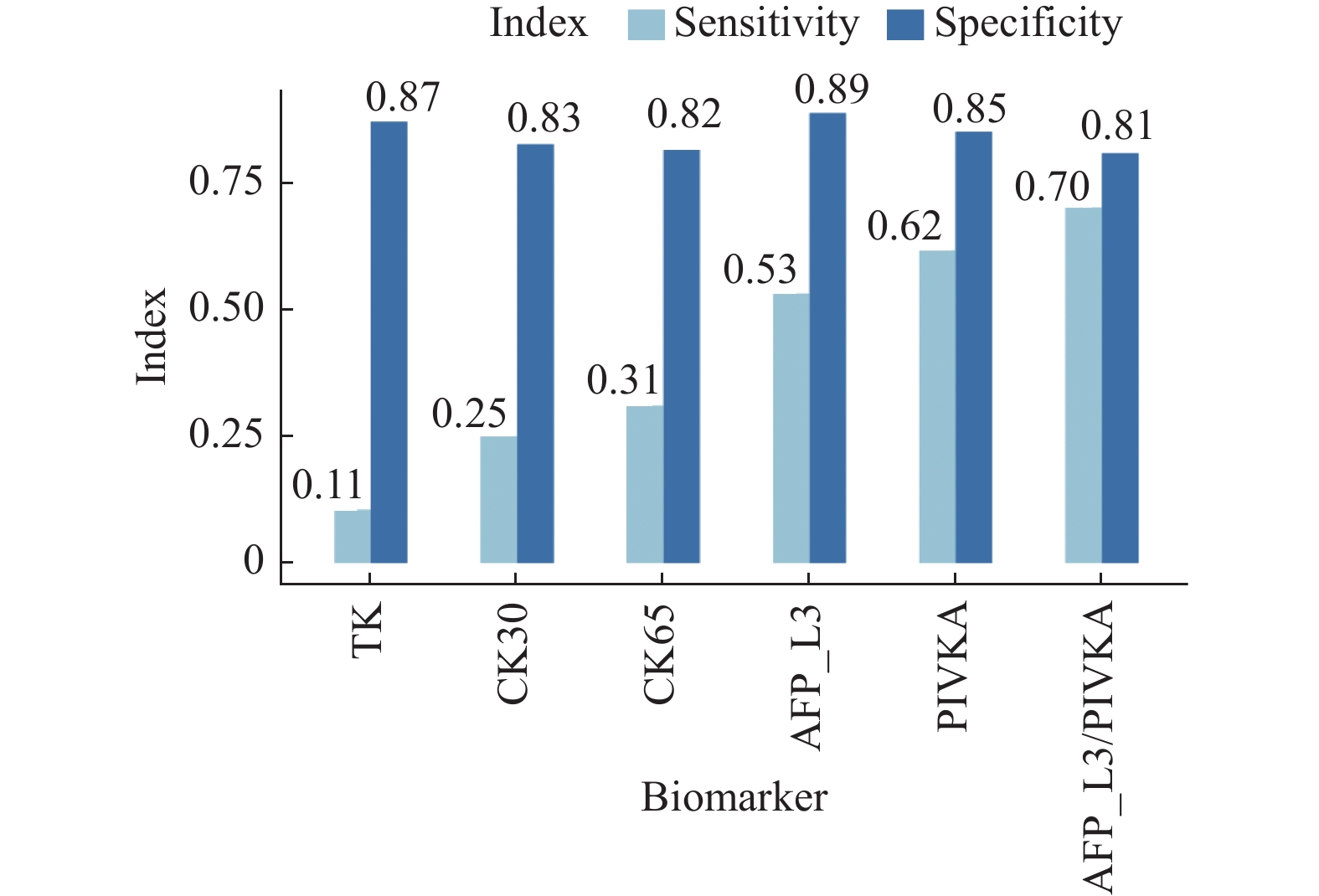
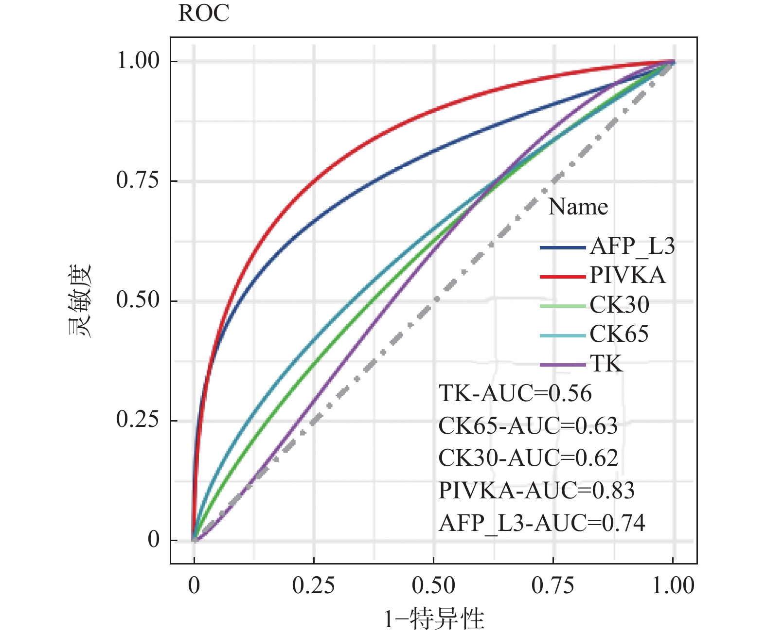
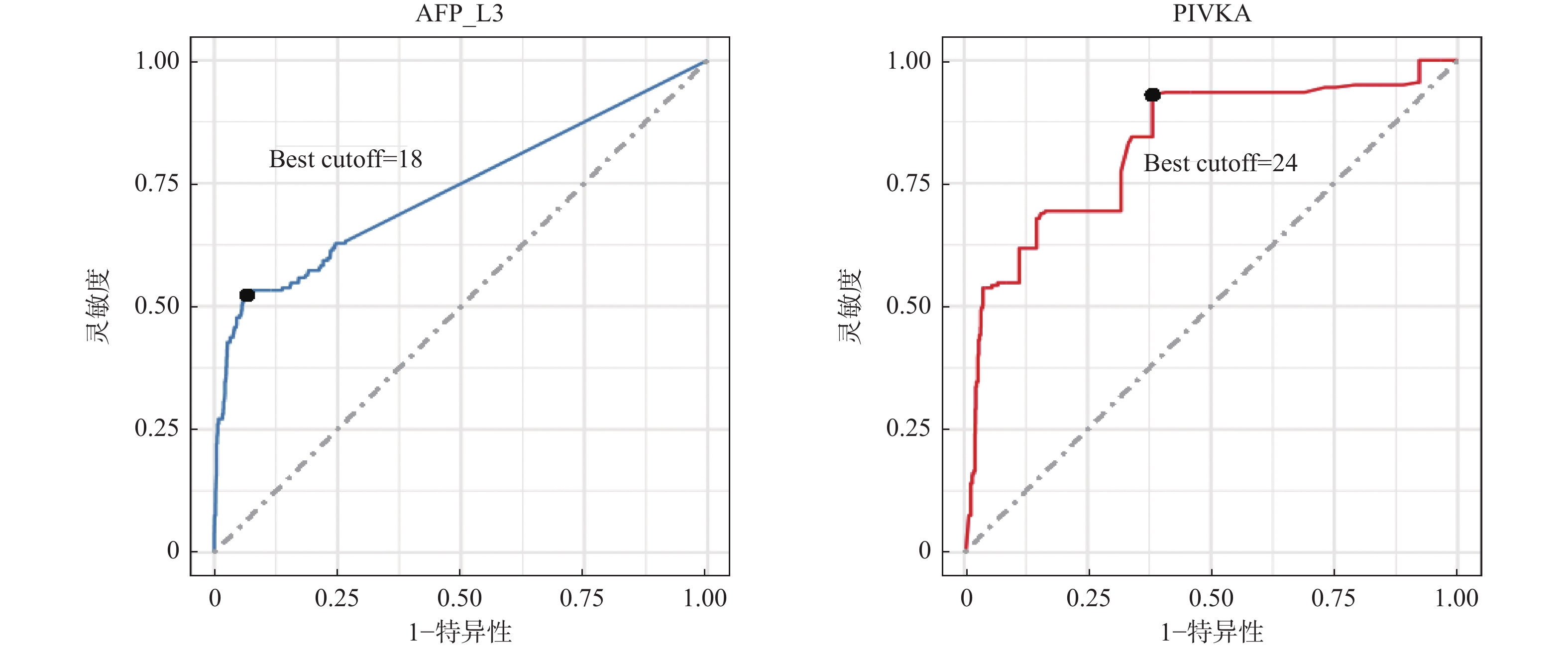
目的 探讨甲胎蛋白异质体L3(alpha-fetoprotein variants L3,AFP-L3)和维生素K缺乏或拮抗剂-Ⅱ诱导的蛋白质(protein induced by vitamin K absence or antagonist II,PIVKAⅡ)在肝癌诊断中的的价值。 方法 收集2019年1月至2022年1月期间青岛市第六人民医院收治的3 066例患有慢性肝脏疾病患者作为研究对象。比较AFP-L3、PIVKAⅡ、CK65、CK30、TK1在肝炎、肝硬化以及肝癌间的水平差异,评估特异性、敏感度,应用受试者工作特征曲线(receiver operating characteristic,ROC)曲线评估生物标志物及提出最优截断点。 结果 AFP-L3和PIVKA在肝硬化组中表达高于肝炎组(P < 0.0001),CK65、CK30和TK在肝硬化组中表达低于肝炎组(P < 0.001)。肝癌组中AFP-L3、CK65、CK30和PIVKA表达显著高于肝硬化组(P < 0.0001),TK在肝癌组中的表达低于肝硬化组(P > 0.05)。在纳入研究的5种生物标志物中,AFP-L3具有最高的特异度(0.89),PIVKAⅡ具有最高的灵敏度(0.62),且联合应用AFP-L3和PIVKAⅡ灵敏度达到0.70,特异度达到0.81,有相较于单个标志物更优的表现。ROC曲线分析表明PIVKAⅡ具有更高的诊断价值(24)。 结论 在肝癌筛查中AFP-L3比PIVKAⅡ特异性高,联合AFP-L3和PIVKAⅡ诊断可其在肝癌中的诊断价值。 -
关键词:
- 肝癌 /
- 甲胎蛋白异质体L3 /
- 维生素K缺乏或拮抗剂-Ⅱ诱导的蛋白质 /
- 诊断价值
Abstract:Objective To investigate the diagnostic value of AFP-L3 and PIVKAⅡ in liver cancer. Methods A total of 3066 patients with chronic liver disease who were admitted to Qingdao Sixth People’s Hospital from January 2019 to January 2022 were selected as objects of the research. The levels of AFP-L3, PIVKAⅡ, CK65, CK30, and TK1 in hepatitis, liver cirrhosis, and liver cancer were compared. The specificity and sensitivity were evaluated, and the ROC curve was used to evaluate the biomarkers and propose the optimal cutoff point. Results The expression of AFP-L3 and PIVKA in the liver cirrhosis group was higher than that in the hepatitis group (P < 0.0001), while the expression of CK65, CK30, and TK in liver cirrhosis group was lower than that in the hepatitis group (P < 0.001). The expression of AFP-L3, CK65, CK30 and PIVKA in the liver cancer group was significantly higher than that in the liver cirrhosis group (P < 0.0001), while the expression of TK in the liver cancer group was lower than that in the liver cirrhosis group (P > 0.05). Among the five biomarkers included in the study, AFP-L3 had the highest specificity (0.89) and PIVKAⅡ had the highest sensitivity (0.62), and the combined application of AFP-L3 and PIVKAⅡ had a sensitivity of 0.70 and a specificity of 0.81, which was better than a single marker. The analysis of ROC curve showed that PIVKAⅡ had a higher diagnostic value (24). Conclusion AFP-L3 is more specific than PIVKAⅡ in liver cancer screening, and the combination of AFP-L3 and PIVKAⅡ diagnosis may have diagnostic value in liver cancer. -

图 1 5种生物标志物在不同肝脏疾病中的测出水平比较
A:AFP-L3;B:CK65;C:CK30;D:PIVKA;E:TK1。图中性别的统计数据表示为[n(%)],年龄、AFP-L3 (%)、PIVKAⅡ (mAU/mL)、CK65 (U/L)、CK30 (U/L)、TK1 (Pmol/L)的统计数据表示为M(P25,P75),*P < 0.05,**P < 0.01,***P < 0.001,****P < 0.0001,ns:差异无统计学意义。
Figure 1. Comparison of measured levels of five biomarkers in different liver diseases

图 2 5种生物标志物诊断肝癌的特异性与灵敏度
Figure 2. Specificity and sensitivity of five biomarkers in diagnosis of liver cancer

图 3 5种生物标志物诊断肝癌的ROC曲线和AUC
Figure 3. ROC curve and AUC of five biomarkers in the diagnosis of liver cancer
表 1 临床信息与生物标志物在不同肝脏疾病中的总结[n(%)/M(P25,P75)]
Table 1. Summary of Clinical Characteristics and Biomarker Levels [n(%)/M(P25,P75)]
项目 肝炎组(n = 1194) 肝硬化组(n = 1673) 肝癌组(n = 199) 共计(n = 3066) Kruskal Wallis P 性别 女 496 (41.54) 582 (34.79) 12 (6.03) 1090 (35.55) / / 男 698 (58.46) 1091 (65.21) 187 (93.97) 1976 (64.45) 年龄(岁) 48.15 (39.00,57.00) 54.80 (48.00,62.00) 55.17 (48.00,62.00) 52.23 (44.75,61.00) 206.20 < 0.001* AFP-L3 2.2576 (0.00,0.00) 5.1586 (0.00,2.25) 32.44 (0.00,70.30) 5.92 (0.00,2.88) 200.40 < 0.001* PIVKAⅡ 124.98 (17.50,27.00) 1239.51 (17.08,31.38) 6357.29 (26.09,9021.65) 1137.65 (17.67,30.04) 185.10 < 0.001* CK65 502.10 (142.38,432.58) 376.23 (137.39,348.73) 876.49 (163.88,602.46) 457.72 (142.32,392.97) 47.41 < 0.001* CK30 222.41 (69.50,191.44) 159.56 (64.52,130.58) 233.51 (88.66,200.63) 188.84 (66.89,160.22) 78.64 < 0.001* TK1 2.10 (0.57,1.32) 1.80 (0.72,1.50) 1.54 (0.82,1.43) 1.90 (0.67,1.42) 41.65 < 0.001* *P < 0.05。  下载: 导出CSV
-
[1] Lee S E,Alcedo K P,Kim H J,et al. Alternative splicing in hepatocellular carcinoma[J]. Cell Mol Gastroenterol Hepatol,2020,10(4):699-712. doi: 10.1016/j.jcmgh.2020.04.018 [2] Wallace M C,Preen D,Jeffrey G P,et al. The evolving epidemiology of hepatocellular carcinoma: A global perspective[J]. Expert Rev Gastroenterol Hepatol,2015,9(6):765-779. doi: 10.1586/17474124.2015.1028363 [3] Anwanwan D,Singh S K,Singh S,et al. Challenges in liver cancer and possible treatment approaches[J]. Biochim Biophys Acta Rev Cancer,2020,1873(1):188314. doi: 10.1016/j.bbcan.2019.188314 [4] Siegel R L,Miller K D,Fuchs H E,et al. Cancer statistics,2021[J]. CA Cancer J Clin,2021,71(1):7-33. doi: 10.3322/caac.21654 [5] Daniele B, Bencivenga A, Megna A S, et al. Alpha-fetoprotein and ultrasonography screening for hepatocellular carcinoma[J]. Gastroenterology, 2004, 127(5 Suppl 1): S108-S112. [6] Liu S,Wang M,Zheng C,et al. Diagnostic value of serum glypican-3 alone and in combination with AFP as an aid in the diagnosis of liver cancer[J]. Clin Biochem,2020,79:54-60. doi: 10.1016/j.clinbiochem.2020.02.009 [7] Ibrahim H M,Elghannam M Z,Elkhawaga O a Y,et al. Evaluation of serum alpha fetoprotein-L3 as an accuracy novel biomarker for the early diagnosis of hepatocellular carcinoma in Egyptian patients[J]. Saudi J Biol Sci,2021,28(10):5760-5764. doi: 10.1016/j.sjbs.2021.06.020 [8] Chaiteerakij R,Zhang X,Addissie B D,et al. Combinations of biomarkers and Milan criteria for predicting hepatocellular carcinoma recurrence after liver transplantation[J]. Liver Transpl,2015,21(5):599-606. doi: 10.1002/lt.24117 [9] Liebman H A,Furie B C,Tong M J,et al. Des-gamma-carboxy (abnormal) prothrombin as a serum marker of primary hepatocellular carcinoma[J]. N Engl J Med,1984,310(22):1427-1431. doi: 10.1056/NEJM198405313102204 [10] 王贵强,王福生,庄辉,等. 慢性乙型肝炎防治指南(2019年版)[J]. 中国病毒病杂志,2020,10(1):1-25. [11] 中国抗癌协会肝癌专业委员会. 中国肿瘤整合诊治指南(CACA)-肝癌部分[J]. 肿瘤综合治疗电子杂志,2022,8(3):31-63. [12] Wei W,Zeng H,Zheng R,et al. Cancer registration in China and its role in cancer prevention and control[J]. Lancet Oncol,2020,21(7):e342-e349. doi: 10.1016/S1470-2045(20)30073-5 [13] Broadfield L A,Duarte J a G,Schmieder R,et al. Fat induces glucose metabolism in nontransformed liver cells and promotes liver tumorigenesis[J]. Cancer Res,2021,81(8):1988-2001. doi: 10.1158/0008-5472.CAN-20-1954 [14] Franssen B,Jibara G,Tabrizian P,et al. Actual 10-year survival following hepatectomy for hepatocellular carcinoma[J]. HPB (Oxford),2014,16(9):830-835. doi: 10.1111/hpb.12206 [15] Zhou J M,Wang T,Zhang K H. AFP-L3 for the diagnosis of early hepatocellular carcinoma: A meta-analysis[J]. Medicine (Baltimore),2021,100(43):e27673. [16] Chen Y,Luo C,Zhao M,et al. Administration of a PTEN inhibitor BPV(pic) attenuates early brain injury via modulating AMPA receptor subunits after subarachnoid hemorrhage in rats[J]. Neurosci Lett,2015,588:131-136. doi: 10.1016/j.neulet.2015.01.005 [17] Li J,Gao T,Gu S,et al. An electrochemical biosensor for the assay of alpha-fetoprotein-L3 with practical applications[J]. Biosens Bioelectron,2017,87:352-357. doi: 10.1016/j.bios.2016.08.071 [18] Wei T,Zhang W,Tan Q,et al. Electrochemical assay of the alpha fetoprotein-l3 isoform ratio to improve the diagnostic accuracy of hepatocellular carcinoma[J]. Anal Chem,2018,90(21):13051-13058. doi: 10.1021/acs.analchem.8b04045 [19] Kotwani P,Chan W,Yao F,et al. DCP and AFP-L3 are complementary to AFP in predicting high-risk explant features: Results of a prospective study[J]. Clin Gastroenterol Hepatol,2022,20(3):701-703.e2. doi: 10.1016/j.cgh.2021.01.043 [20] Fang Y S,Wu Q,Zhao H C,et al. Do combined assays of serum AFP,AFP-L3,DCP,GP73,and DKK-1 efficiently improve the clinical values of biomarkers in decision-making for hepatocellular carcinoma? A meta-analysis[J]. Expert Rev Gastroenterol Hepatol,2021,15(9):1065-1076. doi: 10.1080/17474124.2021.1900731 [21] Abdel-Aziz M M,Elshal M F,Abass A T,et al. Comparison of AFP-L3 and p53 antigen concentration with alpha-fetoprotein as serum markers for hepatocellular carcinoma[J]. Clin Lab,2016,62(6):1121-1129. [22] Hadi H,Wan Shuaib W M A,Raja Ali R A,et al. Utility of PIVKA-II and AFP in differentiating hepatocellular carcinoma from non-malignant high-risk patients[J]. Medicina (Kaunas),2022,58(8):1015. [23] Sun X,Mei J,Lin W,et al. Reductions in AFP and PIVKA-II can predict the efficiency of anti-PD-1 immunotherapy in HCC patients[J]. BMC Cancer,2021,21(1):775. doi: 10.1186/s12885-021-08428-w -

 点击查看大图

计量
- 文章访问数: 3326
- HTML全文浏览量: 2237
- PDF下载量: 34
- 被引次数: 0
