

The Application Value of 2D Ultrasound Combined with SWE Technique in the Evaluation of Rectus Abdominis in Pregnant Women
-
摘要:
目的 采用二维超声联合实时剪切波弹性成像技术( shear wave elastography,SWE)定量评估初产妇孕期、产后早期腹直肌间距(inter-rectus distance,IRD)及硬度,探讨初产妇孕产期IRD与硬度相关性。 方法 选取2021年10月至2022年8月于云南大学附属医院定期产检并足月分娩的初产妇250例,孕期根据孕周分为早孕组、中孕组和晚孕组,产后根据分娩方式分为顺产组和剖宫产组,另选取健康未孕女性50例为对照组。采用二维超声测量IRD,SWE测量腹直肌弹性模量平均值(Emean);比较初产妇孕期及产后IRD及腹直肌硬度的差异,分析二者之间的相关性。 结果 孕期IRD和腹直肌硬度随孕周增加呈上升趋势,差异有统计学意义(P < 0.001);剖宫产组IRD高于顺产组,硬度低于顺产组;孕期BMI与IRD、腹直肌硬度呈正相关(rp = 0.515,0.641,0.564,0.483,0.513,0.462,P < 0.01),IRD与腹直肌硬度呈正相关(rp = 0.559,0.580,0.425,P < 0.01);产后BMI与IRD及硬度不具有相关性(rp = -0.113,-0.071,-0.043,-0.005,-0.086,-0.045,P > 0.05),脐下3cm处IRD与硬度呈微弱负相关(rp = -0.227,P = 0.023)。 结论 二维超声联合SWE技术评估孕产妇IRD及腹直肌硬度具有可行性,可以为产后腹直肌分离的诊断和治疗提供更加直观的临床依据。 Abstract:Objective To evaluate the distance between rectus abdominis muscle (IRD)and the hardness of primipara during the pregnancy and early postpartum by using two-dimensional ultrasound combined with shear wave elastography (SWE), and to explore the correlation between IRD and the hardness of primipara. Methods From October 2021 to August 2022, a total of 250 primipara who received the regular birth check-up in the Affiliated Hospital of Yunnan University and delivered at full term were selected. During the pregnancy, they were divided into the early pregnancy group, the middle pregnancy group and late pregnancy group based on the gestational age.After the birth, they were then were divided into the natural birth group and the cesarean section group based on the delivery mode. Another 50 healthy non-pregnant women were selected as the control group. The mean elastic modulus of rectus abdominis (Emean) was measured by two-dimensional ultrasound and SWE. The differences of IRD and the rectus abdominis hardness during the pregnancy and postpartum were compared, and the correlation between them was analyzed. Results IRD and the hardness increased with the gestational age and the difference was statistically significant (P < 0.01). IRD of the cesarean section group was higher than that of the vaginal delivery group, and the hardness was lower than that of the vaginal delivery group. BMI during the pregnancy IRD was positively correlated with IRD and the hardness of rectus abdominis muscle (rp = 0.515, 0.641, 0.564, 0.483, 0.513, 0.462, P < 0.01) and IRD was positively correlated with the hardness of rectus abdominis muscle (rp = 0.559, 0.580, 0.425, P < 0.01). Postpartum BMI had no correlation with the hardness of rectus abdominis muscle (rp = −0.113, −0.071, −0.043, −0.005, −0.086, −0.045, P > 0.05), and IRD was negatively correlated with the hardness at 3cm below the umbilical cord (rp = −0.227, P = 0.023). Conclusion It is feasible to evaluate IRD and the rectus abdominis hardness by 2D ultrasound combined with SWE, which can provide more intuitive clinical basis for the diagnosis and treatment of postpartum rectus abdominis separation. -
Key words:
- Two-dimensional ultrasound /
- Rectus abdominis /
- Shear wave elastography /
- Pregnancy /
- Postpartum
-

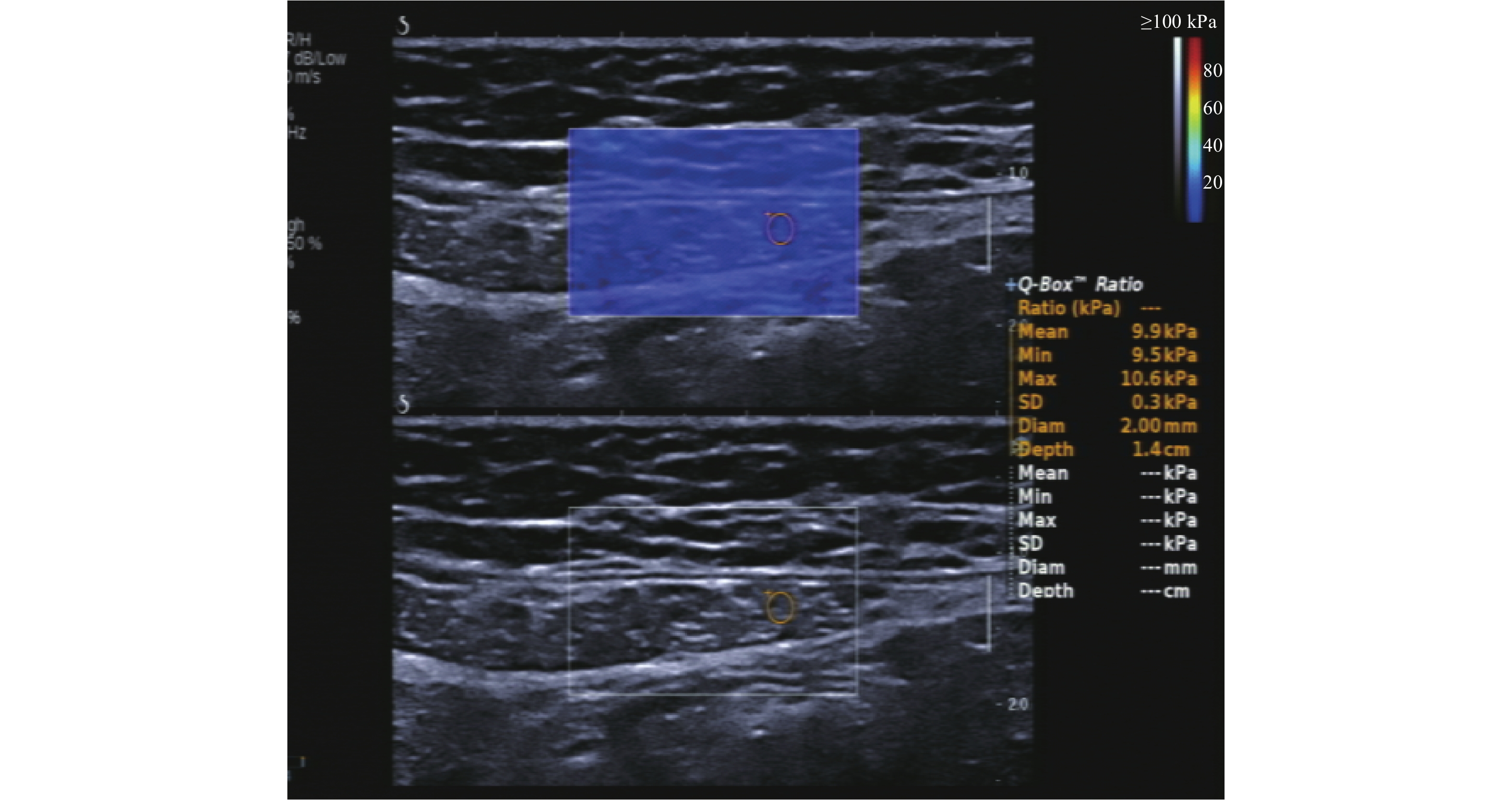
图 2 腹直肌硬度测量
Figure 2. Rectus abdominis hardness measurement
(SWE:Emean 9.9 kPa Emax 10.6 kPa Emin 9.5 kPa)

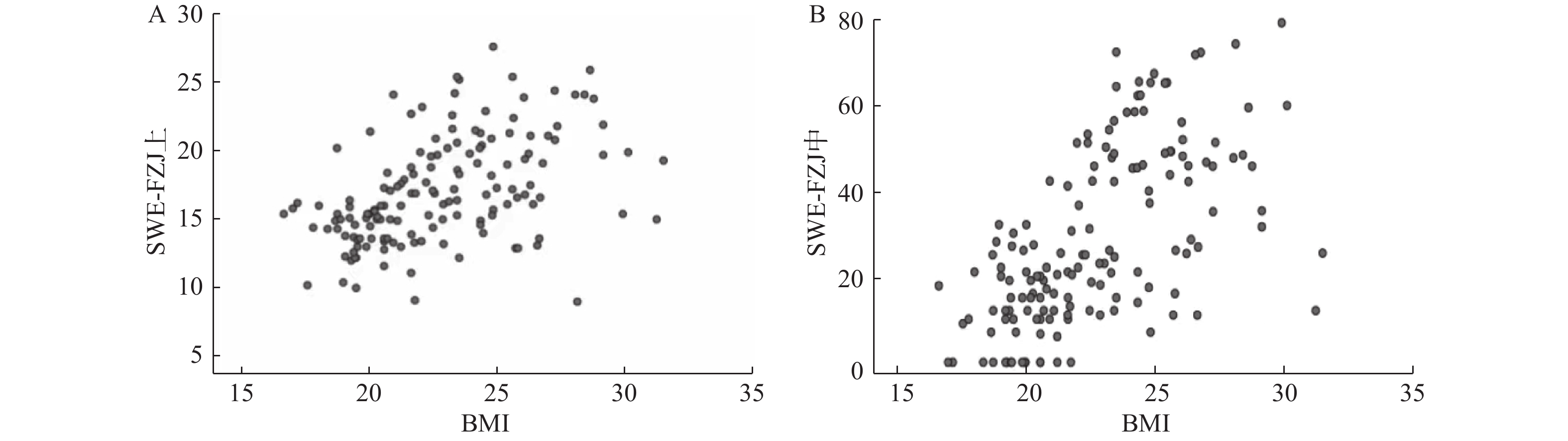
图 3 孕期BMI与腹直肌间距、腹直肌硬度相关性
A:孕期BMI与腹直肌硬度呈正相关(r = 0.513);B:孕期BMI与IRD呈正相关(r = 0.641)。
Figure 3. BMI during pregnancy was correlated with IRD and rectus abdominalis hardness
表 1 孕产妇人口统计学资料比较(
$ \overline x \pm {\text{s}} $ )Table 1. Comparison of maternal demographic data(
$ \overline x \pm {\text{s}} $ )组别 年龄(岁) 身高(cm) 体重(kg) BMI(kg/m²) 产后时间(d) 早孕组 26.74 ± 5.90 158.66 ± 3.54 52.12 ± 6.59 20.70 ± 2.45 − 中孕组 26.98 ± 5.76 160.64 ± 4.99 58.00 ± 11.13 22.38 ± 3.39 − 晚孕组 27.32 ± 3.96 160.24 ± 5.32 65.17 ± 6.19 25.39 ± 2.22 − 对照组 26.34 ± 2.22 161.32 ± 4.18 51.54 ± 5.37 19.79 ± 1.83 − 顺产组 27.28 ± 2.53 159.46 ± 3.72 56.70 ± 4.79 22.30 ± 1.72 42.67 ± 0.83 剖宫产组 27.90 ± 3.22 160.20 ± 4.22 57.20 ± 6.59 22.27 ± 2.26 42.96 ± 1.02 F 1.83 2.78 32.25 40.53 −1.642 P 0.112 0.020* 0.001* 0.001* 0.970 *P < 0.05。  下载: 导出CSV
表 2 不同妊娠期与对照组不同位点IRD及腹直肌弹性值比较(
$\bar x \pm s $ )/M(P25,P75)Table 2. Comparison of IRD and elastic value of rectus abdominalis muscle at different sites between different pregnancy period and control group (
$\bar x \pm s $ )/M(P25,P75)组别 腹直肌间距(mm) 腹直肌Emean(kPa) IRD+3 IRD0 IRD−3 IRD+3 IRD0 IRD−3 早孕组 6.0(0.0,12.0) 10.0(0.0,15.0) 0.0(0.0,2.0) 15.49 ± 2.37 16.16 ± 2.27 14.3 ± 1.97 中孕组 16.0(11.8,23.4) 22.5(17.7,27.5) 1.5(0.0,7.0) 18.01 ± 4.15 19.14 ± 3.17 16.16 ± 2.96 晚孕组 28.3(22.1,41.4) 50.5(45.6,62.0) 21.0(13.7,26.5) 22.45 ± 3.87 23.67 ± 4.33 20.37 ± 3.79 对照组 0.0(0.0,0.0) 0.0(0.0,0.0) 0.0(0.0,0.0) 14.70 ± 2.12 15.05 ± 2.32 13.8 ± 1.89 F/Z 136.894 162.820 137.923 55.678 60.211 43.538 P 0.001* 0.001* 0.001* < 0.001* < 0.001* < 0.001* *P < 0.05。
下载: 导出CSV
表 3 初产妇产后与对照组不同位点IRD及腹直肌弹性值比较(
$\bar x \pm s $ )/M(P25,P75)Table 3. Comparison of IRD and elastic values of rectus abdominalis at different points between primipara and control group (
$\bar x \pm s $ )/M(P25,P75)组别 腹直肌间距(mm) 腹直肌Emean(kPa) IRD+3 IRD0 IRD−3 IRD+3 IRD0 IRD−3 顺产组 18.5(14.8,27.0) 21.5(16.8,28.5) 3.5(0.0,9.8) 13.36 ± 1.73 13.68 ± 1.86 12.70 ± 1.88 剖宫产 24.0(18.0,30.0) 30.0(21.0,34.5) 9.0(4.0,17.0) 11.00 ± 1.86 12.63 ± 1.61 10.32 ± 2.53 对照组 0.0(0.0,0.0) 0.0(0.0,0.0) 0.0(0.0,0.0) 14.70 ± 2.12 15.05 ± 2.32 13.8 ± 1.89 F/Z 95.469 100.845 77.675 45.488 18.847 30.475 P 0.001* 0.001* 0.001* < 0.001* < 0.001* < 0.001* *P < 0.05。
下载: 导出CSV
-
[1] Axer H,Keyserlingk D G,Prescher A. Collagen fibers in linea alba and rectus sheaths. I. General scheme and morphological aspects[J]. J Surg Res,2001,96(1):127-134. doi: 10.1006/jsre.2000.6070 [2] Benjamin D R,Van de water A T,Peiris C L. Effects of exercise on diastasis of the rectus abdominis muscle in the antenatal and postnatal periods: A systematic review[J]. Physiotherapy,2014,100(1):1-8. doi: 10.1016/j.physio.2013.08.005 [3] Fernandes DA Mota P G,Pascoal A G,Carita A I,et al. Prevalence and risk factors of diastasis recti abdominis from late pregnancy to 6 months postpartum,and relationship with lumbo-pelvic pain[J]. Man Ther,2015,20(1):200-205. doi: 10.1016/j.math.2014.09.002 [4] Sperstad J B,Tennfjord M K,Hilde G,et al. Diastasis recti abdominis during pregnancy and 12 months after childbirth: Prevalence,risk factors and report of lumbopelvic pain[J]. Br J Sports Med,2016,50(17):1092-1096. doi: 10.1136/bjsports-2016-096065 [5] 刘雅莉,赵琼蕊,李娟,等. 中国育龄期妇女产后腹直肌分离发生率meta分析[J]. 中国公共卫生,2020,36(10):1507-1509. [6] 吴文静,苏继莲,王军梅. 超声测量育龄期未生育女性腹直肌间隙[J]. 中国医学影像技术,2021,37(9):1382-1385. [7] 刘博姬,徐辉雄. 剪切波弹性成像在肌肉、肌腱、周围神经病变生物力学定量评估中的应用进展[J]. 肿瘤影像学,2022,31(1):11-15. doi: 10.19732/j.cnki.2096-6210.2022.01.003 [8] 钟华,巫燕玲,陈英,等. 二维超声联合实时剪切波弹性成像评估腹直肌分离[J]. 中华医学超声杂志(电子版),2021,18(9):847-853. doi: 10.3877/cma.j.issn.1672-6448.2021.09.006 [9] Fukano M,Tsukahara Y,Takei S,et al. Recovery of abdominal muscle thickness and contractile function in women after childbirth[J]. Int J Environ Res Public Health,2021,18(4):2130. doi: 10.3390/ijerph18042130 [10] Puri J,Sharma S,Samuel A J,et al. Investigate correlation between diastasis of rectus abdominis muscle and low back pain in obese women[J]. J Lifestyle Med,2021,11(1):38-42. doi: 10.15280/jlm.2021.11.1.38 [11] Balasch-Bernat M,Perez-Alenda S,Carrasco J J,et al. Differences in inter-rectus distance and abdominopelvic function between nulliparous,primiparous and multiparous women[J]. Int J Environ Res Public Health,2021,18(23):12396. doi: 10.3390/ijerph182312396 [12] Qu E,Wu J,Zhang M,et al. The ultrasound diagnostic criteria for diastasis recti and its correlation with pelvic floor dysfunction in early postpartum women[J]. Quant Imaging Med Surg,2021,11(2):706-713. doi: 10.21037/qims-20-596 [13] Carlstedt A,Bringman S,Egberth M,et al. Management of diastasis of the rectus abdominis muscles: Recommendations for swedish national guidelines[J]. Scand J Surg,2021,110(3):452-459. doi: 10.1177/1457496920961000 [14] Corvino A,Rosa D,Sbordone C,et al. Diastasis of rectus abdominis muscles: Patterns of anatomical variation as demonstrated by ultrasound[J]. Pol J Radiol,2019,15(84):e542-e548. doi: 10.5114/pjr.2019.91303 [15] Wang X,Hu Y,Zhu J,et al. Effect of acquisition depth and precompression from probe and couplant on shear wave elastography in soft tissue: An in vitro and in vivo study[J]. Quant Imaging Med Surg,2020,10(3):754-765. doi: 10.21037/qims.2020.01.15 [16] Cavalli M,Aiolfi A,Bruni P G,et al. Prevalence and risk factors for diastasis recti abdominis: A review and proposal of a new anatomical variation[J]. Hernia,2021,25(4):883-890. doi: 10.1007/s10029-021-02468-8 [17] Conder R,Zamani R,Akrami M. The biomechanics of pregnancy: A systematic review[J]. J Funct Morphol Kinesiol,2019,4(4):72. doi: 10.3390/jfmk4040072 [18] 王丽芸,邱逦. 超声弹性成像在肌肉硬度评估中的应用进展[J]. 国际医学放射学杂志,2019,42(1):90-93. [19] 刘琛,欧阳征仁. 高频超声联合剪切波弹性成像技术对产后患者腹直肌的评估应用[J]. 中国医药科学,2022,12(7):167-171. [20] 汪亮,黄俊,王小娜. 高频超声及剪切波弹性成像定量评估产后腹直肌分离[J]. 临床超声医学杂志,2023,25(1):44-48. doi: 10.16245/j.cnki.issn1008-6978.2023.01.015 [21] Fan C,Guidolin D,Ragazzo S,et al. Effects of cesarean section and vaginal delivery on abdominal muscles and fasciae[J]. Medicina (Kaunas),2020,56(6):260. doi: 10.3390/medicina56060260 -


 点击查看大图
计量
- 文章访问数: 1591
- HTML全文浏览量: 1280
- PDF下载量: 15
- 被引次数: 0
