Artemisinin Mediates the Aerobic Glycolysis of Clear Cell Renal Cell Carcinomas via Regulating ENO2
-
摘要:
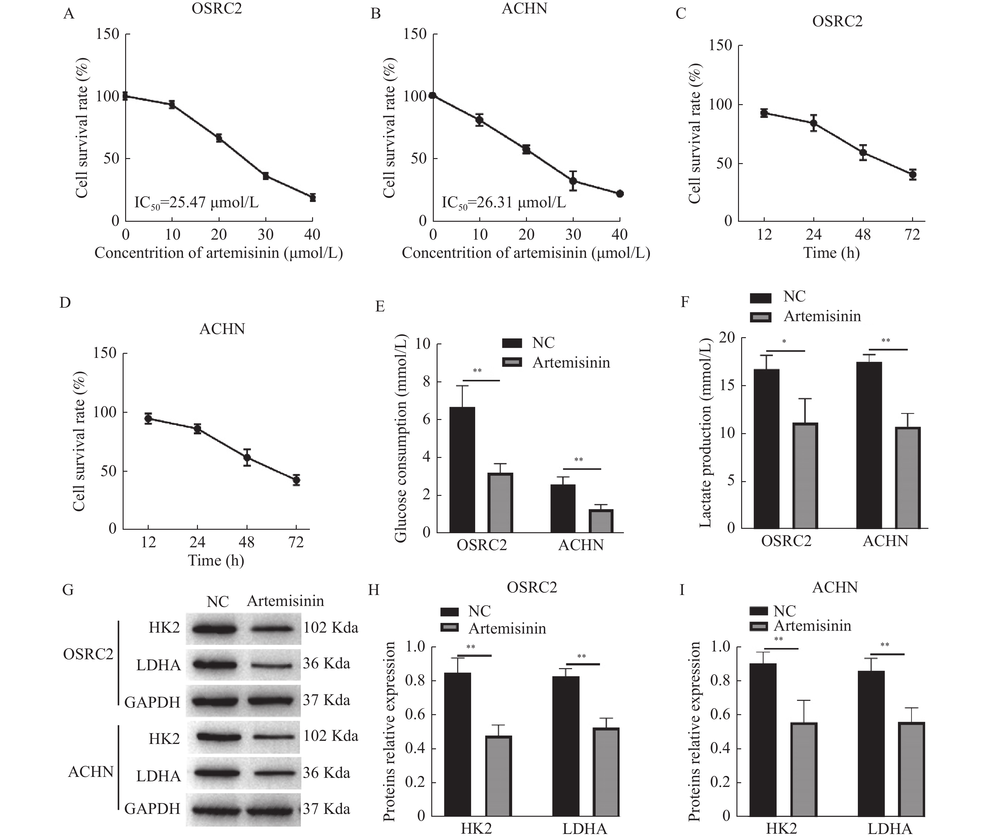
目的 探讨青蒿素(Artemisinin)调控ENO2对肾透明细胞癌(clear cell renal cell carcinomas,ccRCC)细胞增殖和有氧糖酵解的作用及具体调控机制。 方法 分别将ccRCC细胞系OSRC2和ACHN暴露于0、10、20、30、40 μmol/L青蒿素中,OSRC2和ACHN细胞经25 μmol/L青蒿素处理或同时转染si-ENO2,CCK-8检测细胞活力,葡萄糖测试盒和乳酸测试盒检测葡萄糖消耗和乳酸生成量,Western blot检测HK2、LDHA和ENO2的表达,RT-qPCR检测ENO2 mRNA相对表达。 结果 OSRC2和ACHN细胞存活率随青蒿素浓度和暴露时间的增加而降低,OSRC2细胞的IC50值为25.47 μmol/L,ACHN细胞的IC50值为26.31 μmol/L。ENO2在OSRC2和ACHN细胞中表达升高(P < 0.01),青蒿素可下调ENO2在癌细胞中的表达(P < 0.05)。暴露于25 μmol/L青蒿素或敲降ENO2可抑制OSRC2和ACHN细胞的存活(P < 0.001)、葡萄糖消耗(P < 0.05)、乳酸生成(均P < 0.05)以及HK2和LDHA的蛋白表达(P < 0.05)。同时敲降ENO2且暴露于25 μmol/L青蒿素组中OSRC2和ACHN细胞的存活率(P < 0.001)、葡萄糖消耗(P < 0.05)、乳酸生成(P < 0.05)以及HK2和LDHA的表达(均P < 0.05)低于仅敲降ENO2组。 结论 青蒿素可抑制ccRCC细胞的存活率和有氧糖酵解,并通过下调ENO2在ccRCC中的表达而发挥作用。 Abstract:Objective To investigate Artemisinin-affected cell proliferation and aerobic glycolysis on clear cell renal carcinomas(ccRCC) through regulating ENO2 and clarify the mechanism. Methods ccRCC cell lines OSRC2 and ACHN were exposed to 0, 10, 20, 30, and 40 μmol/L Artemisinin, OSRC2 and ACHN cells treated with 25 μmol/L Artemisinin or transfected with si-ENO2 at the same time. CCK-8 assay detected cell proliferation, glucose consumption, and lactate production were detected by glucose test kit and lactate test kit. The expressions of HK2, LDHA, and ENO2 were detected by Western blot, and ENO2 mRNA expression was detected by RT-qPCR. Results The survival rate of OSRC2 and ACHN cells were decreased with the increase of Artemisinin concentration and treatment time, the IC50 of OSRC2 cells was 25.47 μmol/L, and the IC50 of ACHN was 26.31 μmol/L. ENO2 was upregulated in OSRC2 and ACHN cells(P < 0.01), and Artemisinin diminished ENO2 expression in cancer cells(all P < 0.05). Exposure to Artemisinin(25 μmol/L) or knocking down ENO2 significantly inhibited the survival of OSRC2 and ACHN cells(all P < 0.001), glucose consumption(P < 0.05), lactate production(all P < 0.05) and protein expression of HK2 and LDHA(all P < 0.05). Compared with si-ENO2 group, the survival rate(P < 0.001), glucose consumption(P < 0.05), lactate production(all P < 0.05) and the expression of HK2 and LDHA(all P < 0.05) of OSRC2 and ACHN cells in knocking down ENO2 and treated with 25 μmol/L Artemisinin group were decreased. Conclusion Artemisinin can inhibit the survival rate and aerobic glycolysis of ccRCC cells, and plays a role by downregulating the expression of ENO2 in ccRCC. -
Key words:
- Clear cells renal cell carcinomas /
- Aerobic glycolysis /
- Artemisinin /
- ENO2 /
- Survival rate
-
新生儿重症肺炎在床旁超声的监测下发现常合并肺实变[1-2]。新生儿肺实变使患儿处于持续低氧状态、进而影响呼吸循环系统的稳定性,是导致新生儿死亡的重要原因[3-5]。在新生儿重症肺炎的治疗中,氧疗、机械通气,恰当的抗生素的使用等治疗固然占据主导地位,但合适的护理措施也对重症肺炎的恢复起到至关重要的作用[6-8]。在常规的重症肺炎护理中,所有患儿无论肺部病变程度、性质如何,均给以固定时间间隔的翻身、拍背、吸痰等措施,以促进气道分泌物排出[9-10]。但由于新生儿气道清洁能力不足,自主活动能力差,常规护理往往收效甚微[11-12]。因此,为了提高新生儿重症肺炎并肺实变的疗效,需要制定针对不同肺部病变性质的精准护理措施[13]。随着肺脏超声在新生儿领域的广泛应用,该技术为新生儿重症肺炎并肺实变的精准护理提供了一种有效的方法,对缩短病程和提高疗效有重要价值。
1. 资料与方法
1.1 一般资料
选取2021年1月至12月昆明市儿童医院新生儿重症监护室(NICU)收治的60例重症肺炎新生儿为研究对象,该研究获得了昆明市儿童医院伦理委员会的批准(2022-03-130-K01)。纳入标准:新生儿重症肺炎的诊断标准符合第5版《实用新生儿学》中的诊断标准;患儿除咳嗽、鼻塞等呼吸道症状外,合并明显呼吸困难或循环不稳定者定义为重症肺炎[14]。排除标准:(1)伴有先天性疾病;(2)确诊脓毒血症;(3)放弃治疗。采用随机数字法随机分为研究组和对照组2组,每组各30例。研究组男 13 例,女 17例;年龄中位数1.5(8.0,18.5 ) d;体重中位数3.2(2.6,3.8 )kg;胎龄中位数38.5(36.8,40.3)周;剖宫产14例,阴道分娩16例;均有咳嗽咳痰等呼吸道症状,其中机械通气者12例(40%),普通吸氧者18例(60%)。 对照组患儿男 12例,女18例;年龄中位数11( 8.0,21.3) d;体重中位数3.0( 2.5,3.7) kg;胎龄中位数39(37,40)周;剖宫产 10例,阴道分娩 20例;均有咳嗽咳痰等呼吸道症状,其中机械通气者10例(30%),普通吸氧者20例(70%)。2组一般资料差异无统计学意义(P > 0.05),见 表1。
表 1 一般资料比较[M(P25,P75)/n(%)]Table 1. Comparison of general information [M(P25,P75)/n(%)]组别 胎龄(周) 日龄(d) 体重(kg) 咳嗽 腹胀 心衰 休克 中毒性脑病 机械通气 研究组 38.5(36.8,40.3) 11.5
(8.0,18.5 )3.2
(2.6,3.8 )30
(100)6(20) 10(30) 13(43) 2(7) 18(60) 对照组 39
(37,40)11
( 8.0,21.3)3.0
( 2.5,3.7)30
(100)8(27) 12(40 11(37) 1(3) 20(66) Z/χ2 −0.630 −0.220 −0.430 0.570 0.640 0.490 0.680 P 0.530 0.824 0.667 0.678 0.584 0.801 0.433 1.2 研究方法
2组患儿均采取普通吸氧或机械通气、抗感染、雾化、维持循环系统稳定等常规治疗。对照组给予常规护理干预:(1)基础护理: 保持病房清洁、常规口、脐、臀、皮肤护理,发热者及时降温处理,施与袋鼠式护理;(2)呼吸道护理:定时翻身、拍背、根据痰多少给予相应频次的吸痰。
研究组在对照组常规护理的基础上,每天使用床旁超声检查肺脏2~4次,根据超声发现采取不同护理干预措施,具体如下:(1)超声提示肺实变征象,见图1:实变面积较大时予纤维支气管镜下或气管插管下肺泡灌洗术;实变局限于胸膜线下,呈虫蚀样改变时以叩背、机械振动、手法震颤以及吸痰为主,根据严重程度选择一种或多种方式进行排痰,扣背、机械振动、手法震颤等措施位置主要为实变区域,并将患儿体位管理为实变对侧卧位为主。(2)超声提示肺间质综合征征象,见图2:予固定时间翻身,不常规行拍背吸痰。
1.3 观察指标
(1)接受机械通气的患儿撤离呼吸机的时间;(2)接受普通给氧的患儿停氧的时间;(3)咳嗽咳痰、肺部啰音、少吃少动症状体征消失的时间;(4)住院时间。
1.4 统计学处理
应用SPSS 22.0统计软件,计量资料中,对于符合正态分布的数据采用( $\bar x \pm s $)表示,2组间用独立样本t 检验进行比较;对于不符合正态分布的数据采用中位数和四分位间距即M(P25,P75)表示,2组间采用非参数秩和检验(Wilcoxon’s signed rank)比较。计数资料以n(%)表示,行χ2检验,P < 0.05为差异有统计学意义。
2. 结果
2.1 2组超声图像比较
超声下,对照组与研究组均显示肺炎征象,包括胸膜线下虫蚀样改变、实变位置深、至少累及2个肋间,胸腔积液、肺不张和支气管充液征,且以累及2个以上肋间以上的深度实变为主,多合并胸腔积液和肺不张,提示重症肺炎的患儿肺脏感染重,渗出液多,存在因炎性分泌物导致的中小气道堵塞。2组超声图像特征均差异无统计学意义(P > 0.05),见 表2。
表 2 2组超声图像比较[n(%)]Table 2. Comparison of two groups of ultrasound images [n(%)]组别 实变范围 胸腔积液 肺不张 支气管
充液征虫蚀样 深,累及2个及
以上肋间对照组 8(27) 22(73) 19(63) 5(17) 30(100) 研究组 6(20) 24(80) 22(73) 4(13) 30(100) χ2 0.740 0.834 0.830 0.670 P 0.564 0.681 0.632 0.547 2.2 2组临床症状缓解时间及住院时间比较
研究组咳嗽咳痰、肺部啰音、少吃少动症状消失的时间中位数分别为8(6.5,10.0)d及(4.5,10.0)d,住院时间中位数10(8.0,11.0)d;对照组咳嗽咳痰、肺部啰音、少吃少动症状消失的时间中位数分别为11(10.0,12.3)d及15(7.0,18.0)d,住院时间中位数14(12.0,15.3)d。研究组患儿咳嗽咳痰、肺部啰音消、少吃少动消失时间及住院时间均较对照组缩短(P < 0.05),见 表3。
表 3 2组临床征象缓解时间及住院时间比较[M(P25,P75),d]Table 3. Comparison of clinical remission time and hospitalization time between two groups [M(P25,P75),d]组别 临床症状消失 住院时间 咳嗽咳痰 肺部啰音 少吃少动 腹胀 心衰 休克 研究组 8
(6.5,10.0)4
(3.5,11.07
(4.5,10)3(1,5) 2(1,4) 3(1.5,5) 10(8.0,11.0) 对照组 11
(10.0,12.3)10
(9,13)15
(7,18)7(13,9) 5(2,7) 4(2,6) 14(12.0,15.3) Z −4.642 11.031 8.983 9.043 10.031 9.532 −5.642 P < 0.001* < 0.001* < 0.001* < 0.001* < 0.001* < 0.001* < 0.001* *P < 0.05。 2.3 2组撤离呼吸机或停氧时间比较
研究组撤离呼吸机时间中位数为5(4.0,6.3)d,停氧时间中位数7(5.1,9.5)d;对照组撤离呼吸机时间中位数为8(6.0,9.3)d,停氧时间中位数10(8.0,12)d。研究组患儿撤离呼吸机或停氧时间均较对照组短(P < 0.05) ,见 表4。
表 4 2组撤离呼吸机或停氧时间比较[M(P25,P75),d]Table 4. Comparison of extubation time or deaeration time between two groups [M(P25,P75),d]组别 撤离呼吸机 停氧 研究组 5(4.0,6.3) 7(5.1,9.5) 对照组 8(6.0,9.3) 10(8.0,12) Z −4.812 −4.674 P < 0.001* < 0.001* *P < 0.05。 3. 讨论
3.1 新生儿肺实变的特点
新生儿肺实变好发部位与成人和儿童有很大差别 很少有报道新生儿肺实变的好发部位,成人和儿童重症肺炎时发生肺实变常见部位为肺下段,但新生儿发生肺实变的常见部位为脊柱两侧以及肩甲骨区域部位[15]。分析发现与体位有关,成人和儿童当患重症肺炎时,卧床期间,肺下段处于低位,而新生儿卧床期间,一般下肢处于卷曲状态,导致身体处于低位的部位为后备及胸廓段,由于重力的原因,痰液集聚在处于低位的肺段处,故存在新生儿肺实变的区域与成人和儿童不同。
3.2 超声引导精准护理治疗肺实变的价值
新生儿肺实变隐匿且治疗困难 新生儿实变区域多数在脊柱两侧及肩胛骨区域,床旁X-线片很难发现脊柱两侧细小的实变区,但在B超下能明显显示实变[16]。且该区域被骨组织覆盖,在治疗时考虑到施力后导致骨折的原因,导致在进行扣背、手法震颤等护理措施时力度不敢过大,加之震颤的穿透力在透过骨组织时会被大大的削弱,导致治疗效果相对差[17-19]。故发生在新生儿脊柱两侧及肩甲骨区域的肺实变精准护理的时间相对长方能有效。
3.3 体位管理在新生儿肺实变精准护理中的价值
体位引流与儿童及成人差异较大 儿童和成年人建立翻身卡能明确左、右侧卧位以及俯卧位,这类人群能自主维持相应的体位;但新生儿除了俯卧有明确的更换体位外,达到完全的侧卧极为困难,新生儿不能自主维持任何体位,当新生儿处于俯卧位时,如果体位摆放不规范,可能会有“婴儿猝死综合征”的危险[20]。当新生儿摆放侧卧位时,因为新生儿本身处于卷曲状态,侧卧位不能完全实现,导致体位引流效果不如成人及儿童,所以在施以新生儿体位管理时,需要有相应的辅助支撑的用具方能提高体位管理的效果。
床旁肺脏超声在新生儿肺实变临床诊断上有非常大的敏感性[21]。精准化护理措施在超声动态监视下能动态监测肺实变的治疗效果,但在精准化护理措施实施过程中,医护协助极为重要,且治疗过程中力度的掌握、病情的监护极为重要。
-
-
[1] Znaor A,Lortet-Tieulent J,Laversanne M,et al. International variations and trends in renal cell carcinoma incidence and mortality[J]. Eur Urol,2015,67(3):519-530. doi: 10.1016/j.eururo.2014.10.002 [2] Baldewijns M M,Van Vlodrop I J,Schouten L J,et al. Genetics and epigenetics of renal cell cancer[J]. Biochim Biophys Acta,2008,1785(2):133-155. [3] Vaupel P,Schmidberger H,Mayer A. The Warburg effect: Essential part of metabolic reprogramming and central contributor to cancer progression[J]. Int J Radiat Biol,2019,95(7):912-919. doi: 10.1080/09553002.2019.1589653 [4] Nocera G,Jacob C. Mechanisms of Schwann cell plasticity involved in peripheral nerve repair after injury[J]. Cell Mol Life Sci,2020,77(20):3977-3989. doi: 10.1007/s00018-020-03516-9 [5] Reed G H,Poyner R R,Larsen T M,et al. Structural and mechanistic studies of enolase[J]. Curr Opin Struct Biol,1996,6(6):736-743. doi: 10.1016/S0959-440X(96)80002-9 [6] Chen W J,Yang W,Gong M,et al. ENO2 affects the EMT process of renal cell carcinoma and participates in the regulation of the immune microenvironment[J]. Oncol Rep,2023,49(2):33. [7] Tang C,Wang M,Dai Y,et al. Krüppel-like factor 12 suppresses bladder cancer growth through transcriptionally inhibition of enolase 2[J]. Gene,2021,769:145338. [8] Fang L,Ye T,An Y. Circular RNA FOXP1 induced by ZNF263 upregulates U2AF2 expression to accelerate renal cell carcinoma tumorigenesis and warburg effect through sponging miR-423-5p[J]. J Immunol Res,2021,2021:8050993. [9] Wang M,Chen H,He X,et al. Artemisinin inhibits the development of esophageal cancer by targeting HIF-1α to reduce glycolysis levels[J]. J Gastrointest Oncol,2022,13(5):2144-2153. doi: 10.21037/jgo-22-877 [10] Farhan M,Silva M,Xingan X,et al. Artemisinin inhibits the migration and invasion in uveal melanoma via inhibition of the PI3K/AKT/mTOR signaling pathway[J]. Oxid Med Cell Longev,2021,2021:9911537. [11] Wang Z,Li M,Liu Y,et al. Dihydroartemisinin triggers ferroptosis in primary liver cancer cells by promoting and unfolded protein response-induced upregulation of CHAC1 expression[J]. Oncol Rep,2021,46(5):240. doi: 10.3892/or.2021.8191 [12] Cai X,Miao J,Sun R,et al. Dihydroartemisinin overcomes the resistance to osimertinib in EGFR-mutant non-small-cell lung cancer[J]. Pharmacol Res,2021,170:105701. [13] Yu C,Sun P,Zhou Y,et al. Inhibition of AKT enhances the anti-cancer effects of Artemisinin in clear cell renal cell carcinoma[J]. Biomed Pharmacother,2019,118:109383. [14] Zhang M X,Wang J L,Mo C Q,et al. CircME1 promotes aerobic glycolysis and sunitinib resistance of clear cell renal cell carcinoma through cis-regulation of ME1[J]. Oncogene,2022,41(33):3979-3990. doi: 10.1038/s41388-022-02386-8 [15] He Y,Wang X,Lu W,et al. PGK1 contributes to tumorigenesis and sorafenib resistance of renal clear cell carcinoma via activating CXCR4/ERK signaling pathway and accelerating glycolysis[J]. Cell Death Dis,2022,13(2):118. doi: 10.1038/s41419-022-04576-4 [16] Li J,Zhang S,Liao D,et al. Overexpression of PFKFB3 promotes cell glycolysis and proliferation in renal cell carcinoma[J]. BMC Cancer,2022,22(1):83. doi: 10.1186/s12885-022-09183-2 [17] Chen X,Li Z,Yong H,et al. Trim21-mediated HIF-1α degradation attenuates aerobic glycolysis to inhibit renal cancer tumorigenesis and metastasis[J]. Cancer Lett,2021,508:115-126. doi: 10.1016/j.canlet.2021.03.023 [18] Gao L,Yang F,Tang D,et al. Mediation of PKM2-dependent glycolytic and non-glycolytic pathways by ENO2 in head and neck cancer development[J]. J Exp Clin Cancer Res,2023,42(1):1. doi: 10.1186/s13046-022-02574-0 [19] Liu D,Mao Y,Chen C,et al. Expression patterns and clinical significances of ENO2 in lung cancer: an analysis based on Oncomine database[J]. Ann Transl Med,2020,8(10):639. doi: 10.21037/atm-20-3354 [20] Soh M A,Garrett S H,Somji S,et al. Arsenic,cadmium and neuron specific enolase (ENO2,γ-enolase) expression in breast cancer[J]. Cancer Cell Int,2011,11(1):41. doi: 10.1186/1475-2867-11-41 [21] Huang J,Yang M,Liu Z,et al. PPFIA4 promotes colon cancer cell proliferation and migration by enhancing tumor glycolysis[J]. Front Oncol,2021,11:653200. [22] Peng J,Liu F,Zheng H,et al. IncRNA ZFAS1 contributes to the radioresistance of nasopharyngeal carcinoma cells by sponging hsa-miR-7-5p to upregulate ENO2[J]. Cell Cycle,2021,20(1):126-141. doi: 10.1080/15384101.2020.1864128 [23] Liu C C,Wang H,Wang W D,et al. ENO2 promotes cell proliferation,glycolysis,and glucocorticoid-resistance in acute lymphoblastic leukemia[J]. Cell Physiol Biochem,2018,46(4):1525-1535. doi: 10.1159/000489196 [24] Pan J,Jin Y,Xu X,et al. Integrated analysis of the role of enolase 2 in clear cell renal cell carcinoma[J]. Dis Markers,2022,2022:6539203. 期刊类型引用(0)
其他类型引用(1)
-