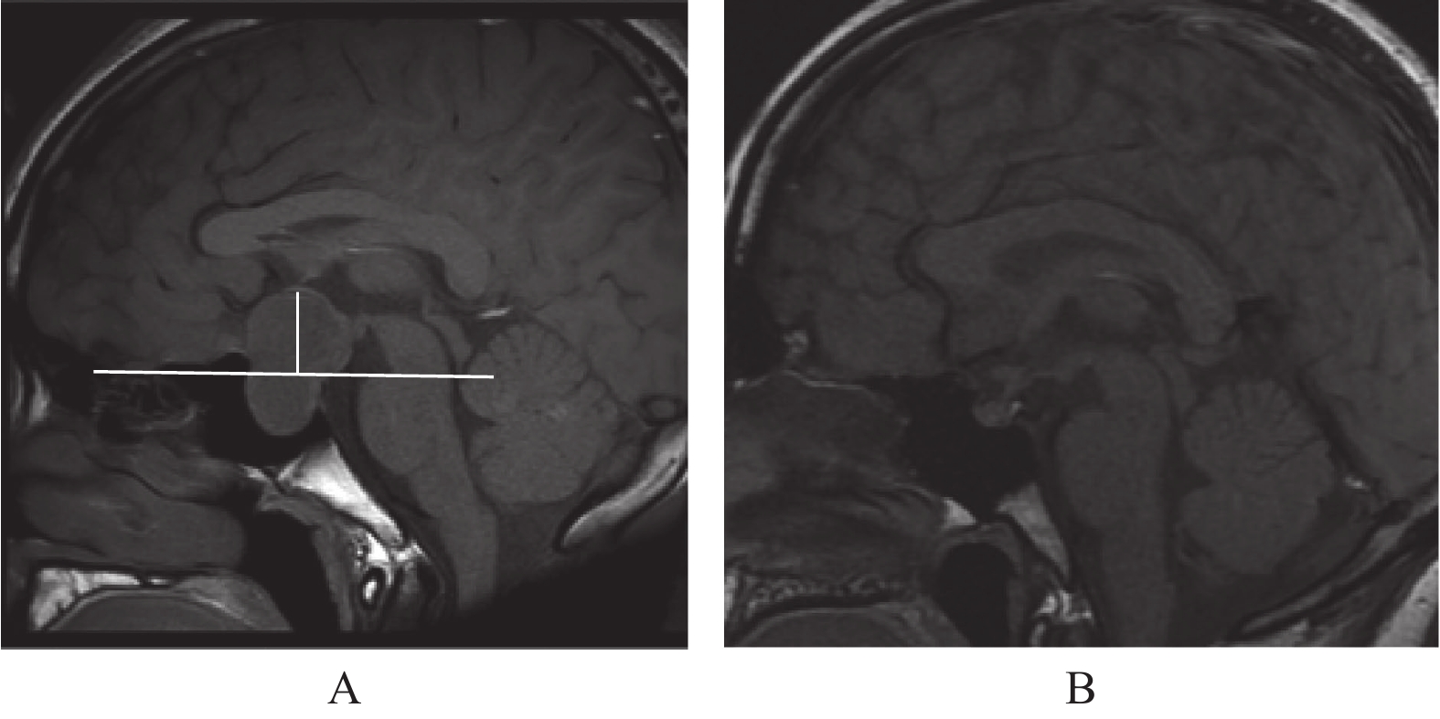
Diffusion Tensor Imaging in the White Matter Changes of Patients with Pituitary Macroadenoma
-
摘要:
目的 利用磁共振弥散张量成像(diffusion tensor imaging,DTI)研究垂体腺瘤患者前视路结构受压后全脑白质纤维束微观结构的变化及其意义。 方法 前瞻性纳入25例伴有视觉功能损伤的垂体腺瘤患者为研究组及相匹配的25例健康志愿者为对照组,2组受试者均进行临床数据及MRI数据采集,获得DTI数据及视功能评价指标。采用基于纤维束的空间统计(tract-based spatial statistics,TBSS)分析2组间脑白质各向异性分数(fractional anisotropy,FA)的差异,并分析其与视功能评价指标的相关性。 结果 研究组双侧视放射及下额枕束的FA值较对照组降低(P < 0.05)。相关性研究显示研究组左侧视放射及下额枕束的FA值与平均缺损、加权视野指数呈正相关(P < 0.05),与模式偏差缺损呈负相关(P < 0.05);研究组双侧视放射及下额枕束的FA值与嗅球-脑球高度呈负相关(P < 0.05)。 结论 垂体腺瘤患者前视路结构受压后双侧视放射及下额枕束发生了损伤,可能影响了患者的视功能,进一步说明DTI可以定量评估垂体腺瘤患者视路微观结构的损伤。 -
关键词:
- 垂体腺瘤 /
- 弥散张量成像 /
- 视觉通路 /
- 脑白质 /
- 基于纤维束的空间统计
Abstract:Objective To investigate the microstructural changes in white matter of pituitary adenoma patients when the anterior visual pathway is compressed using diffusion tensor imaging(DTI) and discuss the clinical significance of these changes. Methods Clinical and MRI data of 25 patients with pituitary adenomas and 25 matched healthy controls were prospectively included. Tract-based spatial statistics(TBSS) was carried out to investigate difference in white matter integrity between 2 groups, which was measured using fractional anisotropy(FA). A correlation between visual function evaluation index and regional FA value was examined using correlation analysis. Results The FA values of bilateral optic radiations and inferior fronto-occipital fasciculus were significantly lower in the research group. The FA values of left optic radiation and inferior fronto-occipital fasciculus were positively correlated to mean deviation and visual field index, negatively correlated to patten standard deviation. The FA values of bilateral optic radiations and inferior fronto-occipital fasciculus were negatively correlated with Bulbo-pontine height. Conclusion Patients with pituitary adenoma may experience damage to the bilateral optic radiation and inferior fronto-occipital fasciculus after compression of the anterior visual pathway, which may affect the patient’ s visual function, further indicating that DTI can quantitatively assess the microstructure damage of the visual pathway in patients with pituitary adenoma. -
图 3 2组间存在差异的白质纤维束簇的FA值与临床指标之间的相关性分析
A:双侧视放射与下额枕束平均FA值与BP-height相关性分析散点图;B:左侧视放射与下额枕束平均FA值与左侧Mean Deviation值相关性分析散点图;C: 左侧视放射与下额枕束平均FA值与左侧VFI值相关性分析散点图;D:左侧视放射与下额枕束平均FA值与左侧PSD值相关性分析散点图。
Figure 3. Correlation analysis of clusters showing significant differences in FA between two groups and clinical index
表 1 研究组与对照组临床资料
Table 1. Clinical data of both study and control group
指标 病例组(n = 25) 对照组(n = 25) t/χ2 P 年龄(岁) 45.480 ± 15.543 43.680 ± 14.522 0.423 0.674 性别(男/女) 14/11 16/9 0.333 0.564 表 2 研究组视野检查结果及BP-height值
Table 2. Perimetry results and BP-height for study group
指标 数值 视野 Mean Deviation -左(dB) −12.69 ± 2.14 PSD-左(dB) 8.46 ± 1.17 VFI-左(%) 62.14 ± 6.65 Mean Deviation -右(dB) −11.89 ± 2.08 PSD-右(dB) 8.08 ± 1.33 VFI-右(%) 66.21 ± 7.13 BP-height(mm) 18.34 ± 1.39 表 3 研究组与对照组FA值存在差异的白质纤维束簇
Table 3. White matter fiber clusters with different FA values between two groups
有差异的白质纤维束簇 簇大小
(体素数目)峰值MNI坐标 X Y Z Cluster1
(右侧视放射+右侧下额枕束)1605 47 85 65 Cluster2
(左侧视放射+左侧下额枕束)161 113 44 74 Cluster3
(左侧下额枕束)75 125 68 69 表 4 2组间存在差异的白质纤维束簇的FA值与临床指标之间的相关性分析
Table 4. Correlation analysis of clusters showing significant differences in FA between two groups and clinical index
指标 Mean Deviation(dB) PSD(dB) VFI(%) r P r P r P Cluster2+3的FA值 0.537 0.048* −0.704 0.005* 0.614 0.020* Cluster1的FA值 0.224 0.442 −0.336 0.240 0.322 0.262 *P<0.05 -
[1] Banskota S,Adamson D C. Pituitary adenomas: From diagnosis to therapeutics[J]. Biomedicines,2021,9(5):494-511. doi: 10.3390/biomedicines9050494 [2] Dekkers O M,Karavitaki N,Pereira A M. The epidemiology of aggressive pituitary tumors (and its challenges)[J]. Rev Endocr Metab Disord,2020,21(2):209-212. doi: 10.1007/s11154-020-09556-7 [3] Danesh-Meyer H V,Yoon J J,Lawlor M,et al. Visual loss and recovery in chiasmal compression[J]. Prog Retin Eye Res,2019,11(73):100765-100791. [4] Pal A,Leaver L,Wass J. Pituitary adenomas[J]. BMJ,2019,6(365):l2091-l2097. [5] Yu S Y,Du Q,Yao S Y,et al. Outcomes of endoscopic and microscopic transsphenoidal surgery on non-functioning pituitary adenomas: A systematic review and meta-analysis[J]. J Cell Mol Med,2018,22(3):2023-2027. doi: 10.1111/jcmm.13445 [6] Chung Y S,Na M,Yoo J,et al. Optical coherent tomography predicts long-term visual outcome of pituitary adenoma surgery: New perspectives from a 5-year follow-up study[J]. Neurosurgery,2020,88(1):106-112. [7] Baliyan V,Das C J,Sharma R,et al. Diffusion weighted imaging: Technique and applications[J]. World J Radiol,2016,8(9):785-798. doi: 10.4329/wjr.v8.i9.785 [8] Chavhan G B,Alsabban Z,Babyn P S. Diffusion-weighted imaging in pediatric body MR imaging: Principles,technique,and emerging applications[J]. Radiographics,2014,34(3):E73-88. doi: 10.1148/rg.343135047 [9] Lilja Y,Gustafsson O,Ljungberg M,et al. Visual pathway impairment by pituitary adenomas: Quantitative diagnostics by diffusion tensor imaging[J]. J Neurosurg,2017,127(3):569-579. doi: 10.3171/2016.8.JNS161290 [10] Liang L,Lin H,Lin F,et al. Quantitative visual pathway abnormalities predict visual field defects in patients with pituitary adenomas: A diffusion spectrum imaging study[J]. Eur Radiol,2021,31(11):8187-8196. doi: 10.1007/s00330-021-07878-x [11] Frisén L,Jensen C. How robust is the optic chiasm? Perimetric and neuro-imaging correlations[J]. Acta Neurol Scand,2008,117(3):198-204. doi: 10.1111/j.1600-0404.2007.00927.x [12] Sullivan E V,Rohlfing T,Pfefferbaum A. Quantitative fiber tracking of lateral and interhemispheric white matter systems in normal aging: Relations to timed performance[J]. Neurobiol Aging,2010,31(3):464-481. doi: 10.1016/j.neurobiolaging.2008.04.007 [13] Friedrich P,Fraenz C,Schlüter C,et al. The relationship between axon density,myelination,and fractional anisotropy in the human corpus callosum[J]. Cereb Cortex,2020,30(4):2042-2056. doi: 10.1093/cercor/bhz221 [14] Lazari A,Lipp I. Can MRI measure myelin? Systematic review,qualitative assessment,and meta-analysis of studies validating microstructural imaging with myelin histology[J]. Neuroimage,2021,4(230):117744. [15] 王春节,王宇桐,翟方兵,等. 不同类型原发性青光眼的扩散张量成像研究[J]. 磁共振成像,2022,13(1):114-117. doi: 10.12015/issn.1674-8034.2022.01.023 [16] Puthenparampil M,Federle L,Poggiali D,et al. Trans-synaptic degeneration in the optic pathway. A study in clinically isolated syndrome and early relapsing-remitting multiple sclerosis with or without optic neuritis[J]. PLoS One,2017,12(8):e0183957. doi: 10.1371/journal.pone.0183957 [17] Burge W K,Griffis J C,Nenert R,et al. Cortical thickness in human V1 associated with central vision loss[J]. Sci Rep,2016,24(6):23268. [18] Dinkin M. Trans-synaptic retrograde degeneration in the human visual system: Slow,silent,and real[J]. Curr Neurol Neurosci Rep,2017,17(2):16. doi: 10.1007/s11910-017-0725-2 [19] Rutland J W,Padormo F,Yim C K,et al. Quantitative assessment of secondary white matter injury in the visual pathway by pituitary adenomas: A multimodal study at 7-Tesla MRI[J]. J Neurosurg,2019,132(2):333-342. [20] Song G,Qiu J,Li C,et al. Alterations of regional homogeneity and functional connectivity in pituitary adenoma patients with visual impairment[J]. Sci Rep,2017,7(1):13074. doi: 10.1038/s41598-017-13214-5 [21] Chouinard P A,Striemer C L,Ryu W H,et al. Retinotopic organization of the visual cortex before and after decompression of the optic chiasm in a patient with pituitary macroadenoma[J]. J Neurosurg,2012,117(2):218-224. doi: 10.3171/2012.4.JNS112158 [22] Qian H,Wang X,Wang Z,et al. Altered vision-related resting-state activity in pituitary adenoma patients with visual damage[J]. PLoS One,2016,11(8):e0160119. doi: 10.1371/journal.pone.0160119 [23] Phal PM,Steward C,Nichols AD,et al. Assessment of optic pathway structure and function in patients with compression of the optic chiasm: A correlation with optical coherence tomography[J]. Invest Ophthalmol Vis Sci,2016,57(8):3884-3890. doi: 10.1167/iovs.15-18734 [24] 张权,张云亭,张敬,等. 垂体大腺瘤对后视路影响的功能磁共振成像和弥散张量成像联合研究[J]. 中华生物医学工程杂志,2009,15(1):9-13. [25] Martino J,De Lucas E M. Subcortical anatomy of the lateral association fascicles of the brain: A review[J]. Clin Anat,2014,27(4):563-569. doi: 10.1002/ca.22321 [26] Yasmin H,Aoki S,Abe O,et al. Tract-specific analysis of white matter pathways in healthy subjects: A pilot study using diffusion tensor MRI[J]. Neuroradiology,2009,51(12):831-840. doi: 10.1007/s00234-009-0580-1 [27] Tsai T H,Su H T,Hsu Y C,et al. White matter microstructural alterations in amblyopic adults revealed by diffusion spectrum imaging with systematic tract-based automatic analysis[J]. Br J Ophthalmol,2019,103(4):511-516. doi: 10.1136/bjophthalmol-2017-311733 [28] Zikou A K,Kitsos G,Tzarouchi L C,et al. Voxel-based morphometry and diffusion tensor imaging of the optic pathway in primary open-angle glaucoma: A preliminary study[J]. AJNR Am J Neuroradiol,2012,33(1):128-134. doi: 10.3174/ajnr.A2714 -


 下载:
下载:


