Role of HK2 and VDAC1 in Diacetylmorphine-induced Cardiomyocyte Apoptosis
-
摘要:
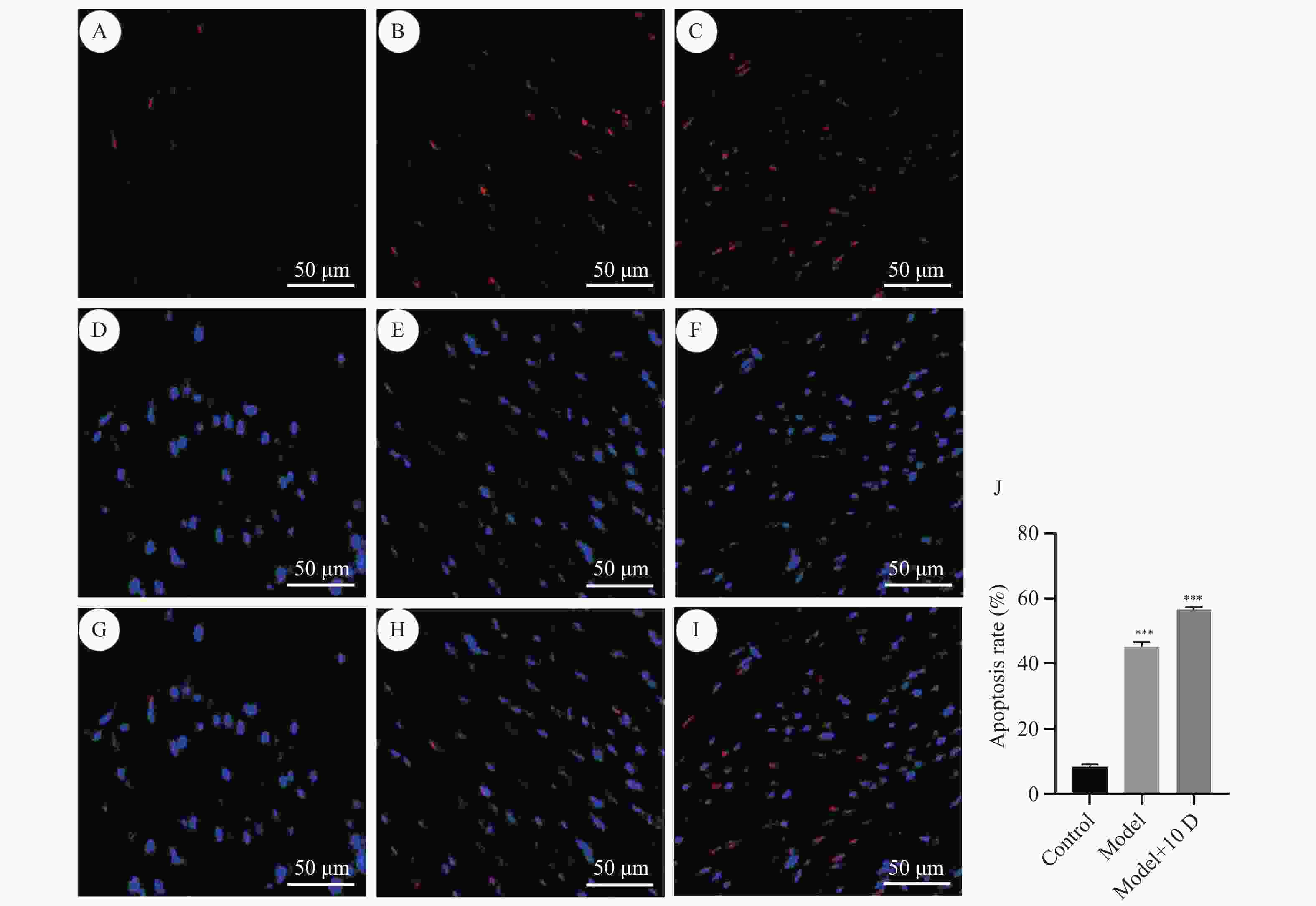
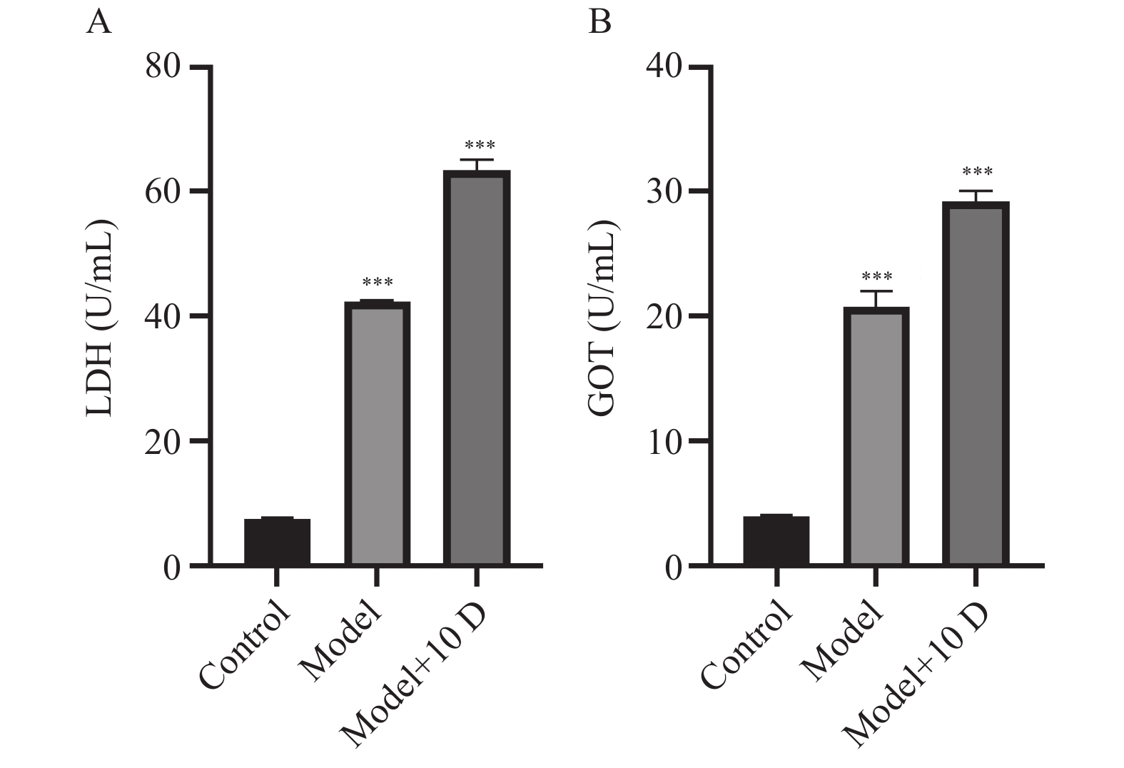
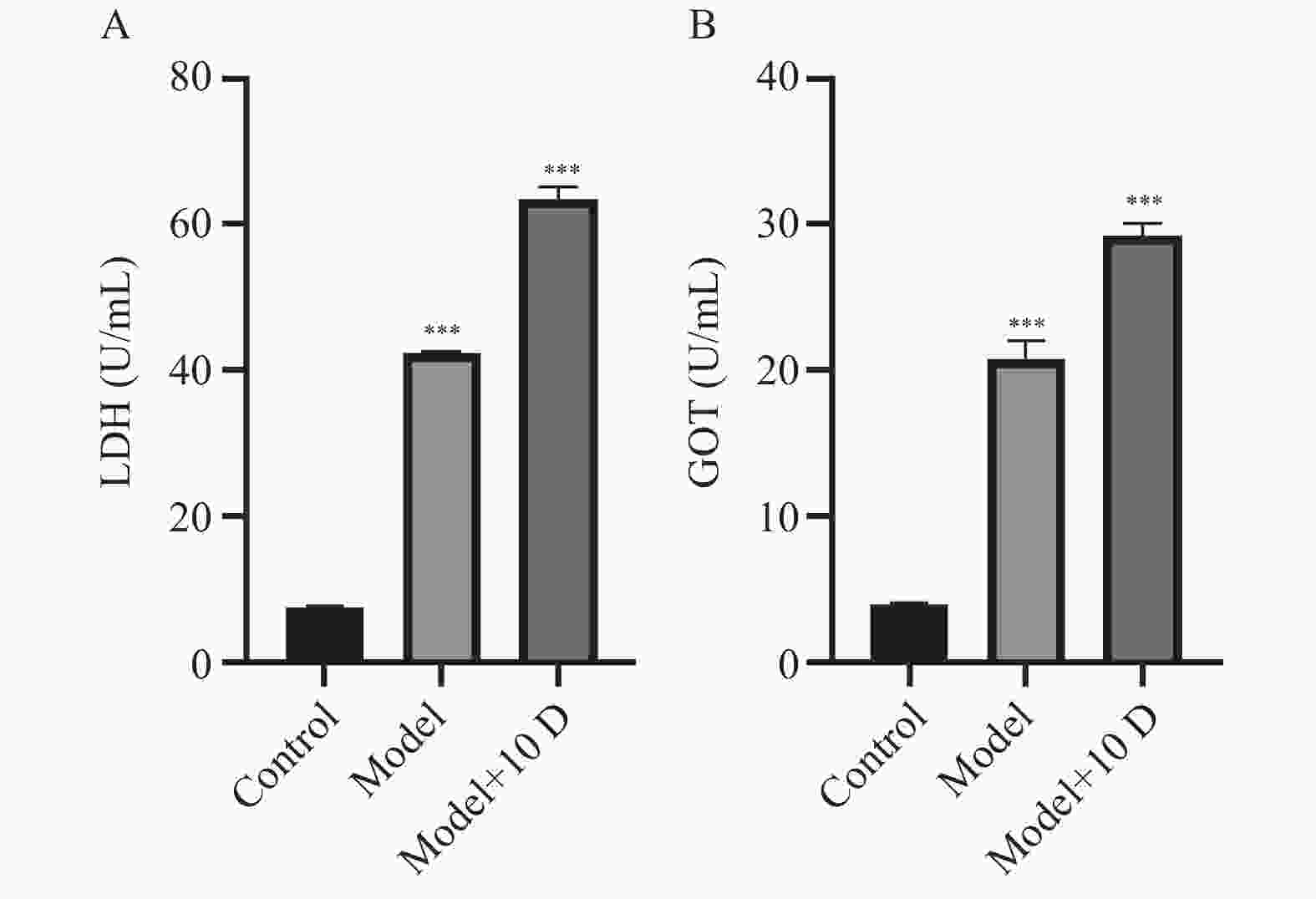
目的 探讨HK2和VDAC1在二乙酰吗啡致心肌细胞凋亡中的作用。 方法 采用剂量递增方法,建立二乙酰吗啡成瘾大鼠模型。将40只SD大鼠随机分为3组,正常组(Control,n=10)皮下注射等量生理盐水;模型组(Model,n=15)首次皮下注射二乙酰吗啡剂量为 5 mg/kg,以后逐日递增剂量 2.5 mg/(kg·d),连续注射20 d;模型+10 D组(Model+10D,n=15)在模型组基础上继续递增剂量至第10天。采用ELISA法检测乳酸脱氢酶(lactate dehydrogenase,LDH)、谷草转氨酶(glutamic oxaloacetic transaminase,GOT)含量;采用 HE 染色观察各组心肌组织病理变化;采用TUNEL染色检测各组心肌组织细胞凋亡;采用免疫组化、RT-qPCR和Western blot检测HK2、VDAC1及凋亡相关因子的mRNA和蛋白的表达变化。 结果 HE染色发现随着二乙酰吗啡干预时间的延长,心肌组织表现出不同程度的损伤。与正常组相比,模型组中血清LDH、GOT含量及心肌凋亡率升高,HK2和抗凋亡因子Bcl-2的mRNA和蛋白水平下降,VDAC1和促凋亡因子Bax、Caspase-3的mRNA和蛋白水平升高,Clevead Caspase-3蛋白水平升高;与正常组相比,模型+10 D组中以上指标,差异有统计学意义(P < 0.05)。 结论 二乙酰吗啡可导致心肌细胞凋亡,HK2和VDAC1可能参与了二乙酰吗啡致心肌细胞凋亡的过程。 -
关键词:
- 二乙酰吗啡 /
- 心肌凋亡 /
- 己糖激酶2 /
- 电压依赖性阴离子通道1 /
- 裂解半胱氨酸天冬氨酸蛋白酶-3
Abstract:Objective To investigate the role of HK2 and VDAC1 in diacetylmorphine-induced cardiomyocyte apoptosis. Methods A dose-escalation method was used to establish a rat model of diacetylmorphine addiction. Forty SD rats were randomly divided into three groups, the normal group (n=10) was injected with an equal amount of saline subcutaneously, the model group (n=15) was injected with 5 mg/kg of diacetylmorphine for the first time, and then the dose was increased by 2.5 mg/ (kg·d) day by day for 20 days, and the group of model + 10 D (n=15) continued to increase the dose based on the model group up to the 10th day. Lactate dehydrogenase (LDH) and glutamic oxaloacetic transaminase (GOT) were detected by ELISA; HE staining was used to observe the pathological changes of myocardial tissues in each group; TUNEL staining was used to detect apoptosis in myocardial tissues in each group; and immunohistochemistry, RT-q-analysis, and immunochemistry were used to detect apoptosis in myocardial tissues in each group. Immunohistochemistry, RT-qPCR and Western blot were used to detect the mRNA and protein expression of HK2, VDAC1 and apoptosis-related factors. Results HE staining revealed that myocardial tissues exhibited different degrees of damage with the prolongation of diacetylmorphine intervention. Compared with the normal group, serum LDH, GOT content and myocardial apoptosis rate increased in the model group, mRNA and protein levels of HK2 and anti-apoptotic factor Bcl-2 decreased, mRNA and protein levels of VDAC1 and pro-apoptotic factors Bax and Caspase-3 increased, and the protein level of Clevead Caspase-3 increased; in the model + 10 D group the above indexes, there was a statistically significant difference (P < 0.05). Conclusion Diacetylmorphine can cause cardiomyocyte apoptosis, and VDAC1 may be involved in the process of cardiomyocyte apoptosis caused by diacetylmorphine. -
Key words:
- Diacetylmorphine /
- Cardiac apoptosis /
- HK2 /
- VDAC1 /
- Clevead Caspase-3
-
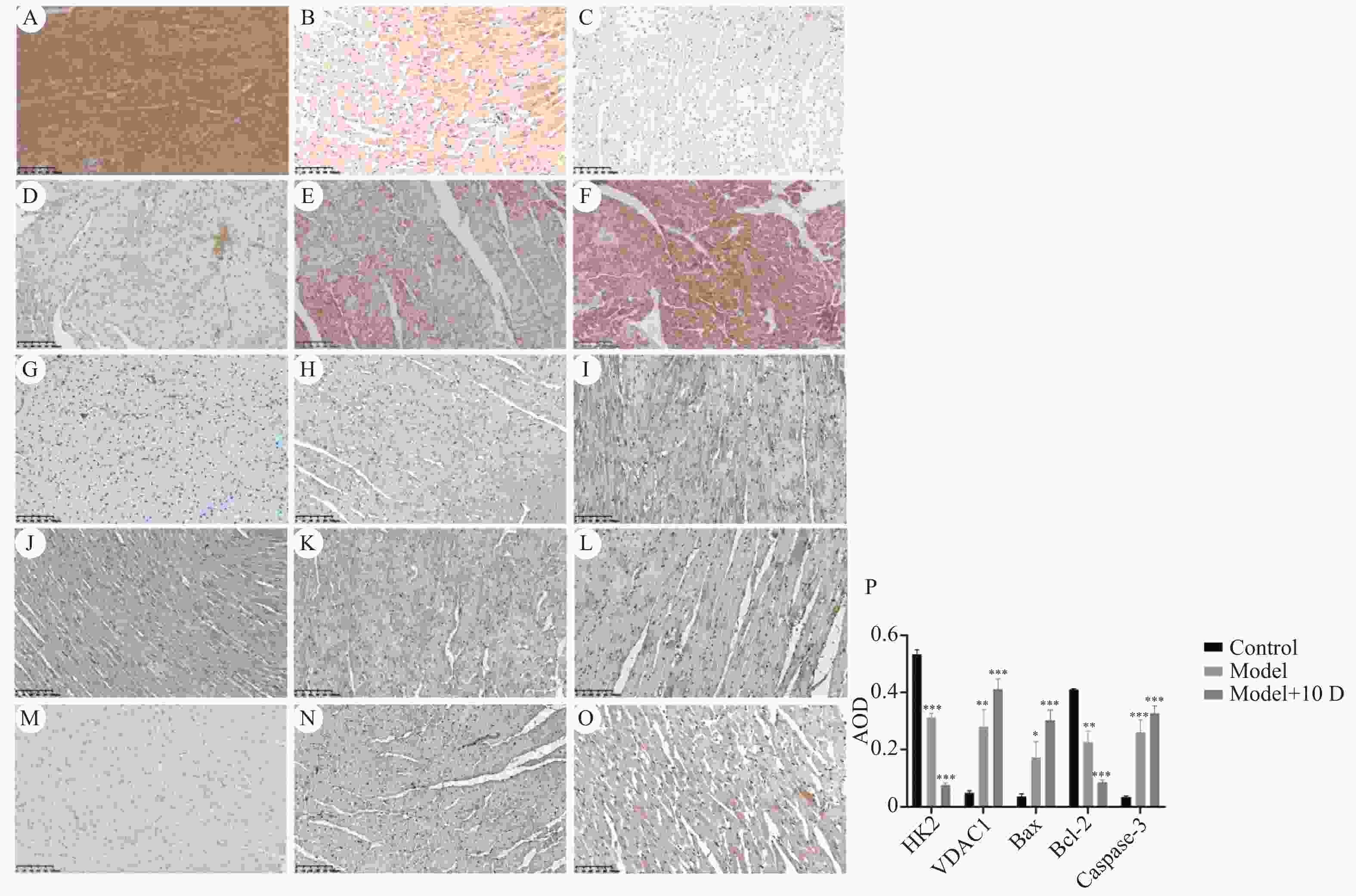
图 4 大鼠心肌组织HK2、VDAC1、Bax、Bcl-2、Caspase-3蛋白表达(×200)
A:正常组HK2表达;B:模型组HK2表达;C:模型+10 D组HK2表达;D:正常组VDAC1表达;E:模型组VDAC1表达;F:模型+10 D组VDAC1表达;G:正常组Bax表达;H:模型组Bax表达;I:模型+10 D组Bax表达;J:正常组Bcl-2表达;K:模型组Bcl-2表达;L:模型+10 D组Bcl-2表达;M:正常组Caspase-3表达;N:模型组Caspase-3表达;O:模型+10 D组Caspase-3表达;P:大鼠心肌组织HK2、VDAC1、Bax、Bcl-2、Caspase-3蛋白AOD表达情况($ \bar x \pm s $,n=6)。与正常组比较,*P<0.05,**P<0.01,***P<0.001。
Figure 4. Expression of HK2,VDAC1,Bax,Bcl-2 and Caspase-3 protein in rat myocardial tissue (×200)
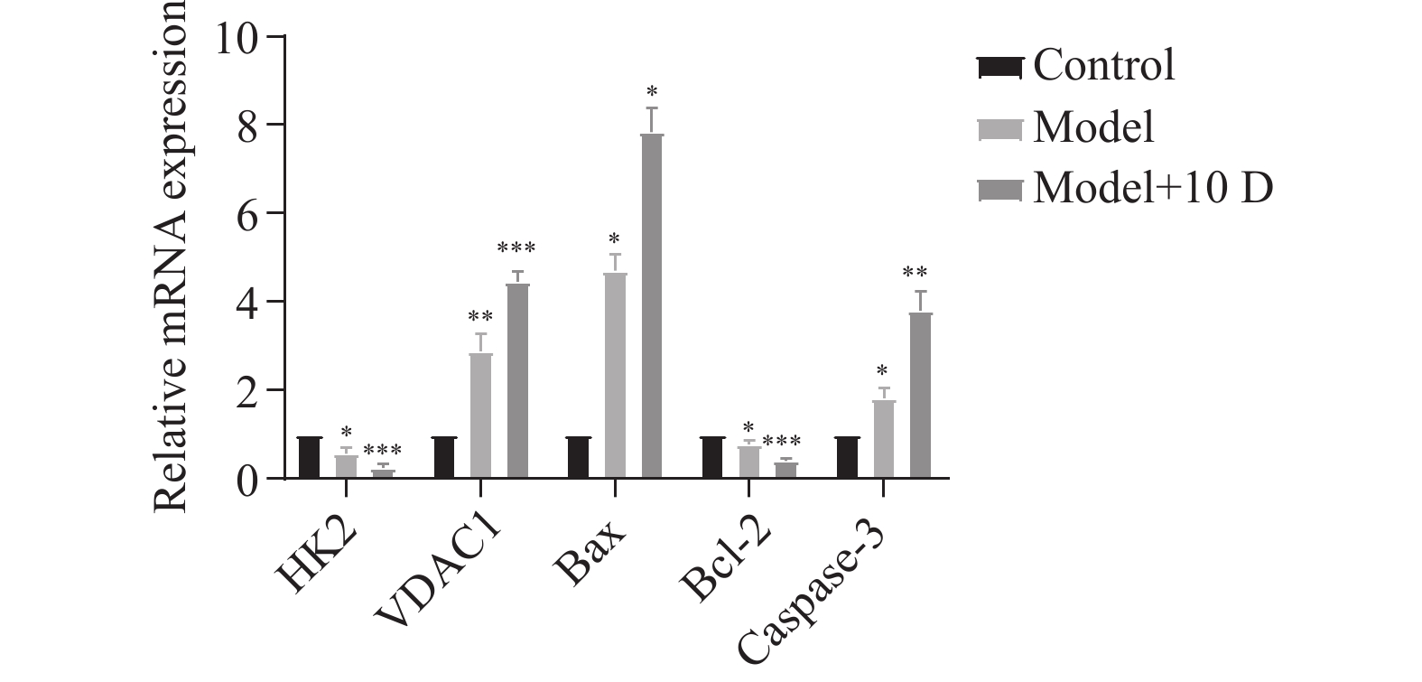
表 1 二乙酰吗啡对心肌组织中HK2、VDAC1、Bax、Bcl-2、Caspase-3 mRNA表达的影响($ \bar x \pm s $,n=6)
Table 1. Effects of diamorphine on the mRNA expression of HK2,VDAC1,Bax,Bcl-2 and Caspase-3 in myocardium($ \bar x \pm s $,n=6)
基因 正常组 模型组 模型+10 D组 F P HK2 1.00±0.00 0.57±0.14* 0.24±0.09*** 48.77 0.009# VDAC1 1.00±0.00 3.00±0.61** 4.46±0.25*** 32.62 0.027# Bax 1.00±0.00 4.88±0.17* 7.64±0.44* 50.32 0.001# Bcl-2 1.00±0.00 0.75±0.10* 0.39±0.05*** 34.86 0.020# Caspase-3 1.00±0.00 1.91±0.11* 3.78±0.38** 45.67 0.020# 与正常组比较,*P < 0.05,**P < 0.01,***P < 0.001。组间比较,#P < 0.05。 -
[1] Milella M S,D'Ottavio G,De Pirro S,et al. Heroin and its metabolites: relevance to heroin use disorder[J]. Transl Psychiatry,2023,13(1):120. doi: 10.1038/s41398-023-02406-5 [2] 王珺,李光伟,汪娜,等. Kelch样ECH相关蛋白1/核因子E2相关因子2对2型糖尿病心肌细胞凋亡的影响[J]. 中国临床药理学杂志,2023,39(5):635-638. [3] 刘小山,陈玉川,李朝晖,等. 海洛因成瘾大鼠心电图及心肌超微结构改变的研究[J]. 法医学杂志,2004,20(3):129-132,135. [4] Ciscato F,Ferrone L,Masgras I,et al. Hexokinase 2 in cancer: A prima donna playing multiple characters[J]. Int J Mol Sci,2021,22(9):4716. doi: 10.3390/ijms22094716 [5] Bao C,Zhu S,Song K,et al. HK2: A potential regulator of osteoarthritis via glycolytic and non-glycolytic pathways[J]. Cell Commun Signal.,2022,20(1):132. doi: 10.1186/s12964-022-00943-y [6] Shoshan-Barmatz V,Maldonado E N,Krelin Y. VDAC1 at the crossroads of cell metabolism,apoptosis and cell stress[J]. Cell Stress,2017,1(1):11-36. doi: 10.15698/cst2017.10.104 [7] 周晓昕,张玉静,肖芳,等. 六价铬诱导的肝细胞线粒体依赖性凋亡中VDAC1的作用[J]. 生态毒理学报,2017,12(6):91-98. [8] Fan K,Fan Z,Cheng H,et al. Hexokinase 2 dimerization and interaction with voltage-dependent anion channel promoted resistance to cell apoptosis induced by gemcitabine in pancreatic cancer[J]. Cancer Med,2019,8(13):5903-5915. doi: 10.1002/cam4.2463 [9] Ji M,Su L,Liu L,et al. CaMKII regulates the proteins TPM1 and MYOM2 and promotes diacetylmorphine-induced abnormal cardiac rhythms[J]. Sci Rep,2023,13(1):5827. doi: 10.1038/s41598-023-32941-6 [10] Rosen D,Smith M L,Reynolds C F 3rd. The prevalence of mental and physical health disorders among older methadone patients[J]. Am J Geriatr Psychiatry,2008,16(6):488-497. doi: 10.1097/JGP.0b013e31816ff35a [11] 杜磊,夏海发,吴映辉,等. 吗啡对急性心肌缺血大鼠的作用及机制研究[J]. 中国临床药理学杂志,2021,37(18):2451-2455. [12] Xi H,Wang S,Wang B,et al. The role of interaction between autophagy and apoptosis in tumorigenesis (Review)[J]. Oncol Rep,2022,48(6):208. [13] 苟陶然,李重阳,欧锐,等. 海洛因对小鼠视网膜结构和抗氧化物酶的影响[J]. 解剖学报,2017,48(6):663-668. [14] Bernstein H G,Trübner K,Krebs P,et al. Increased densities of nitric oxide synthase expressing neurons in the temporal cortex and the hypothalamic paraventricular nucleus of polytoxicomanic heroin overdose victims: possible implications for heroin neurotoxicity[J]. Acta Histochem,2014,116(1):182-190. doi: 10.1016/j.acthis.2013.07.006 [15] 刘小山,刘水平,李朝晖,等. 海洛因成瘾大鼠心肌超微结构改变及凋亡检测[J]. 中山大学学报(医学科学版),2005,26(1):61-64,83. [16] Roberts D J,Miyamoto S. Hexokinase II integrates energy metabolism and cellular protection: Akting on mitochondria and TORCing to autophagy [published correction appears in cell death differ. 2015 Feb;22(2): 364][J]. Cell Death Differ,2015,22(2):248-257. doi: 10.1038/cdd.2014.173 [17] Song G,Lu Q,Fan H,et al. Inhibition of hexokinases holds potential as treatment strategy for rheumatoid arthritis[J]. Arthritis Res Ther,2019,21(1):87. doi: 10.1186/s13075-019-1865-3 [18] Kim K W,Kim S W,Lim S,et al. Neutralization of hexokinase 2-targeting miRNA attenuates the oxidative stress-induced cardiomyocyte apoptosis[J]. Clin Hemorheol Microcirc,2021,78(1):57-68. doi: 10.3233/CH-200924 [19] Shoshan-Barmatz V,Shteinfer-Kuzmine A,Verma A. VDAC1 at the intersection of cell metabolism,apoptosis,and diseases[J]. Biomolecules,2020,10(11):1485. doi: 10.3390/biom10111485 [20] Shoshan-Barmatz V,Nahon-Crystal E,Shteinfer-Kuzmine A,Gupta R. VDAC1,mitochondrial dysfunction,and Alzheimer’s disease[J]. Pharmacol Res,2018,131:87-101. doi: 10.1016/j.phrs.2018.03.010 [21] 宋阿北. VDAC1参与调控BCG感染的巨噬细胞凋亡的机制研究[D]. 银川: 宁夏大学, 2021. [22] Li Y,Kang J,Fu J,et al. PGC1α Promotes cisplatin resistance in ovarian cancer by regulating the HSP70/HK2/VDAC1 signaling pathway[J]. Int J Mol Sci,2021,22(5):2537. doi: 10.3390/ijms22052537 [23] Yuan S,Fu Y,Wang X,et al. Voltage-dependent anion channel 1 is involved in endostatin-induced endothelial cell apoptosis[J]. FASEB J,2008,22(8):2809-2820. doi: 10.1096/fj.08-107417 [24] Shoshan-Barmatz V,Ben-Hail D,Admoni L,et al. The mitochondrial voltage-dependent anion channel 1 in tumor cells[J]. Biochim Biophys Acta,2015,1848(10 Pt B):2547-2575. -


 下载:
下载:

