

Efficacy of Conventional Ultrasound and Contrast-enhanced Ultrasound in Preoperative Diagnosis of Lateral Cervical Lymph Node Metastasis from Papillary Thyroid Carcinoma
-
摘要:
目的 探讨常规超声(US)与超声造影(CEUS)在甲状腺乳头状癌(PTC)患者侧颈区淋巴结转移(LCLNM)术前诊断的超声特征,评价其在PTC侧颈区淋巴结转移的术前诊断效能。 方法 选取80例在昆明医科大学第二附属医院接受PTC手术及侧颈区淋巴结清扫术患者并获得126枚淋巴结超声检查资料。经术后病理证实,81枚为PTC转移性淋巴结,45枚为未转移性淋巴结。观察常规超声(US)及超声造影(CEUS)检查淋巴结转移声像图特征。与病理结果对比,计算US、CEUS及联合应用对PTC患者LCLNM的术前诊断的敏感度、特异度、准确度、阳性预测值、阴性预测值。 结果 US检查转移性淋巴结声像图特征多表现为淋巴门消失或偏移、皮质回声为非均匀低回声、内部存在团状高回声及钙化,血流类型多表现为周围型和混合型,血流分级多为Adler2级和3级,与非转移组比较,差异有统计学意义(P < 0.05);CEUS检查转移性淋巴结声像图特征多表现为向心型和Ⅱ型,其次见于Ⅲ型; US与CEUS联合应用对于诊断PTC侧颈区淋巴结转移的敏感度、特异度、准确度、阳性预测值、阴性预测值均优于US或CEUS单独应用。 结论 术前US与CEUS联合应用能提高PTC患者侧颈区淋巴结转移的诊断效能,可以在临床上为超声引导下淋巴结细针穿刺细胞学检查和术前制定淋巴结清扫术及手术治疗策略提供参考依据。 Abstract:Objective To investigate the ultransonic characteristics of conventional ultrasound (US)combined with Contrast-enhanced ultrasound (CEUS) in the pre-operative diagnosis effectiveness of lateral cervical lymph node metastasis (LCLNM)from papillary thyroid carcinoma (PTC), and to evaluate its efficacy in preoperative diagnosis of PTC lateral cervical lymph node metastasis. Methods A total of 80 patients who received PTC surgery and lateral cervical lymph node dissection in the Second Affiliated Hospital of Kunming Medical University were selected, and 126 lymph node ultrasonography data were obtained. Postoperative pathology confirmed that 81 lymph nodes were metastatic PTC and 45 lymph nodes were non-metastatic. The ultrasonographic features of lymph node metastasis were observed by conventional ultrasound (US) and contrast-enhanced ultrasound (CEUS). Compared with the pathological results, the sensitivity, specificity, accuracy, positive predictive value and negative predictive value of US, CEUS and combined application in the preoperative diagnosis of LCLNM in PTC patients were calculated. Results The ultrasonographic features of metastatic lymph nodes were mostly lymphatic portal disappearance or deviation. Cortical echo was non-uniform hypoecho, internal mass hyperecho and calcification, blood flow types were mostly peripheral type and mixed type, and blood flow grades were mostly Adler grade 2 and grade 3; compared with the non-metastatic group, the difference was statistically significant (P < 0.05). The ultrasonographic characteristics of metastatic lymph nodes detected by CEUS were mostly centriotype and type Ⅱ, followed by type Ⅲ. The sensitivity, specificity, accuracy, positive predictive value and negative predictive value of the combination of US and CEUS were superior to those of US or CEUS alone in the diagnosis of PTC lateral cervical lymph node metastasis. Conclusion Preoperative conventional ultrasound combined with CEUS can improve the diagnostic efficacy of lateral cervical lymph node metastasis from PTC. -

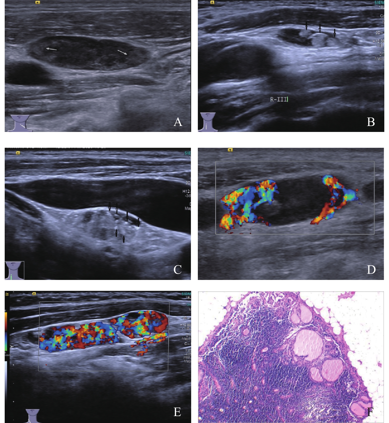
图 1 PTC患者侧颈区转移性淋巴结US声像图
A:淋巴门消失,皮质呈非均匀低回声(箭头);B:淋巴结内见团状高,回声(箭头所指); C:淋巴结内见钙化(箭头);D:周围型血流,Adler 3级;E:混合型血流,Adler 3级;F:手术病理:甲状腺乳头状癌侵犯淋巴结被膜及皮质成分(HE×100)。
Figure 1. The conventional ultrasonographic images of lateral cervical metastatic lymph node in PTC patients

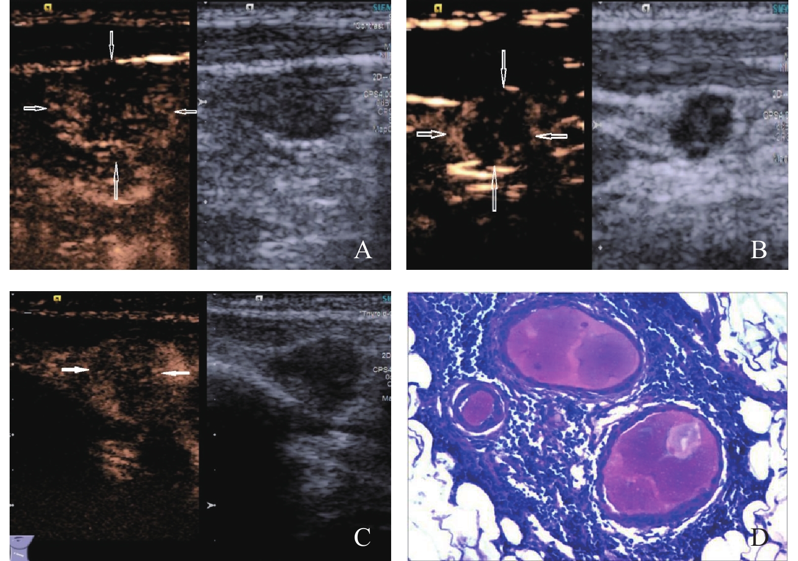
图 2 PTC患者侧颈区转移性淋巴结CEUS声像图
A:向心型增强(箭头所指);B:不均匀增强,内有无增强区(Ⅱ型)(箭头所指);C:不均匀微弱增强(Ⅲ型)(箭头所指);D:手术病理:淋巴结内可见甲状腺滤泡上皮及胶原成分(HE×200)。
Figure 2. The CEUS images of lateral cervical metastatic lymph node in PTC patients
表 1 PTC患者侧颈区淋巴结US声像特征[n(%)]
Table 1. The conventional ultrasonographic image features of lateral cervical lymph node status in PTC patients [n(%)]
US检查 超声检查特征 未转移淋巴结(n = 45) 转移淋巴结(n = 81) t/χ2 P 长短径比 < 2 13(28.9) 31(38.3) 1.618 0.157 ≥2 32(71.1) 50(61.7) 淋巴门 存在 19(42.2) 4(4.9) 4.776 0.028* 消失或偏移 26(57.8) 77(95.1) 皮质回声 均匀低回声 30(66.7) 23 (28.4) 0.453 0.015* 非均匀低回声 15(33.3) 58(71.6) 团状高回声 无 37(82.2) 32(39.5) 4.706 0.027* 有 8(17.8) 49(60.5) 钙 化 无 43(95.6) 33(40.7) 4.248 0.034* 有 2(4.4) 48(59.3) 囊变区 无 43(95.6) 60(74.1) 2.585 0.089 有 2(4.4) 21(25.9) 血流分布类型 无血流 12(26.7) 2(2.5) −3.990 < 0.001* 中央型 24(53.3) 6(7.4) 周围型 3(6.7) 42(51.8) 混合型 6(13.3) 31(38.3) 血流分级 0级 12(26.7) 2(2.5) −5.605 < 0.001* 1级 23(51.1) 9(11.1) 2级 6(13.3) 31(38.3) 3级 4(8.9) 39(48.1) US:常规超声; *P < 0.05。  下载: 导出CSV
表 2 PTC患者侧颈区淋巴结CEUS声像图特征 [n(%)]
Table 2. The contrast-enhanced ultrasound image features of lateral cervical lymph node status in PTC patients [n(%)]
CEUS检查 CEUS特征 未转移淋巴结(n = 45) 转移淋巴结(n = 81) t/χ2 P 增强模式 离心型 32(71.1) 2(2.5) 向心型 3(6.7) 59(72.8) −3.150 0.002* 混合型 10(22.2) 20(24.7) 增强类型 Ⅰ 35(77.8) 3(3.7) Ⅱ 4(8.9) 43(53.1) Ⅲ 6(13.3) 33(40.7) 4.876 0.027* Ⅳ 0(0.0) 2(2.5) CEUS:超声造影;*P < 0.05。
下载: 导出CSV
表 3 US、CEUS及US+CEUS联合应用对PTC患者侧颈区淋巴结转移的诊断效能 (%)
Table 3. Diagnostic efficacy of US,CEUS and US+CEUS combined in patients with PTC patients with lateral cervical lymph node metastasis (%)
检查方法 检查结果 病理结果(n) 敏感度 特异度 阳性预测值 阴性预测值 准确度 有转移 未转移 US 有转移 67 16 82.7 64.4 80.7 67.4 76.2 未转移 14 29 CEUS 有转移 62 5 76.5 88.9 92.5 67.8 81.0 未转移 19 40 US + CEUS 有转移 78 4 96.3 91.1 95.1 93.2 94.4 未转移 3 41 注:US: 常规超声;CEUS: 超声造影 ; US+CEUS:常规超声与超声造影联合应用。
下载: 导出CSV
-
[1] Siegel R L,Miller K D,Fuchs H E,et al. Cancer statistics,2021[J]. CA Cancer J Clin.,2021,71(1):7-33. doi: 10.3322/caac.21654 [2] Kim Y,Roh J L,Gong G,et al. Risk factors for lateral neck recurrence of N0/N1a papillary thyroid cancer[J]. Ann Surg Oncol,2017,24(12):3609-3616. doi: 10.1245/s10434-017-6057-2 [3] Haugen B R,Alexander E K,Bible K C,et al. 2015 American thyroid association management guidelines for adult patients with thyroid nodules and differentiated thyroid cancer:The American thyroid association guidelines task force on thyroid nodules and differentiated thyroid cancer[J]. Thyroid,2016,26(1):1-133. doi: 10.1089/thy.2015.0020 [4] Wu J H,Zeng W,Wu R G,et al. Comparison of ultrasonography and CT for determining the preoperative benign or malignant nature of thyroid nodules:Diagnostic performance according to calcification[J]. Technol Cancer Res Treat,2020,19(1):1-6. [5] Brauckhoff K,Biermann M. Multimodal imaging of thyroid cancer[J]. Curr Opin Endocrinol Diabetes Obes,2020,27(5):335-344. doi: 10.1097/MED.0000000000000574 [6] Poanta L,Serban O,Pascu I,et al. The place of CEUS in distinguishing benign from malignant cervical lymph nodes:A prospective study[J]. Med Ultrason,2014,16(1):7-14. doi: 10.11152/mu.2014.2066.161.lp1os2 [7] Cui Q L,Yin S S,Fan Z H,et al. Diagnostic value of contrast-enhanced ultrasonography and time-intensity curve in differential diagnosis of cervical metastatic and tuberculous lymph nodes[J]. J Ultrasound Med,2018,37(1):83-92. doi: 10.1002/jum.14311 [8] Grégoire V,Ang K,Budach W,et al. Delineation of the neck node levels for head and neck tumors:A 2013 update. DAHANCA,EORTC,HKNPCSG,NCIC CTG,NCRI,RTOG,TROG consensus guidelines[J]. Radiother Oncol,2014,110(1):172-181. doi: 10.1016/j.radonc.2013.10.010 [9] Adler D D,Carson P L,Rubin J M,Quinn-Reid D. Doppler ultrasound color flow imaging in the study of breast cancer:Preliminary findings[J]. Ultrasound Med Biol,1990,16(6):553-559. doi: 10.1016/0301-5629(90)90020-D [10] 张雪云. 超声造影及弹性成像在浅表淋巴结病变鉴别诊断中的价值[D]. 兰州: 兰州大学硕士学位论文, 2019. [11] Luster M,Aktolun C,Amendoeira I,et al. European perspective on 2015 American thyroid association management guidelines for adult patients with thyroid nodules and differentiated thyroid cancer:Proceedings of an interactive international symposium[J]. Thyroid,2019,29(1):7-26. doi: 10.1089/thy.2017.0129 [12] 周倩,许萍. 超声评估甲状腺癌颈部转移淋巴结的研究进展[J]. 中国医学影像技术,2019,35(11):1752-1756. doi: 10.13929/j.1003-3289.201906103 [13] Zhao B H,Huang Z H,Huang Y C,et al. Preliminary study of superselective lymph node dissection in regional lateral cervical lymph node metastasis of papillary thyroid carcinoma[J]. Zhonghua Zhong Liu Za Zhi [Chinese Journal of Oncology],2021,43(4):484-489. [14] 任玲,费翔,张艳,等. 高帧率超声造影对颈部浅表淋巴结病变良恶性的鉴别诊断[J]. 中国医学影像学杂志,2021,29(10):989-992,997. doi: 10.3969/j.issn.1005-5185.2021.10.007 [15] 王晓荣,梁奎,艾迪拜·木合买提,等. 剪切波弹性成像及超声造影鉴别颈部不典型反应性增生淋巴结及淋巴瘤的价值[J]. 中国医学影像学杂志,2020,28(8):566-570. doi: 10.3969/j.issn.1005-5185.2020.08.002 [16] 何守伟,王知力. 恶性浅表淋巴结病变的常规超声及超声造影特征[J]. 中国医学影像学杂志,2021,29(1):9-13. doi: 10.3969/j.issn.1005-5185.2021.01.003 [17] 卜锐,杨娜,夏春娟,等. 高频超声术前诊断甲状腺乳头状癌患者颈部转移性淋巴结[J]. 中国医学影像技术,2019,35(1):55-58. doi: 10.13929/j.1003-3289.201803009 [18] 王磊,李海,唐嘉阅,等. 不同病理性钙化类型甲状腺乳头状癌微血管密度及淋巴结转移比较研究[J]. 肿瘤研究与临床,2018,30(8):531-535. doi: 10.3760/cma.j.issn.1006-9801.2018.08.006 [19] 田菊,勇强,郑超,等. 甲状腺结节微小强回声与组织沙粒体的相关性分析[J]. 中国超声医学杂志,2020,36(4):302-305. doi: 10.3969/j.issn.1002-0101.2020.04.004 [20] Machado M R,Tavares M R,Buchpiguel C A,et al. Ultrasonographic evaluation of cervical lymph nodes in thyroid cancer[J]. Otolaryngol Head Neck Surg,2017,156(2):263-271. [21] 任玲,罗渝昆. 经静脉超声造影在浅表淋巴结诊断中的应用[J]. 中国医学影像学杂志,2019,27(8):626-629. [22] 张琦,庄连婷,李家兴,等. 常规超声联合超声造影对颈部实性淋巴结良恶性的鉴别诊断[J]. 中国医学影像学杂志,2021,29(1):14-18. -

 点击查看大图
计量
- 文章访问数: 5113
- HTML全文浏览量: 2584
- PDF下载量: 22
- 被引次数: 0
