 Email
Email RSS
RSSCurrent Issue
2026, Volume 47, Issue 6
2026,
47(6):
1-10.
doi: 10.12259/j.issn.2095-610X.S20260601
Abstract:
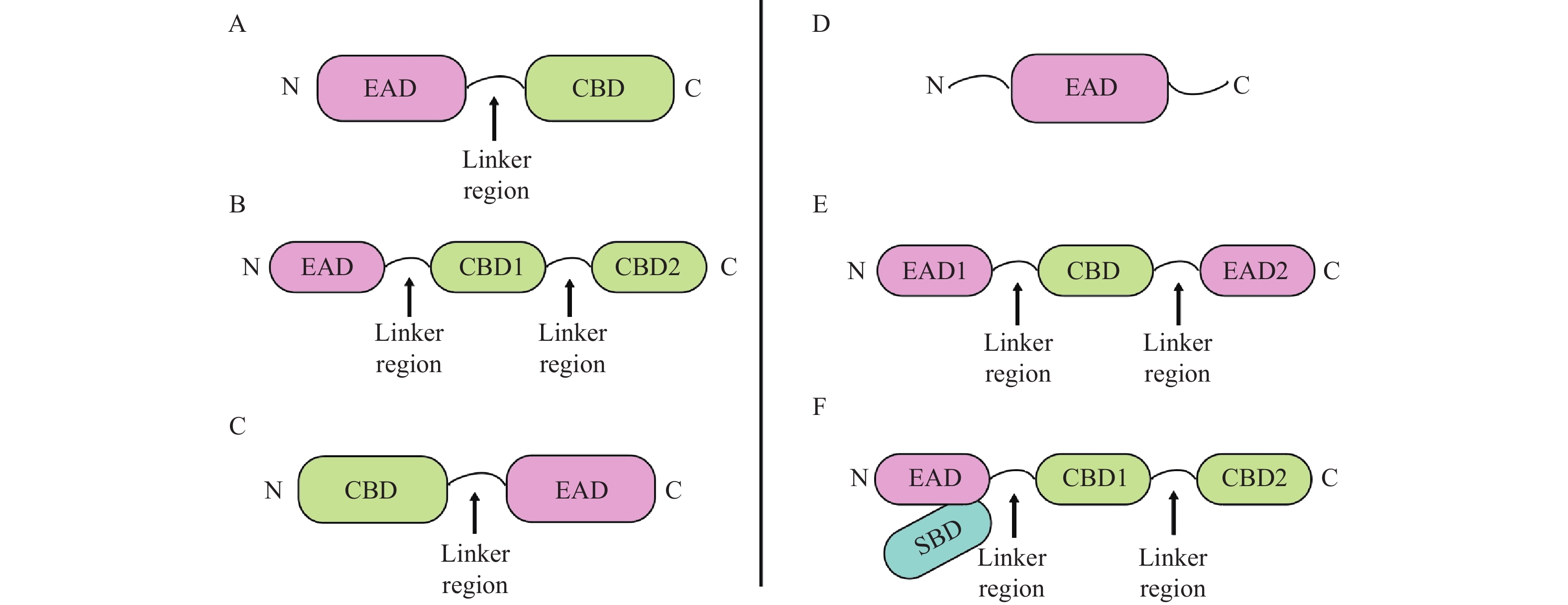
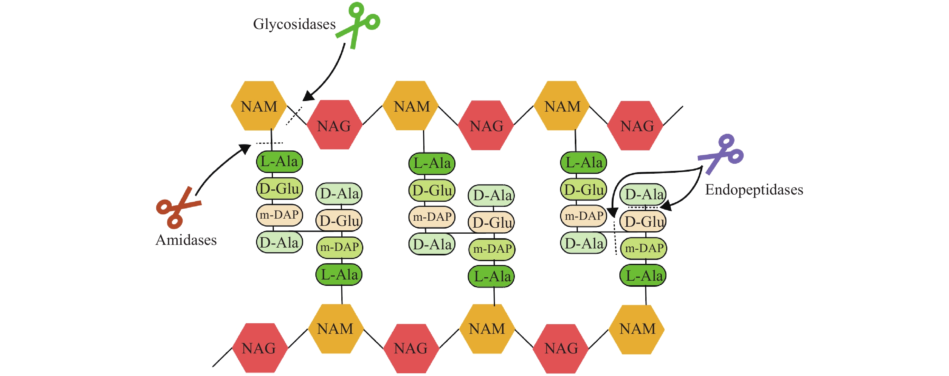
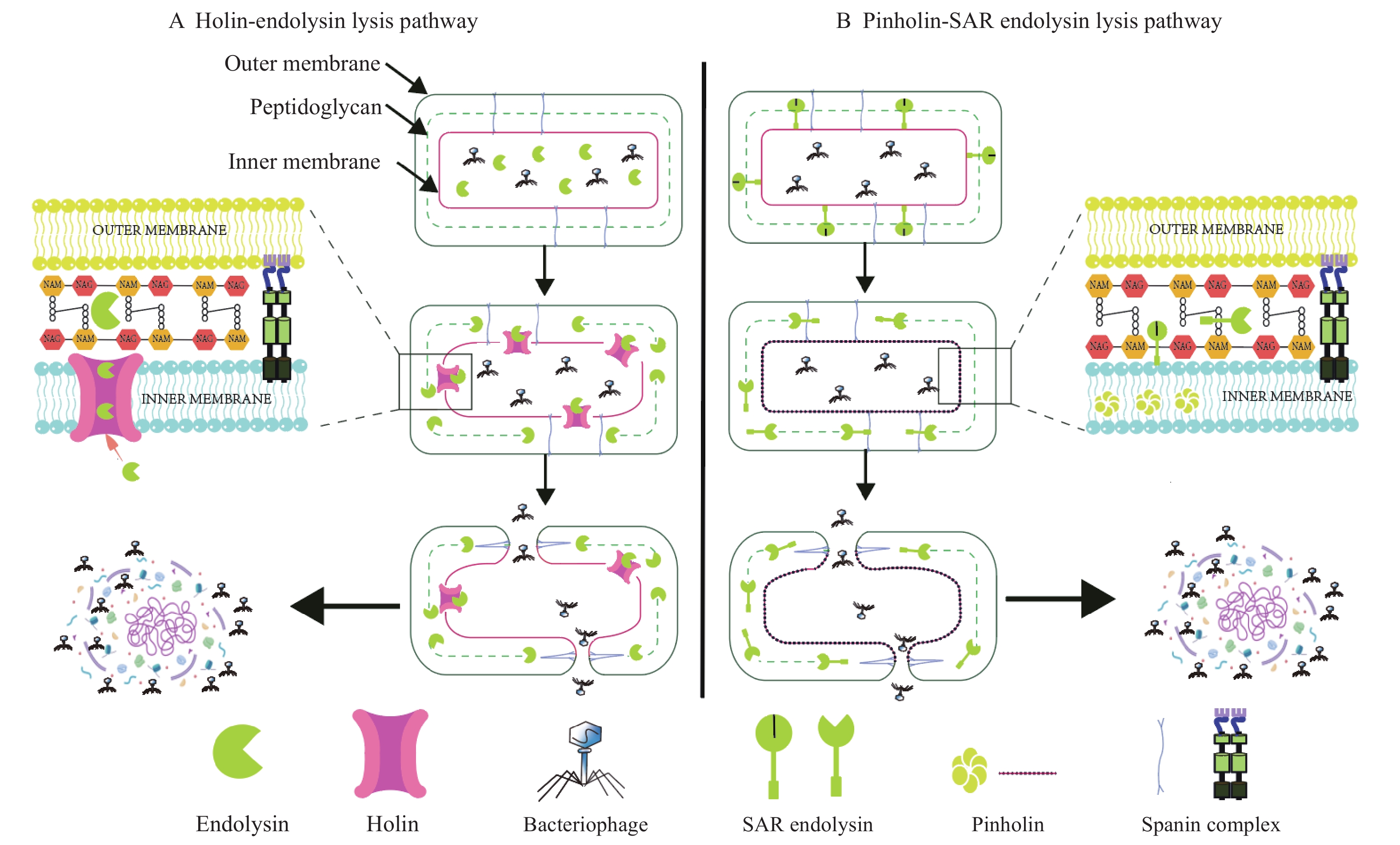
The continued spread of bacterial antibiotic resistance has become a major challenge in global public health. Endolysins derived from bacteriophages, with their unique antimicrobial mechanisms, are gradually emerging as novel antibacterial agents to replace conventional antibiotics. Endolysins are peptidoglycan hydrolases encoded by bacteriophages that exert bactericidal effects by lysing bacterial cell walls, with advantages such as strong host specificity, low propensity for resistance development, minimal disruption to the microbiota, and synergistic multi-target mechanisms. Endolysins have demonstrated potent lytic activity against various antibiotic-resistant bacteria in human medicine, veterinary medicine, and the food industry. When combined with conventional antibiotics, endolysins can produce synergistic effects, significantly reducing the minimum inhibitory concentration of antibiotics and improving survival rates in infection models. This review summarizes the application and mechanisms of action of endolysins as novel antimicrobial agents, with the aim of providing reference for future research on the application of bacteriophage endolysins in treating bacterial antibiotic-resistant infections.
The continued spread of bacterial antibiotic resistance has become a major challenge in global public health. Endolysins derived from bacteriophages, with their unique antimicrobial mechanisms, are gradually emerging as novel antibacterial agents to replace conventional antibiotics. Endolysins are peptidoglycan hydrolases encoded by bacteriophages that exert bactericidal effects by lysing bacterial cell walls, with advantages such as strong host specificity, low propensity for resistance development, minimal disruption to the microbiota, and synergistic multi-target mechanisms. Endolysins have demonstrated potent lytic activity against various antibiotic-resistant bacteria in human medicine, veterinary medicine, and the food industry. When combined with conventional antibiotics, endolysins can produce synergistic effects, significantly reducing the minimum inhibitory concentration of antibiotics and improving survival rates in infection models. This review summarizes the application and mechanisms of action of endolysins as novel antimicrobial agents, with the aim of providing reference for future research on the application of bacteriophage endolysins in treating bacterial antibiotic-resistant infections.
2026,
47(6):
11-22.
doi: 10.12259/j.issn.2095-610X.S20260602
Abstract:
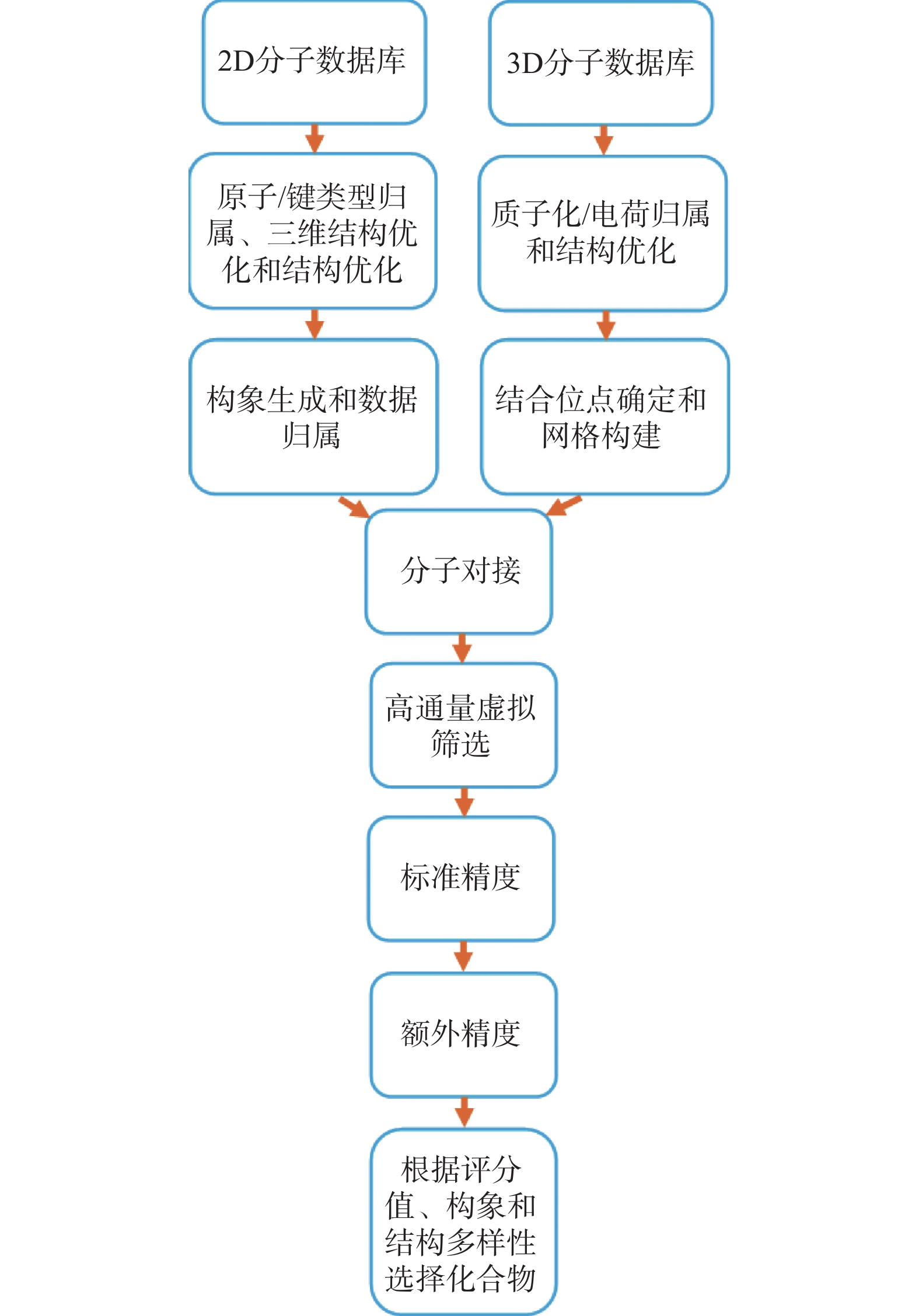
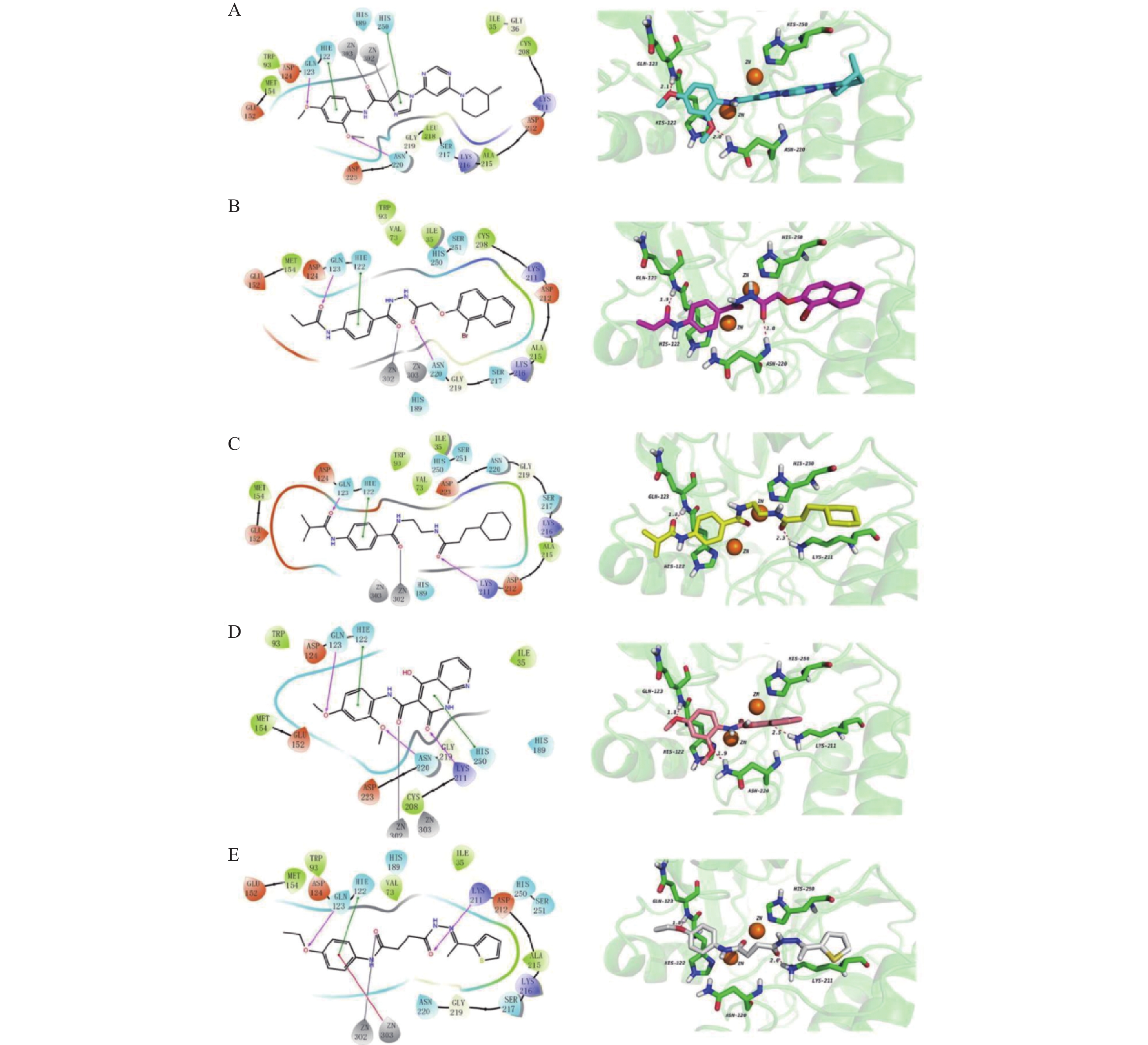
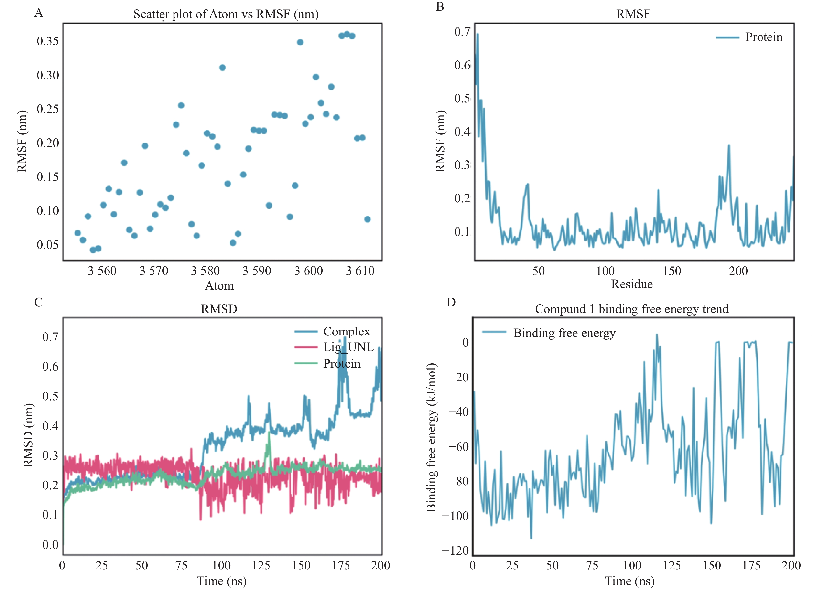
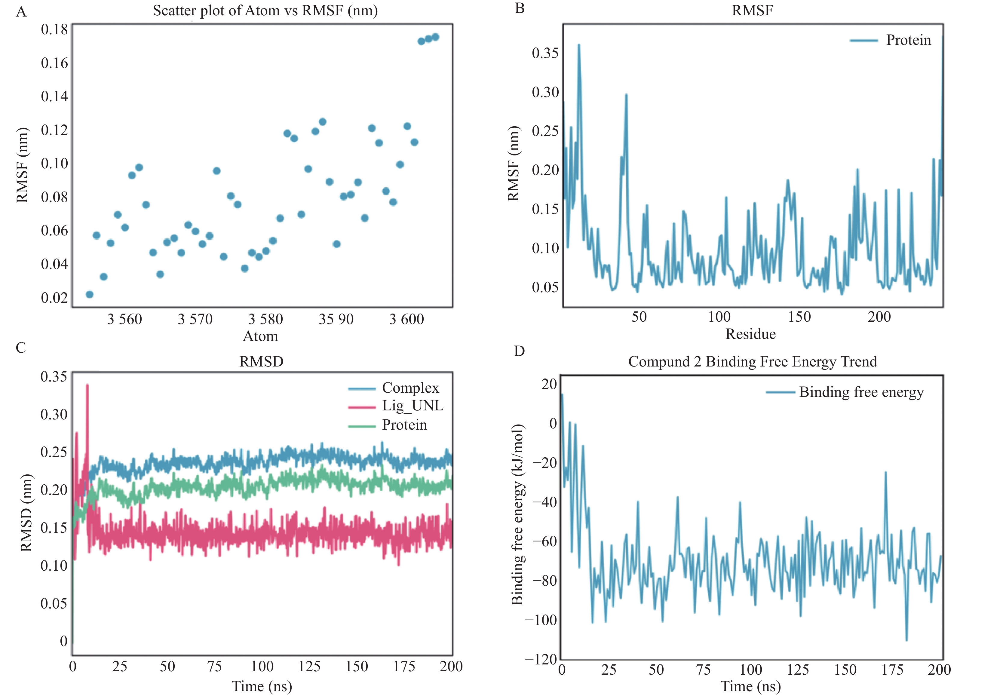
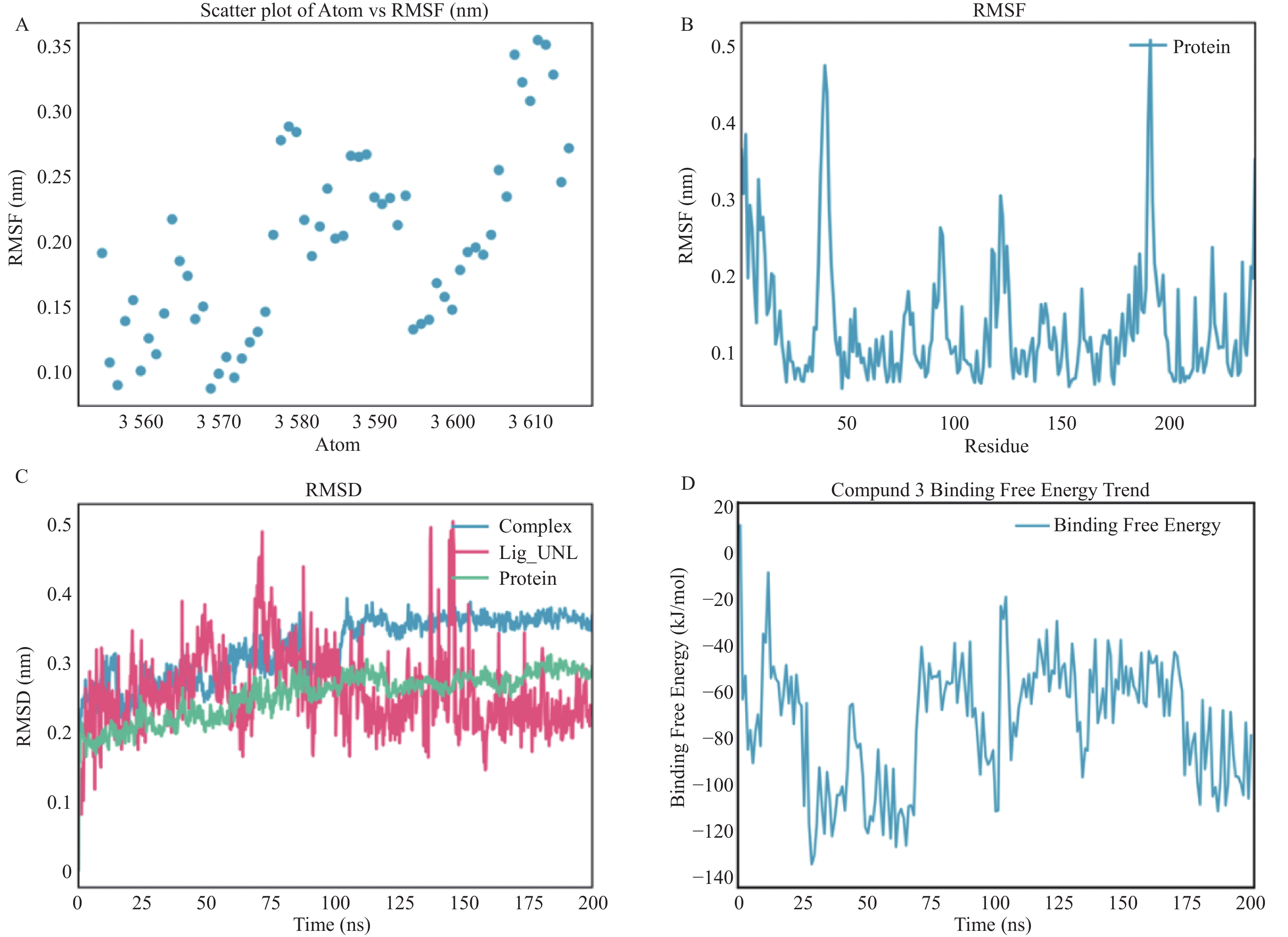
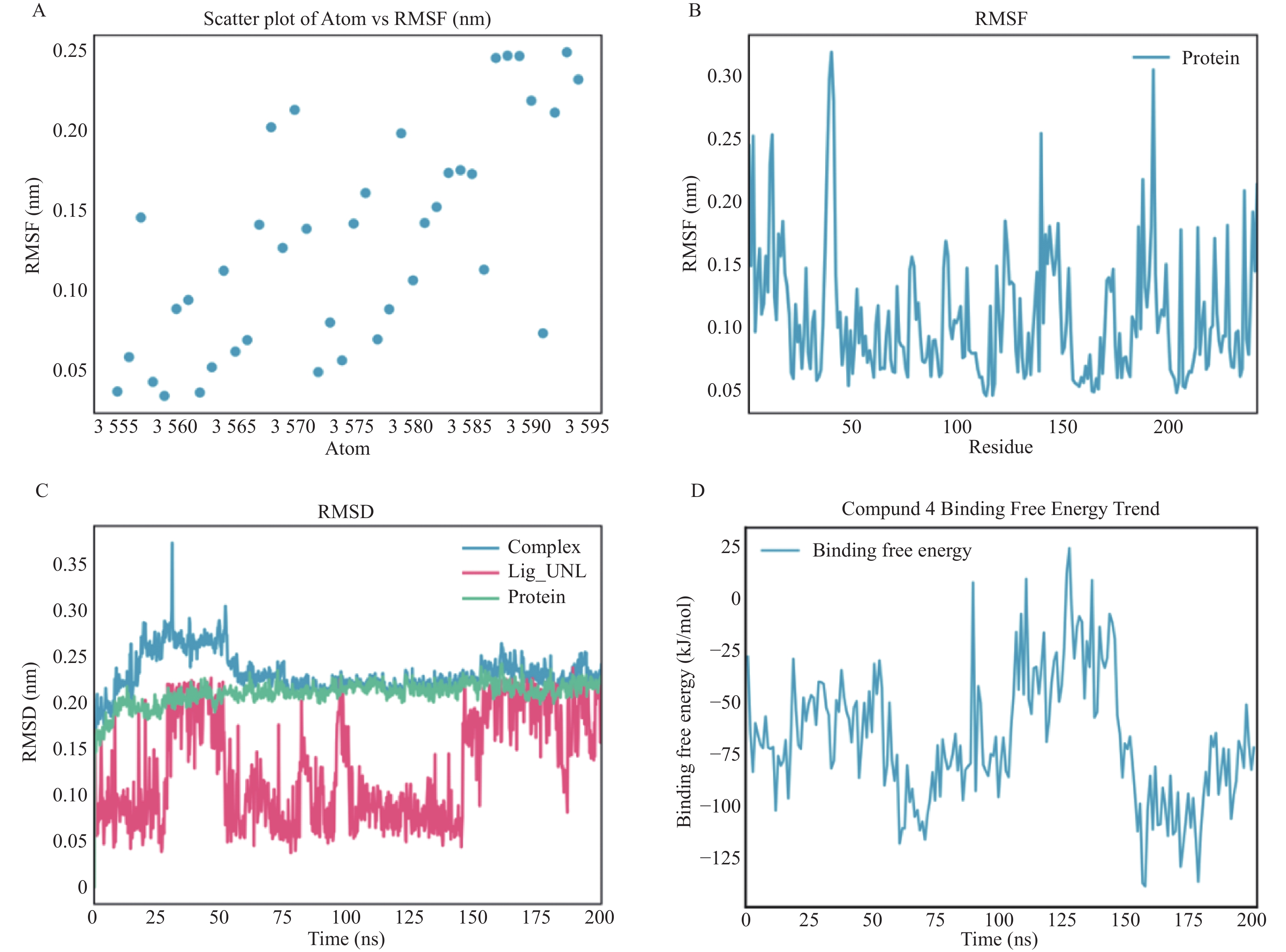
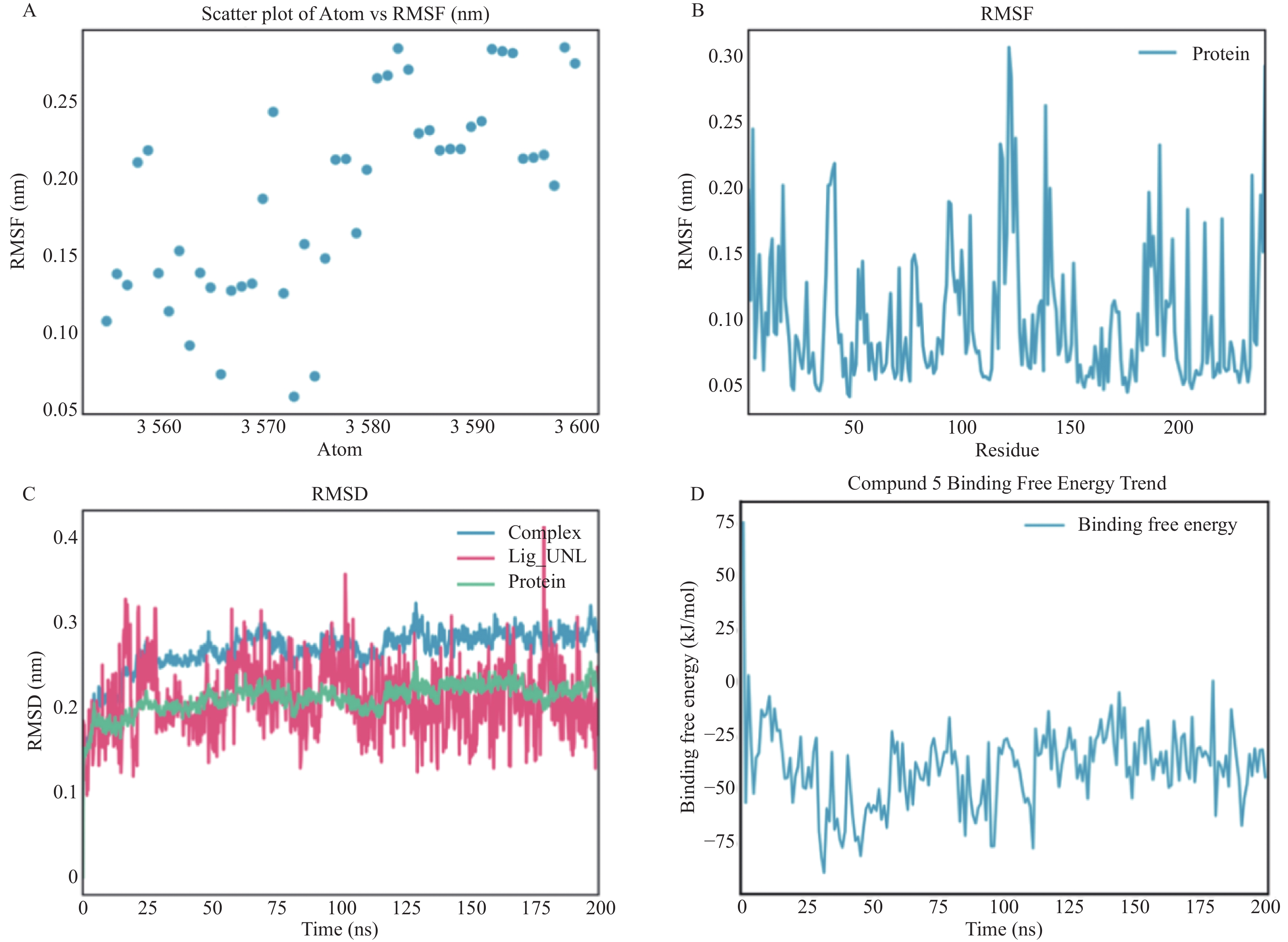
Objective To screen potential inhibitors based on the three-dimensional structure of NDM-1 enzyme and evaluate their pharmacological activity. Methods Computer-aided drug design methods were employed to screen the top 5 candidate compounds with the highest molecular docking scores from the 50K Diversity Library. The minimum inhibitory concentration (MIC) was further determined in combination with meropenem, imipenem, and ceftazidime to assess their inhibitory activity against NDM-1. The stability of the candidate compounds binding to NDM-1 was analyzed through molecular dynamics simulation. Results When the top 5 compounds with the highest molecular docking scores were combined with three β-lactam antibiotics, the MIC values showed no significant reduction, suggesting limited inhibitory effects on NDM-1. Molecular dynamics simulation revealed that compounds 1, 3, 4, and 5 exhibited large root-mean-square deviation (RMSD) and free energy fluctuations in the reaction system, indicating unstable conformations; although compound 2 showed no significant RMSD fluctuation, its binding free energy value was relatively low, potentially resulting in insufficient binding affinity. Conclusion Differences in binding stability between the five candidate compounds and NDM-1 protein led to their overall limited inhibitory activity.
2026,
47(6):
23-32.
doi: 10.12259/j.issn.2095-610X.S20260603
Abstract:
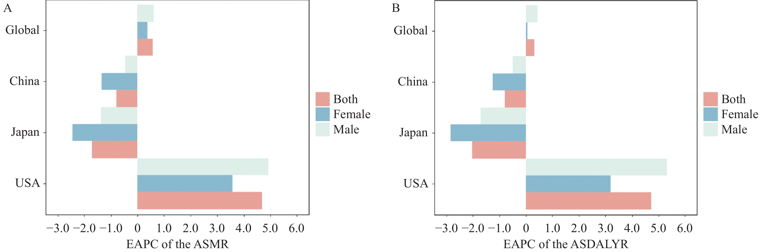
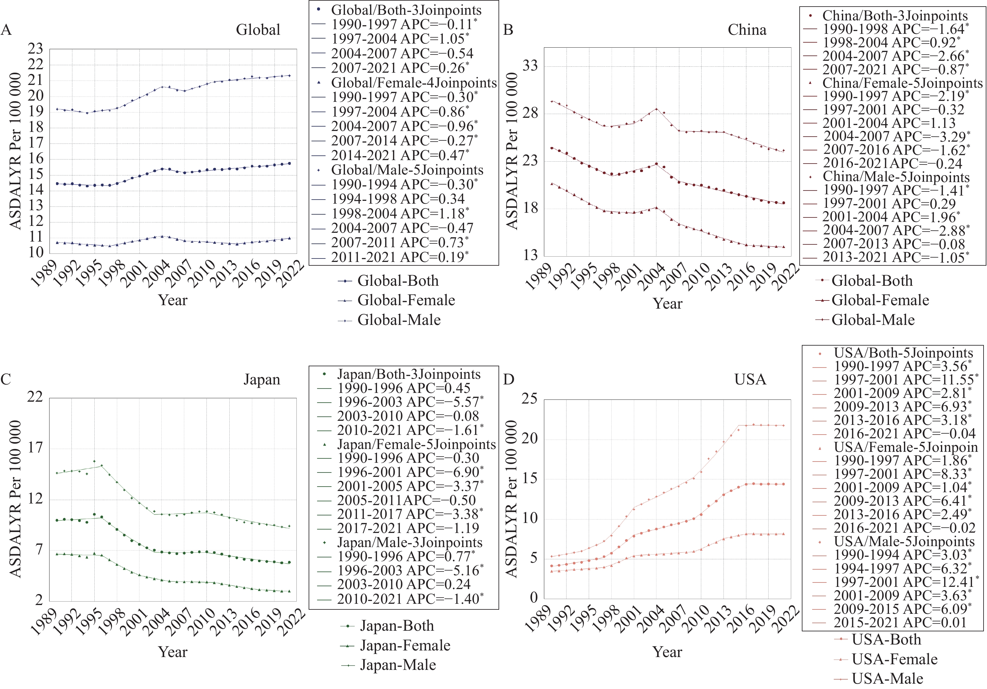
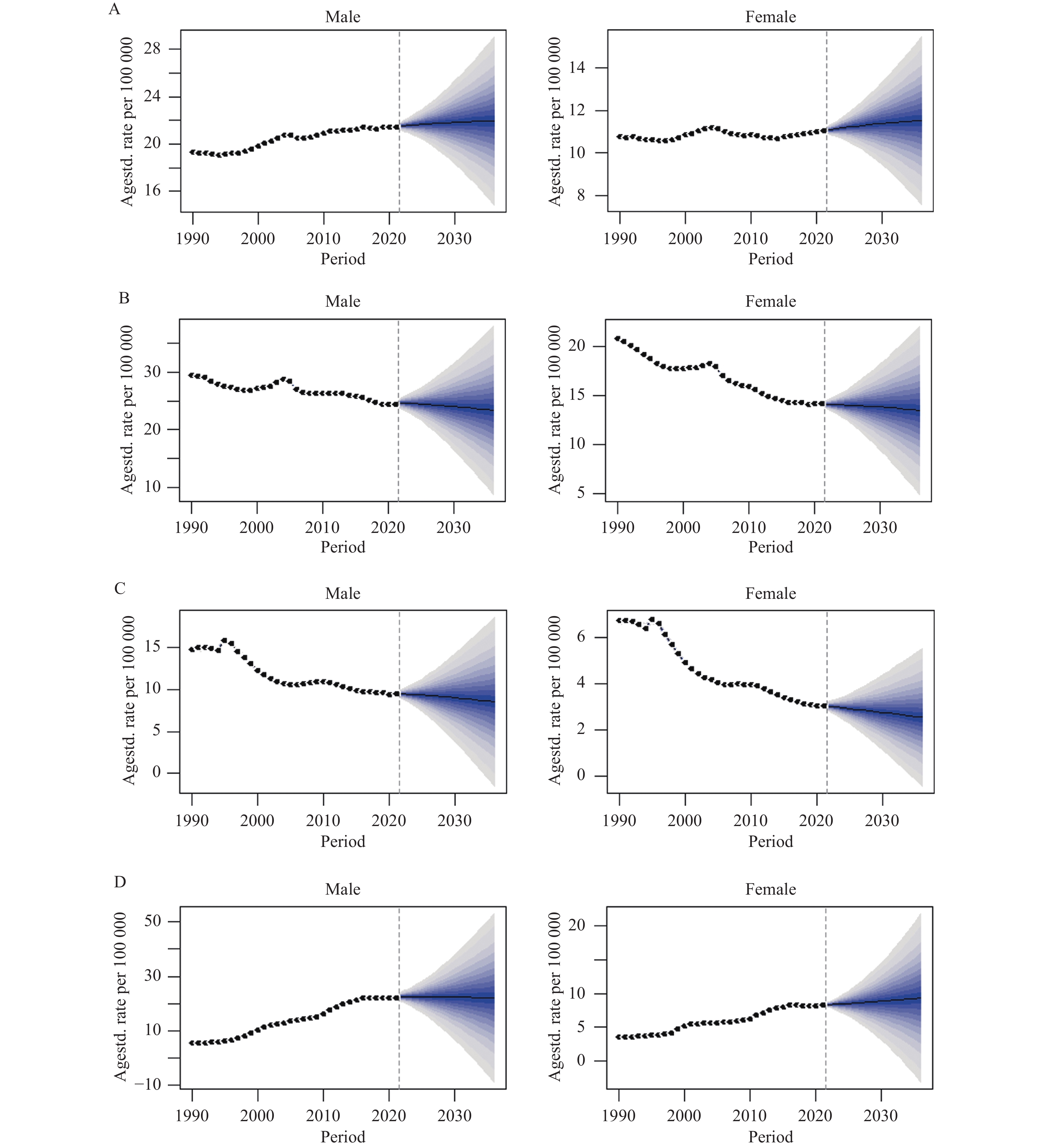
Objective To analyze the disease burden and trends of hypertensive nephropathy attributed to high sodium diet in China, Japan, and the United States from 1990 to 2021, and to project trends from 2022 to 2036. Methods Data were extracted from the 2021 Global Burden of Disease (GBD 2021) database, including age-standardized mortality rates (ASMR) and age-standardized disability-adjusted life year rates (ASDALYR) for hypertensive nephropathy attributed to high sodium diet. Estimated annual percentage change (EAPC) and Joinpoint regression models were used to analyze historical trends, while the Bayesian age-period-cohort (BAPC) model was employed to project future disease burden. Results Globally, ASMR and ASDALYR for hypertensive nephropathy attributed to high sodium diet showed significant upward trends, with EAPC of 0.58 (95%CI: 0.53 to 0.62) and 0.32 (95%CI: 0.28 to 0.37), respectively. Stratified analysis revealed statistically significant differences in ASDALYR among males (EAPC = 0.44, 95%CI: 0.39 to 0.48). Among the three countries, China bore the heaviest disease burden, though with a declining trend (EAPC: -1.35 [95%CI: -1.43 to -1.27] for ASMR and -1.26 [95%CI: -1.36 to -1.16] for ASDALYR), with more pronounced decline in females. Japan had the lightest disease burden with the most significant declining trends (EAPC: -1.72 [95%CI: -2.01 to -1.42] for ASMR and -2.02 [95%CI: -2.26 to -1.79] for ASDALYR), particularly among females. The United States showed upward trends (EAPC: 4.68 [95%CI: 4.40 to 4.97] for ASMR and 4.72 [95%CI: 4.38 to 5.50] for ASDALYR), with males showing significantly higher increases than females. Joinpoint analysis revealed that ASDALYR declined after 2004 globally in females and in both sexes in China, while Japan demonstrated declining trends after 1995. BAPC projections for 2022 to 2036 indicated that ASDALYR in both males and females in China and Japan would show steady decreases, with rates declining from 24.65 per 100,000 and 14.06 per 100,000 to 23.38 per 100,000 and 13.46 per 100,000 in Chinese males and females, respectively, and from 9.47 per 100,000 and 3.03 per 100,000 to 8.56 per 100,000 and 2.55 per 100,000 in Japanese males and females, respectively. The United States showed sex-specific divergence, with male ASDALYR slightly declining from 22.53 per 100,000 to 22.10 per 100,000, while female rates continued to increase from 8.34 per 100,000 to 9.34 per 100,000. Conclusion The global disease burden of hypertensive nephropathy attributed to high sodium diet remains inadequately controlled with significant national and gender disparities. Strict sodium intake control represents a key potential intervention target for preventing hypertensive nephropathy. Countries should adopt effective strategies from Japan's experience and develop precision-based prevention and control measures.
2026,
47(6):
33-43.
doi: 10.12259/j.issn.2095-610X.S20260604
Abstract:
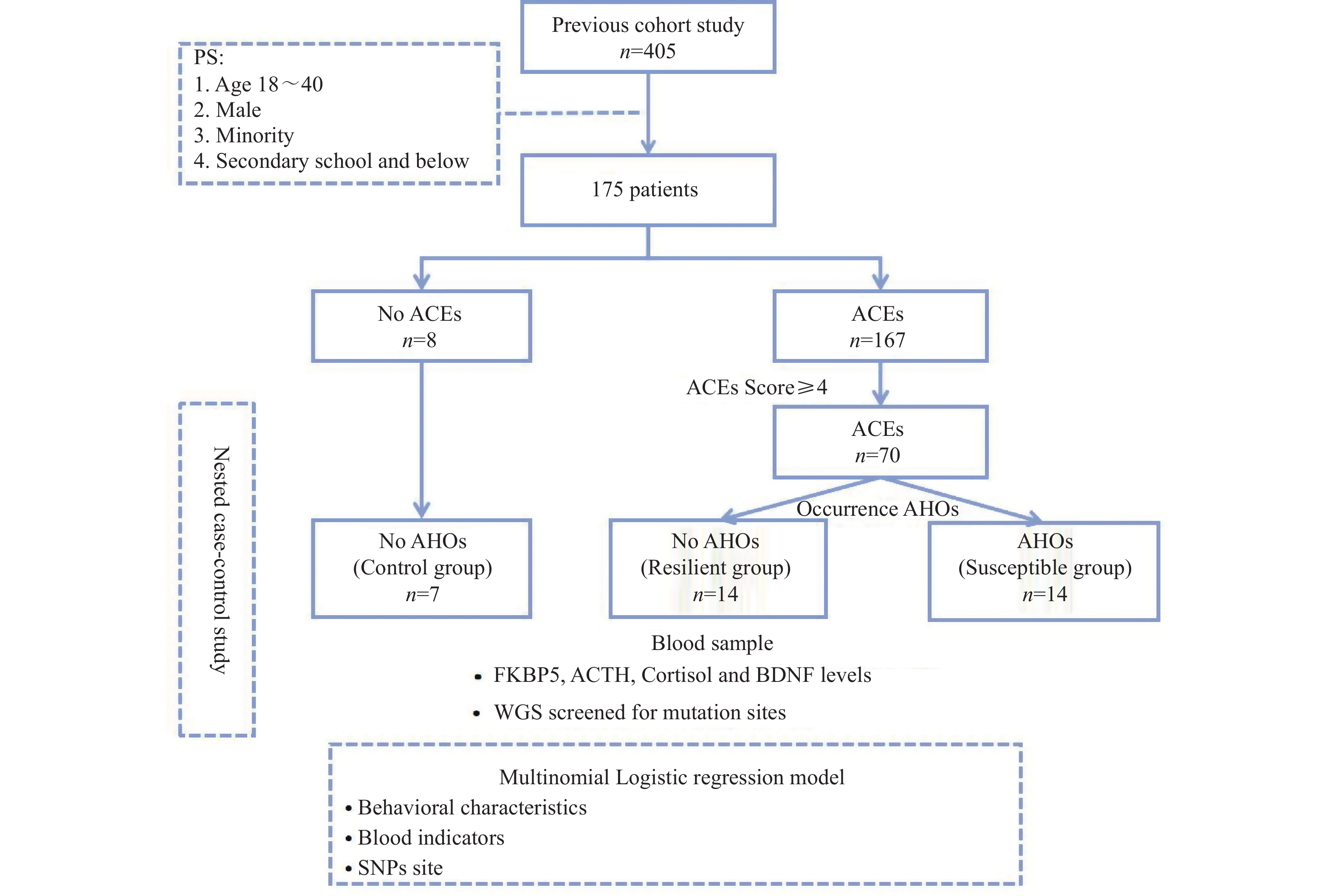
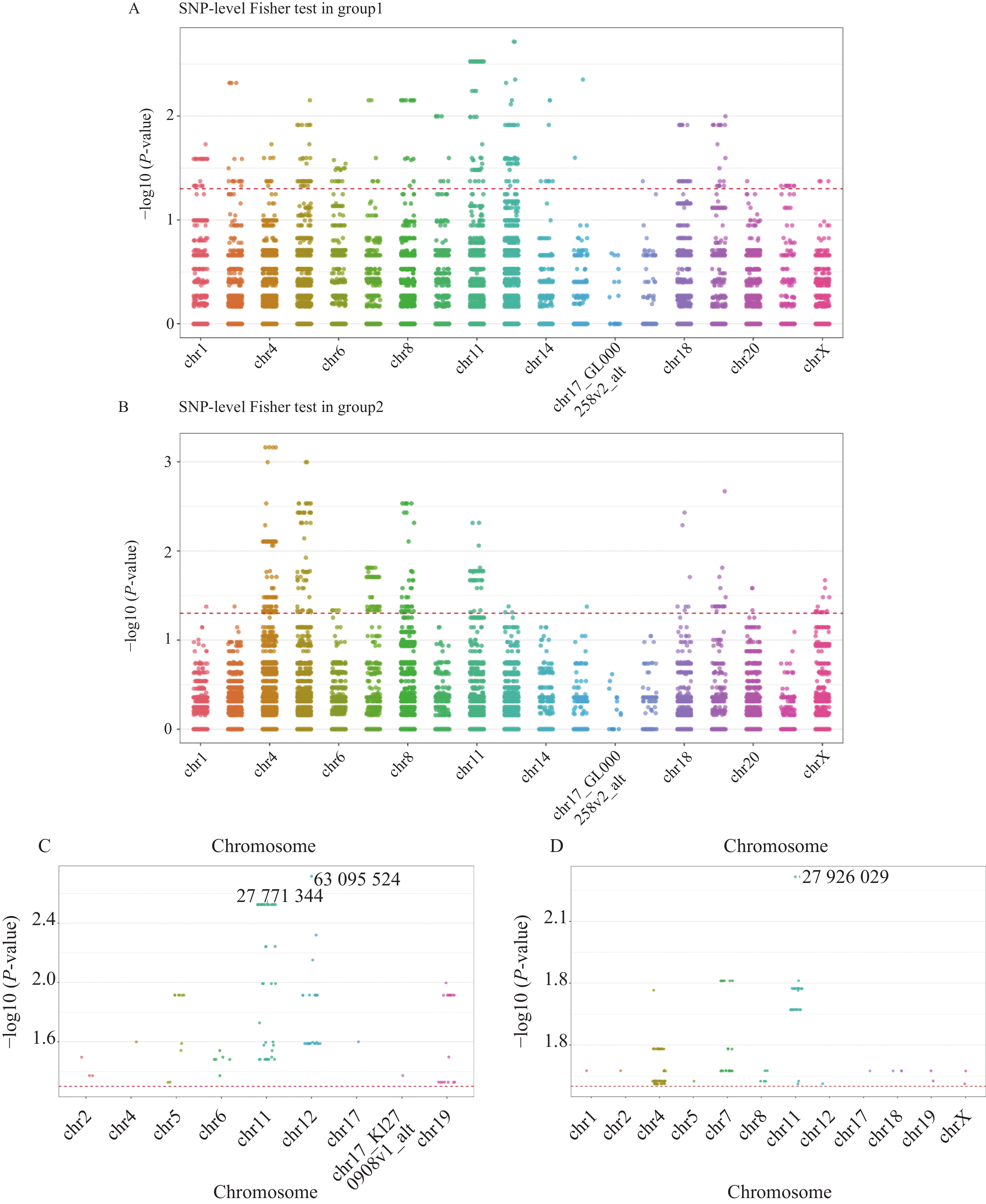
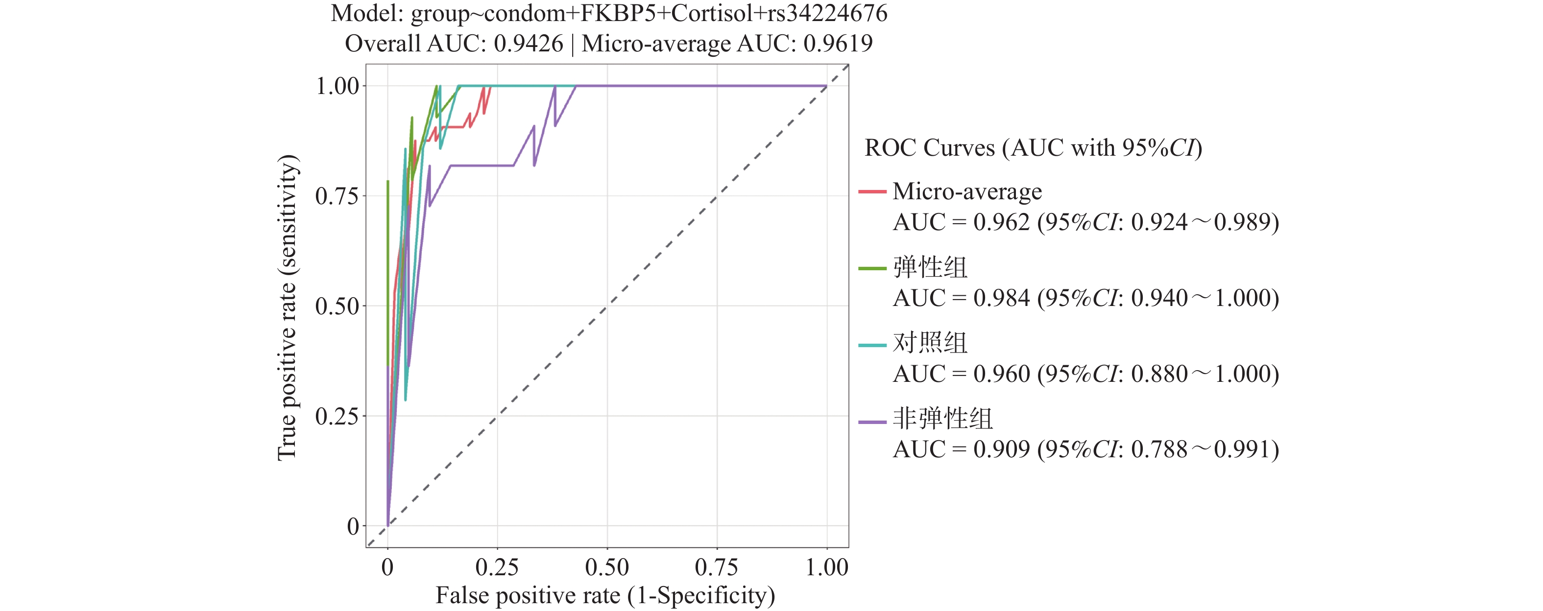
Objective To investigate the association between adverse childhood experiences (ACEs) and adverse health outcomes (AHOs), and to compare the levels of FKBP5, ACTH, cortisol, and BDNF between susceptible and resilient individuals exposed to ACEs during childhood. Methods Based on a cohort of 405 individuals with substance use disorder in Yunnan Province established from January to July 2021, a nested case-control study was conducted. FKBP5, ACTH, cortisol, and BDNF levels were detected in the control group, resilient group and susceptible group. Whole-genome sequencing (WGS) technology was used to identify mutation sites Descriptive, univariate and multivariate analyses were conducted using SPSS 24.0 and R Studio version 4.3.1.n Results Significant differences in FKBP5 levels were found between the control group (n = 7) and resilient groups (n = 14) [(4.67±1.08) vs. (6.86±1.87) ng/mL, P < 0.05]. Compared with the control group, ACTH levels in the susceptible group (n = 14) were elevated [(54.05±8.75) pg/mL, P < 0.05]; compared with the susceptible group, ACTH levels in the resilient group were elevated [(67.28±8.36) pg/mL, P < 0.05]. Cortisol levels showed significant differences between the control and susceptible groups [(254.92±70.46) vs. (278.50±49.60) nmol/L, P < 0.05] and between the susceptible and resilient groups [(278.50±49.60) vs. (406.27±72.07) nmol/L, P < 0.05]. Multivariate logistic regression analysis showed that, with the control group as reference, the resilient group had significant differences in FKBP5, cortisol, and rs34224676 genotype levels (OR = 3.62, 95%CI: 1.36~9.64, P < 0.05; OR = 1.02, 95%CI: 1.00~1.04, P < 0.05; OR = 185.34, 95%CI: 69.56~493.83, P < 0.05). Conclusion Individuals with substance use disorder exposed to ACEs exhibited higher levels of FKBP5, ACTH, and cortisol compared to those without ACEs exposure. Under ACEs exposure, the expression of FKBP5, cortisol, and the SNP locus rs34224676 in individuals with substance use disorder may be associated with the resilience phenomena.
2026,
47(6):
44-55.
doi: 10.12259/j.issn.2095-610X.S20260605
Abstract:
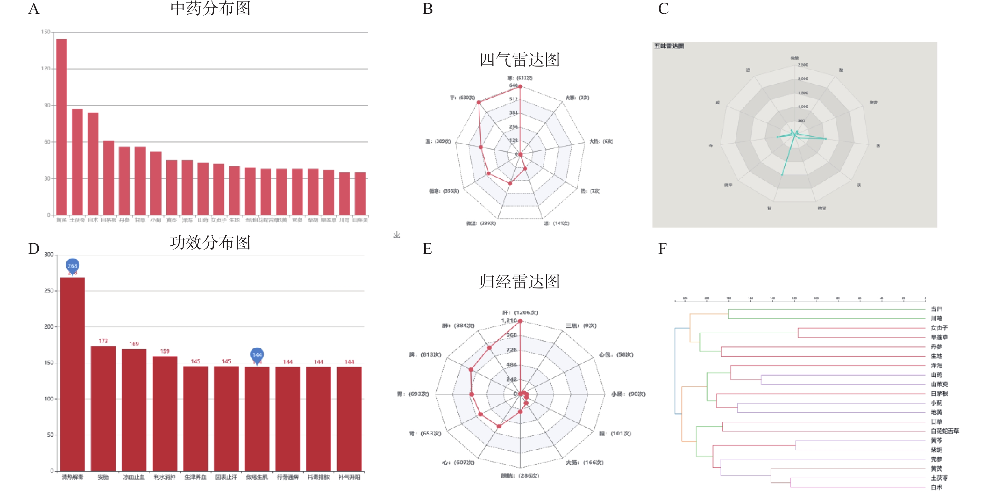
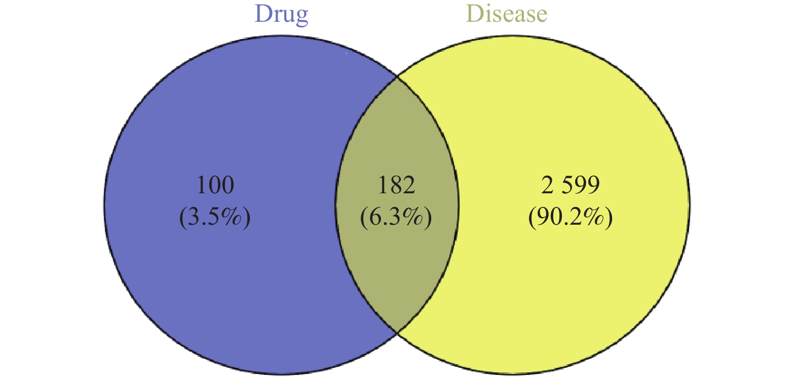
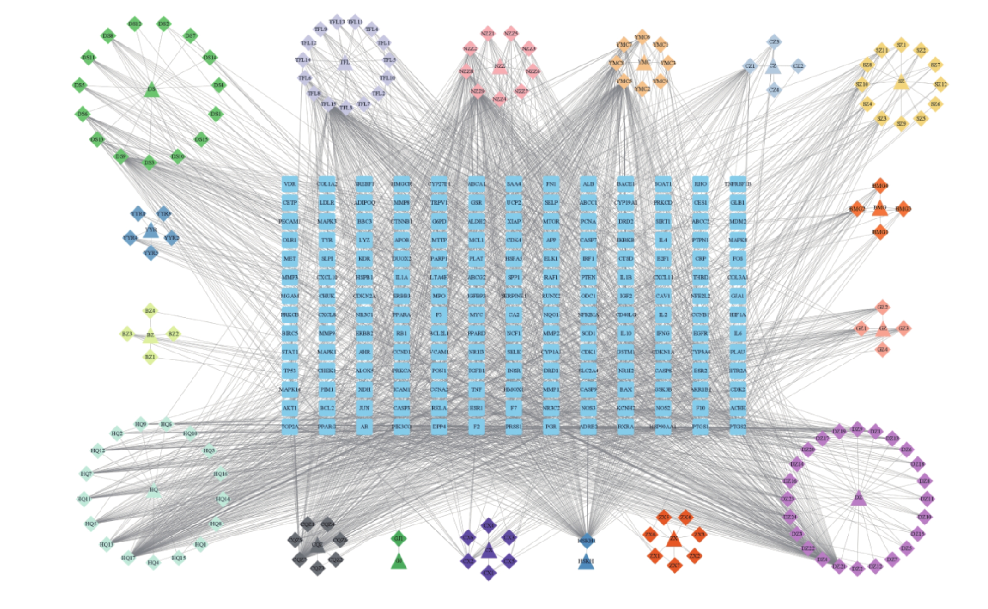
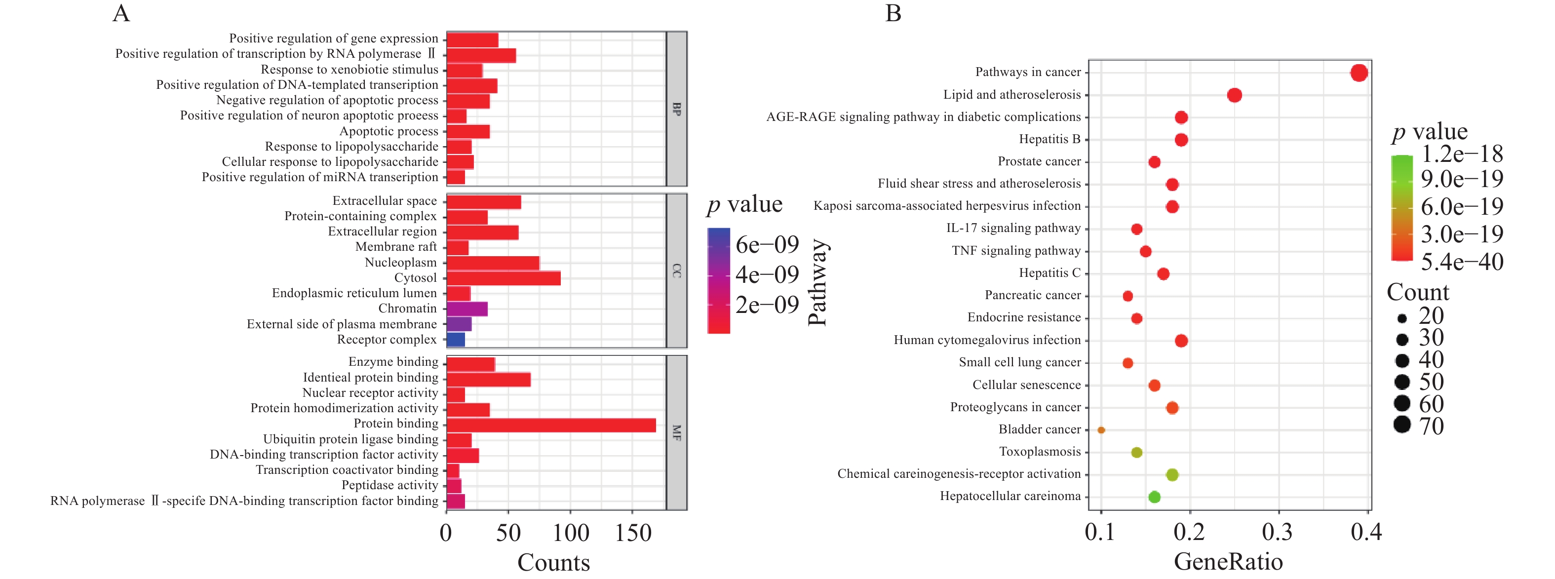
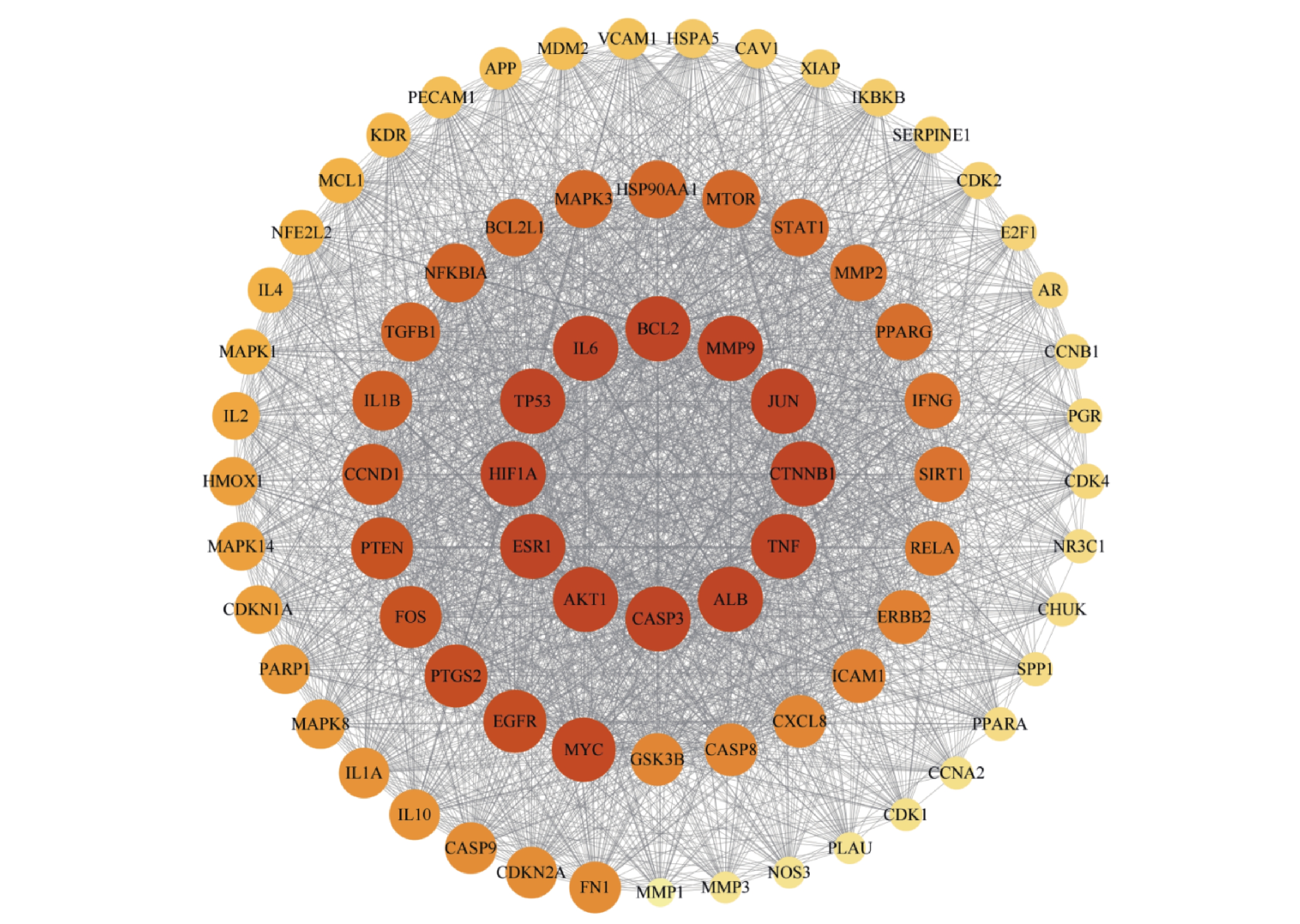
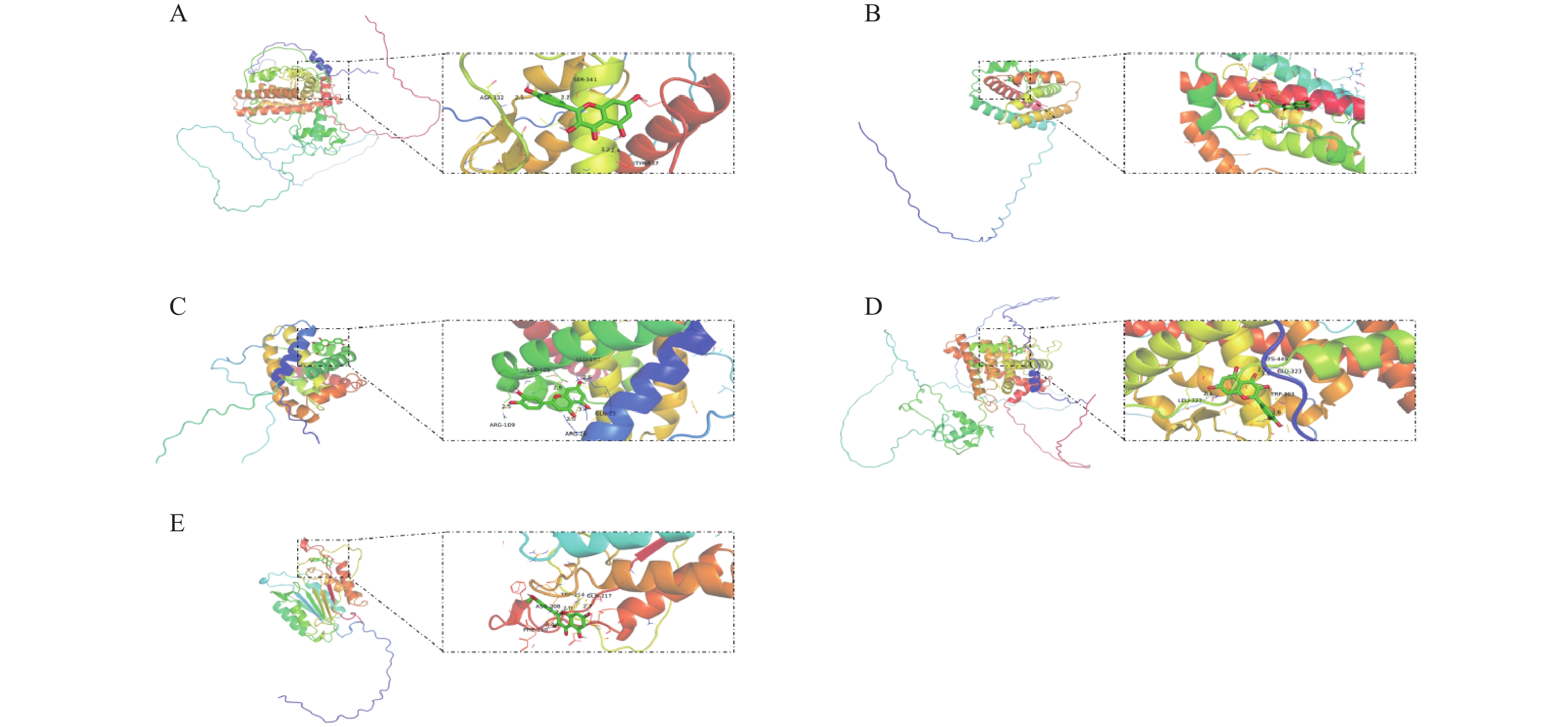
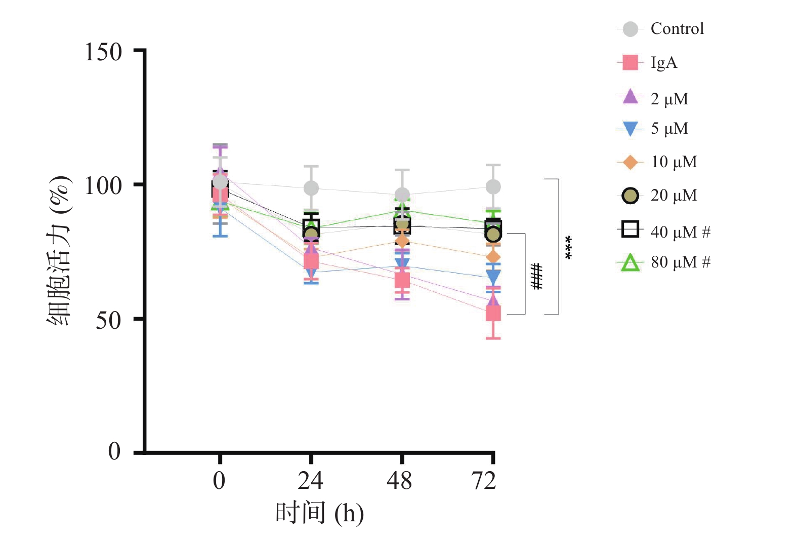
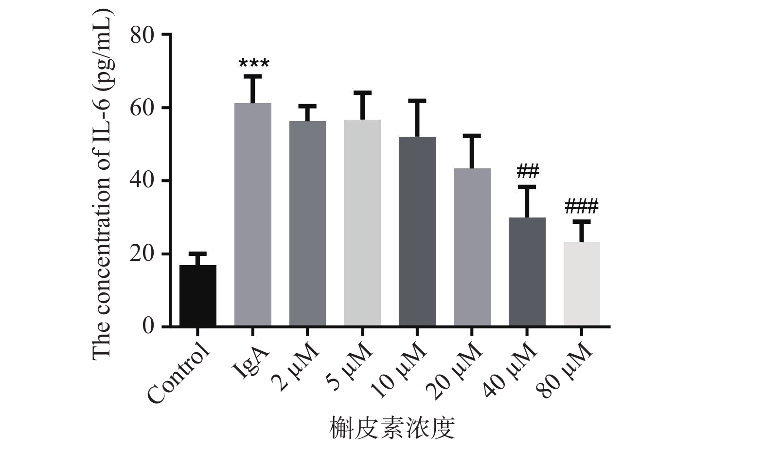
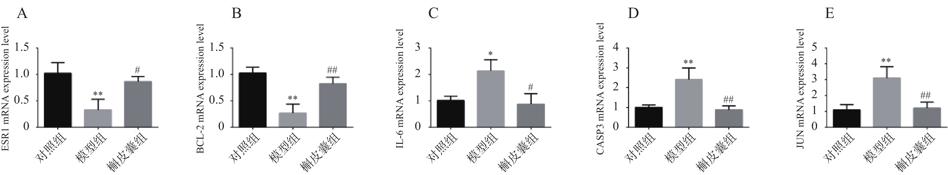
Objective To predict the mechanism of traditional Chinese medicine (TCM) in treating IgA nephropathy (IgAN) based on data mining and network pharmacology, and to verify findings through in vitro experiments. Methods Medical case records for IgAN treatment were retrieved from the Ancient and Modern Medical Case Cloud Platform to screen core drugs. Drug targets were searched in databases including TCMSP, and IgAN targets were searched in databases such as Genecards. Venn diagrams were used to identify intersecting targets, establishing disease-component-target network maps. PPI networks were constructed, followed by GO and KEGG analysis, with molecular docking validation. An IgAN cell model was established with control groups, model groups (IgA), and drug-treated groups at different concentrations (quercetin). Cell viability was detected using CCK-8 assays; IL-6 secretion levels were measured by ELISA; target gene expression levels were detected by RT-qPCR. Results Data mining revealed Astragalus had the highest frequency at 144 occurrences; TCM properties were predominantly cold, sweet, hepatic-targeting, and heat-clearing with detoxifying effects. Network pharmacology analysis identified core substances as Quercetin、Luteolin、Kaempferol、Wogonin and Isorhamnetin. PPI analysis identified core targets as ESR1, IL-6, BCL-2, JUN, and CASP3. Molecular docking verified strong binding affinity between core substances and targets. GO analysis included positive regulation of gene expression; KEGG enrichment pathways included AGE-RAGE signaling and IL-17 signaling pathways. Experimental verification showed that quercetin intervention increased cell viability (P < 0.05); suppressed IL-6 expression (P < 0.01), and upregulated ESR1, BCL-2 mRNA levels (P < 0.05), and downregulating IL-6, JUN, and CASP3 levels (P < 0.05). Conclusion This study identified core therapeutic substances and corresponding targets, and experimentally validated that quercetin enhances cell viability under IgA stimulation, suppresses inflammatory cytokine secretion, and modulates target gene expression, providing valuable insights for investigating molecular mechanisms of IgAN therapy.
2026,
47(6):
56-63.
doi: 10.12259/j.issn.2095-610X.S20260606
Abstract:
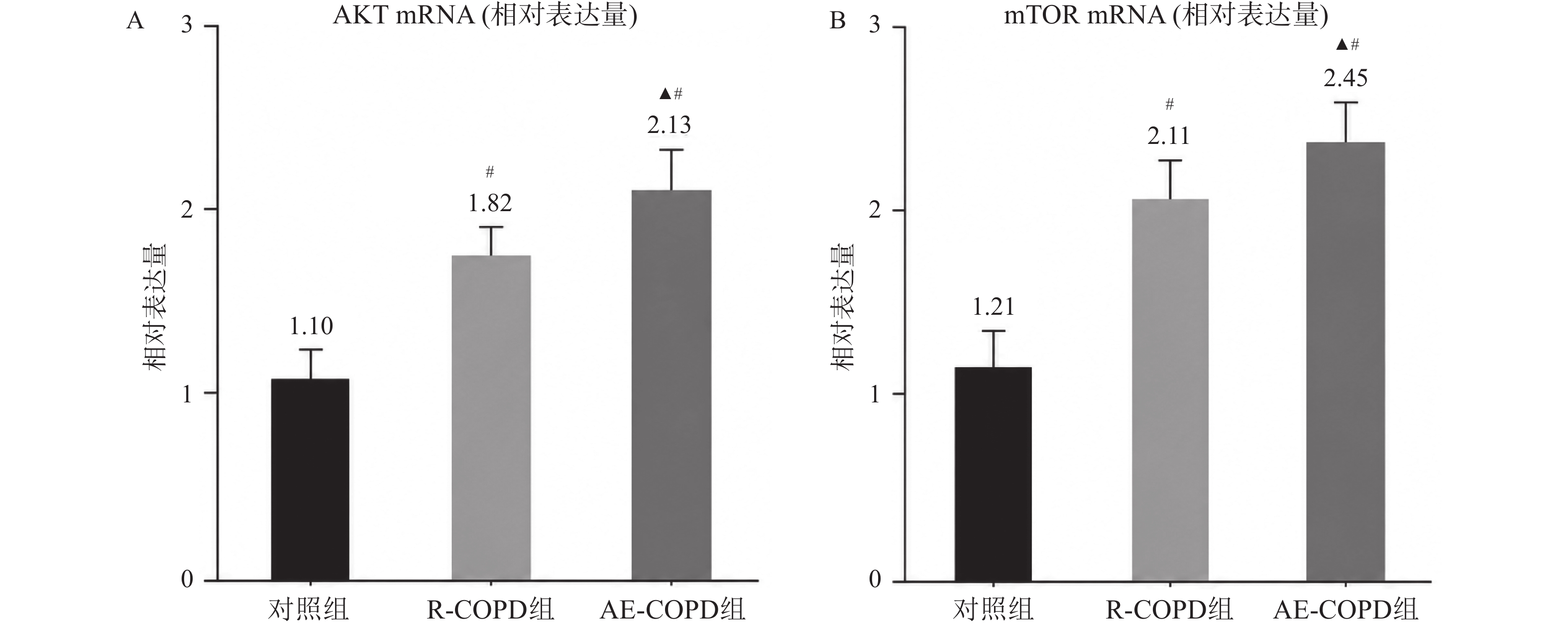
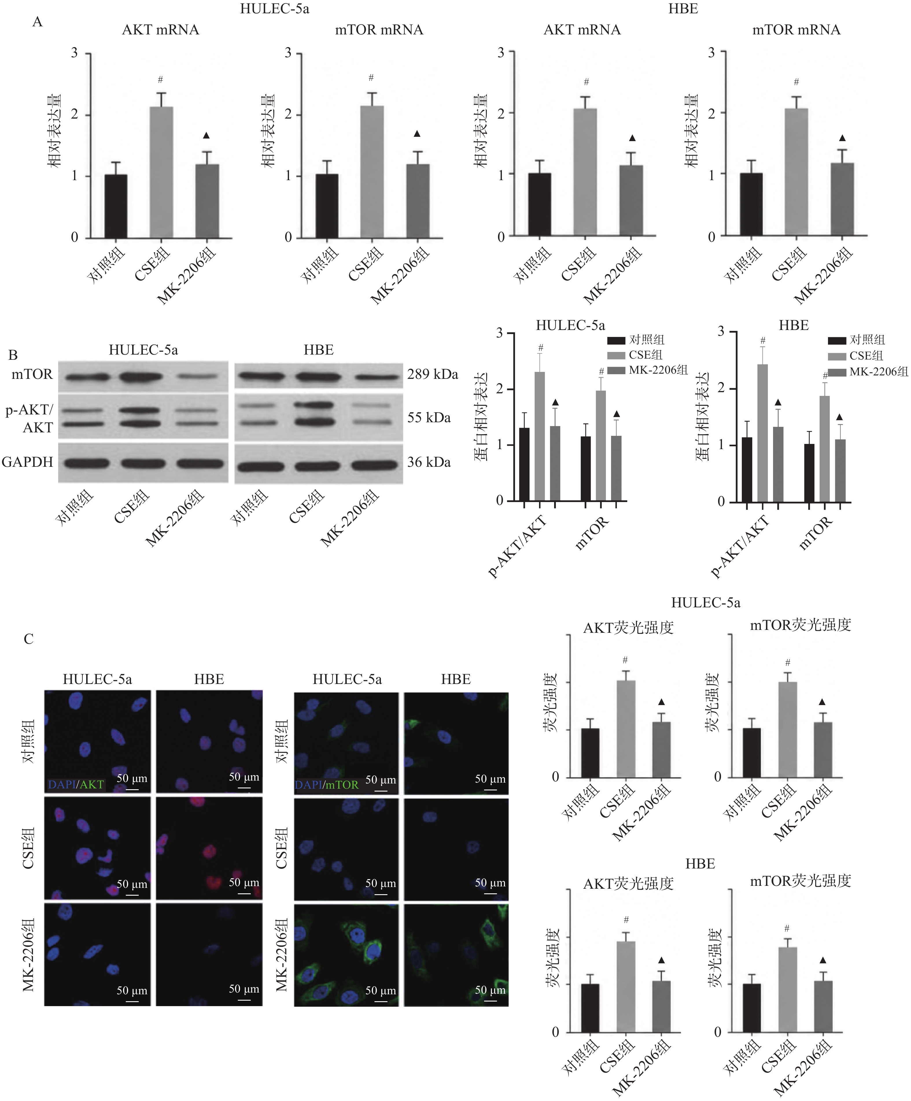
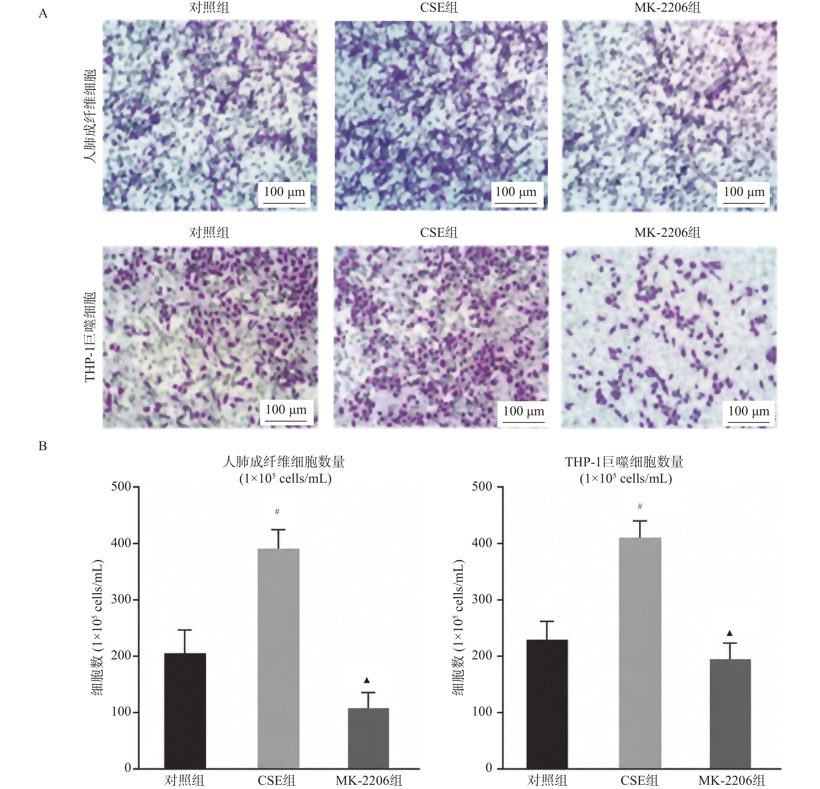
Objective To investigate the research value of AKT/mTOR signaling pathway expression in patients with chronic obstructive pulmonary disease (COPD) . Methods From January 2023 to June 2024, 20 healthy controls, 20 stable COPD (R-COPD) patients, and 20 acute exacerbation COPD (AE-COPD) patients were recruited from the Central Hospital of Ordos City. Peripheral venous blood samples were collected from all participants, with serum and peripheral blood mononuclear cells (PBMCs) isolated. Quantitative reverse transcription polymerase chain reaction (qRT-PCR) was employed to assess the expression levels of the AKT/mTOR signaling pathway in PBMCs. In vitro experiments involved human lung microvascular endothelial cells (HULEC-5a) and human bronchial epithelial cells (HBE), which were categorized into control, cigarette smoke extract (CSE), and AKT inhibitor MK-2206 groups. qRT-PCR, Western blot, and immunofluorescence techniques were utilized to evaluate the expression of the AKT/mTOR signaling pathway in each group. Through conditional medium co-culture experiments, HULEC-5a cell supernatant containing MK-2206 was introduced to the culture wells of lung fibroblasts or THP-1 cell line-derived macrophages, and cell migration ability was assessed using the Transwell method. Results No statistically significant differences were observed in gender, age, weight, height, and BMI among the three groups (P > 0.05). In terms of lung function, the AE-COPD group exhibited lower FVC (72.08±10.78, % pred.), FEV1 (47.21±11.32, % pred.), and FEV1/FVC (58.67±11.68, %) compared to the R-COPD and healthy control groups (P < 0.05). qRT-PCR analysis indicated higher AKT mRNA and mTOR mRNA expression in PBMCs of the AE-COPD group relative to the R-COPD and healthy control groups (F = 10.31, 13.23, P = 0.001). In vitro experiments revealed that the CSE group had elevated AKT/mTOR mRNA and protein expression in HULEC-5a and HBE cells compared to the control group, while the MK-2206 group exhibited lower levels (P < 0.05). Immunofluorescence quantification demonstrated increased AKT/mTOR fluorescence intensity in the CSE group and significantly reduced intensity in the MK-2206 group (F = 18.632, 17.853, P = 0.000). Transwell experiments showed higher numbers of lung fibroblasts [(391.31±23.21) cells/field] and THP-1 macrophages [(411.25±32.65) cells/field] in the CSE group, with the MK-2206 group displaying reduced numbers compared to the CSE group (F = 24.389, 36.645, P = 0.001). Conclusion AKT/mTOR signaling pathway is upregulated during COPD exacerbation. AKT inhibitor MK-2206 reduces AKT/mTOR pathway expression and may affect lung fibroblast and monocyte-derived macrophage migration, potentially playing a crucial role in COPD exacerbation process.
2026,
47(6):
64-75.
doi: 10.12259/j.issn.2095-610X.S20260607
Abstract:
Objective To explore the current status and influencing factors of depression, anxiety, and suicidal behaviors among children and adolescents in Jinghong City, Yunnan Province, analyze the correlation between suicidal behaviors and symptoms of depression and anxiety, and propose practical prevention and control measures. Methods A cross-sectional study was conducted in December 2023 using two-stage random cluster sampling to recruit 6,014 school-attending students aged 10~17 years from Jinghong, Yunnan Province. Psychological health status was assessed using the Patient Health Questionnaire-9 (PHQ-9), Generalized Anxiety Disorder-7 (GAD-7), and Suicidal Behaviors Questionnaire Revised (SBQ-R). Personal, family, and school-related information was collected using a self-designed questionnaire. Chi-square tests and binary logistic regression analysis were employed to identify risk factors for depression and anxiety, and to explore the associations between suicidal behaviors and these conditions. Results A total of 5,857 valid questionnaires were collected with a response rate of 97.4%; 49.3% were male and 50.7% were female. The overall detection rates of depression and anxiety symptoms were 17.7% and 10.5%, respectively. Univariate analysis revealed significant differences in depression symptoms across different genders, grades, school transfer history, household population size, parental illness, parental marital status, household income stability, and parental migration patterns (P < 0.05). Anxiety symptoms showed significant differences across gender, household population size, parental illness, parental marital status, household income stability, and parental migration patterns (P < 0.05). Multivariate analysis identified female gender (OR = 3.194, 95%CI: 2.741~3.720), junior high school (OR = 1.85, 95%CI: 1.435~2.385), senior high school (OR = 2.055, 95%CI: 1.550~2.724), history of school transfer (OR = 1.318, 95%CI: 1.084~1.603), paternal illness (OR = 1.611, 95%CI: 1.327~1.956), maternal illness (OR = 1.701, 95%CI: 1.352~2.141), parental divorce (OR = 1.290, 95%CI: 1.033~1.612), parental remarriage (OR = 1.658, 95%CI: 1.314~2.092), and unstable family income (OR = 1.348, 95%CI: 1.004~1.809) as risk factors for depression. Boarding school residence (OR = 0.814, 95%CI: 0.681~0.973), larger household size (2~4 persons, ≥5 persons) (OR1 = 0.647, 95%CI: 0.483~0.867; OR2 = 0.693, 95%CI: 0.497~0.966), and father not engaged in migrant work (OR = 0.755, 95%CI: 0.575~0.990) were protective factors against depression. Female gender (OR = 2.688, 95%CI: 2.230~3.240), paternal illness (OR = 1.692, 95%CI: 1.345~2.129), and parental divorce (OR = 1.450, 95%CI: 1.094~1.922) were risk factors for anxiety. Larger household size (2~4 persons) (OR = 0.676, 95%CI: 0.478~0.956) was a protective factor against anxiety. Both depression and anxiety were independent risk factors for suicide planning and suicide attempts among children and adolescents. Conclusion The detection rate of depression and anxiety symptoms among children and adolescents in Jinghong, Yunnan exceed the national average. Gender, grade level, school enrollment type, school transfer history, parental marital and health status, household income stability, and household population size are major influencing factors. Depression and anxiety symptoms significantly increase suicide risk in children and adolescents.
2026,
47(6):
76-83.
doi: 10.12259/j.issn.2095-610X.S20260608
Abstract:
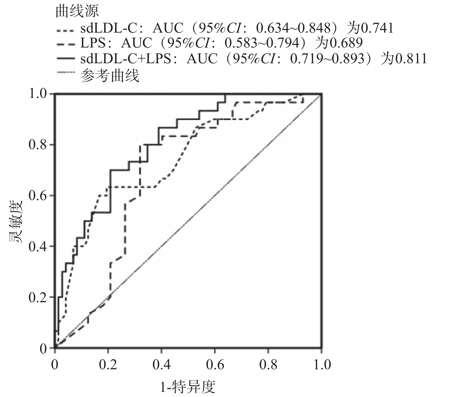
Objective To explore the correlations between gut microbiota, serum small and dense low-density lipoprotein cholesterol (sdLDL-C), lipopolysaccharide (LPS), and diabetic retinopathy (DR). Methods A total of 102 patients with type 2 diabetes (T2DM) who attended the First People's Hospital of Kashgar from October 2021 to October 2023 were selected. According to the presence or absence of diabetic retinopathy, patients were divided into non-DR group (n = 72) and DR group (n = 30). General information, laboratory indices, intestinal flora, and serum levels of sdLDL-C and LPS were compared between the two groups. Multivariate Logistic regression analysis was conducted to identify risk factors for DR. Spearman's correlation analysis was used to analyze the rank correlation between intestinal flora, serum sdLDL-C, LPS and DR. Receiver operating characteristic curves (ROC) was used to evaluate the predictive performance of serum sdLDL-C and LPS levels for DR. Results Compared with the non-DR group, the DR group exhibited significantly elevated Bacteroides abundance (31.28±7.70) (t = 9.905, P < 0.05); significantly elevated serum sdLDL-C (1.51±0.37) mmol/L and LPS (117.45±9.39) pg/mL levels (t = 4.422, 25.160, P < 0.05); and significantly reduced Bifidobacterium and Prevotella abundance (t = 19.886, 11.883, P < 0.05). There were no significant differences in Lactobacillus, Plasmodicoccus, Enterococcus, Eubacterium rectale, Veillonella, Clostridium tenellum, and Roseburia species (P > 0.05). Univariate analysis showed that the duration of diabetes, systolic pressure (SBP), serum creatinine (SCr), fasting blood glucose (FPG), triglyceride (TG), low density lipoprotein cholesterol (LDL-C), glycosylated hemoglobin (HbA1c), and history of hypertension in the DR group were significantly higher than those in the non-DR group (P < 0.05). Age, body mass index (BMI), gender, diastolic pressure (DBP), smoking history, drinking history, total cholesterol (TC), high density lipoprotein cholesterol (HDL-C) showed no statistically significant difference (P > 0.05). Multivariate Logistic regression showed that the duration of diabetes, SCr, TG, HbA1c, sdLDL-C, LPS, and elevated Bacteroides were risk factors for DR (P < 0.05), while reduced Prevotella was a protective factor for DR (P < 0.05). Correlation analysis showed that Bacteroides abundance, sdLDL-C, and LPS levels were positively correlated with DR (P < 0.05); while Bifidobacterium and Prevotella abundance were negatively correlated with DR (P < 0.05). ROC curve showed that combination of sdLDL-C and LPS had an area under the curve(AUC) (95%CI: 0.719~ 0.893), with a sensitivity of 86.72% and a specificity of 79.83%. Conclusion Patients with T2DM complicated by DR exhibit gut dysbiosis and abnormally elevated serum sdLDL-C and LPS levels. sdLDL-C, LPS, and elevated Bacteroides are risk factors for DR, while Prevotella is a protective factor.
2026,
47(6):
84-92.
doi: 10.12259/j.issn.2095-610X.S20260609
Abstract:
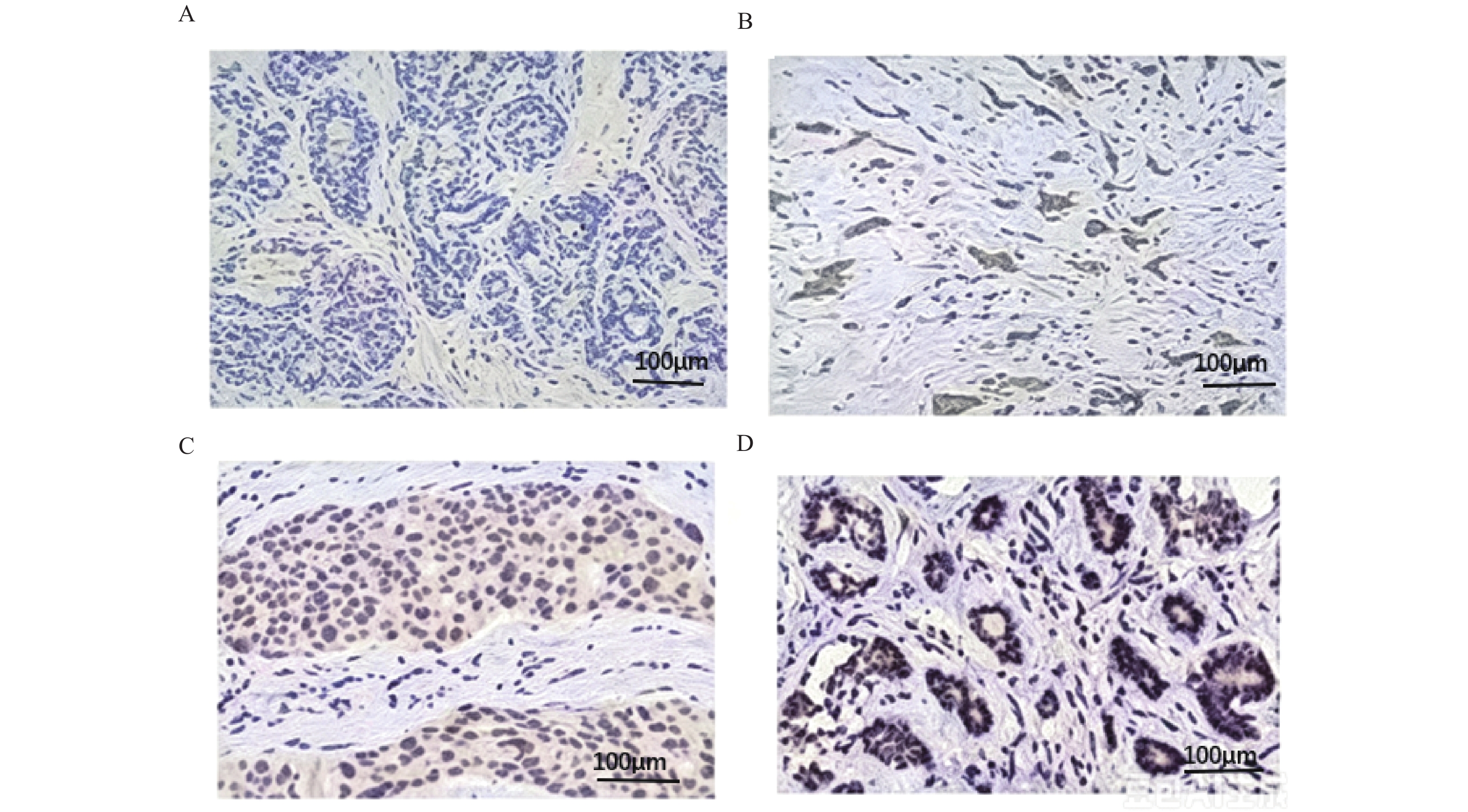
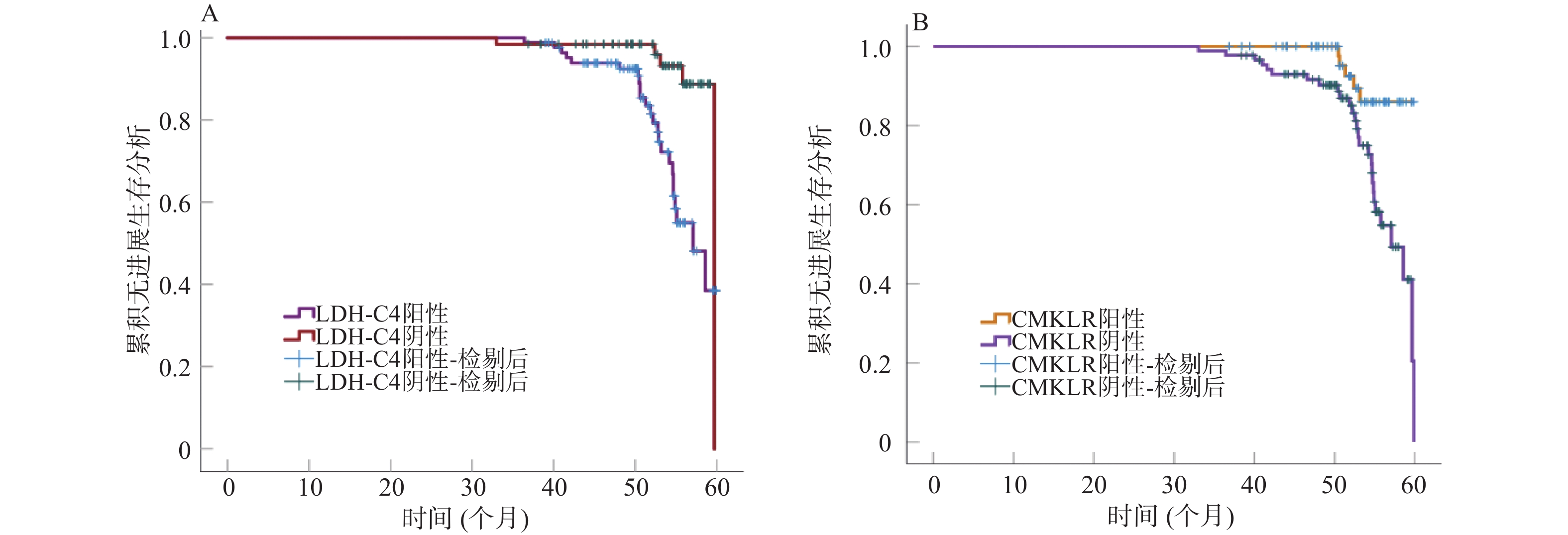
Objective To investigate the expression of lactate dehydrogenase C4 (LDH-C4) and chemokine-like receptor 1 (CMKLR1) in breast cancer tissues and their relationship with clinical pathological characteristics, tumor markers and prognosis. Methods A prospective cohort study was conducted on 150 patients with breast cancer admitted to the First People’ s Hospital of Shuangliu District, Chengdu from January 2016 to June 2020. All patients underwent radical mastectomy and cancer tissues and adjacent normal tissues were collected intraoperatively. Immunohistochemical staining was used to detect LDH-C4 and CMKLR1 expression. LDH-C4 and CMKLR1 expression in different tissues was compared, and the relationship between the expression of LDH-C4 and CMKLR1 in cancer tissues and the pathological characteristics, serum tumor markers [carcinoembryonic antigen (CEA), cancer antigen 153 (CA153)] of breast cancer patients was analyzed. Follow-up was conducted for 6~60 months. Kaplan-Meier method analysis was performed to analyze the relationship between LDH-C4 and CMKLR1 expression and postoperative progression-free survival (PFS) prognosis. Stratified Cox regression analysis was used to identify factors influencing PFS prognosis, and subgroup analysis of LDH-C4 and CMKLR1 expression with PFS prognosis were performed. Results The positive expression rate of LDH-C4 in breast cancer tissue was 53.33%, significantly higher than the 26.67% in adjacent normal tissue, while the positive expression rate of CMKLR1 was 41.33%, significantly lower than the 89.33% in adjacent normal tissue (χ2 = 22.218, 76.083, all P < 0.05). In patients with pathological stage II–III, lymph node metastasis, CEA > 5.39 μg/L, and CA153 > 26.57 U/mL, the positive expression of LDH-C4 was higher, and CMKLR1 positive expression was lower compared to patients with stage I disease, no lymph node metastasis, CEA ≤ 5.39 μg/L, and CA153 ≤ 26.57 U/mL (P < 0.05). During the 6~60 month follow-up, 33 patients experienced tumor progression. Kaplan-Meier survival analysis showed that the 5-year overall progression-free survival rate in LDH-C4 positive patients was 65.85%, lower than the 92.06% in negative patients, while CMKLR1 positive patients had a rate of 91.67%, higher than the 67.06% in negative patients (Log-rank χ2 = 11.748, 8.832, P < 0.05). Cox regression analysis showed that pathological stage, lymph node metastasis, CEA, CA153 and LDH-C4 expression were independent risk factors for PFS prognosis, while CMKLR1 expression was an independent protective factor (P < 0.05). Stratified Cox proportional hazard regression analysis showed that after adjusting for pathological stage, lymph node metastasis status, CEA and CA153 levels, LDH-C4 positive positively ramained a risk factor for PFS in breast cancer patients (HR = 3.082, 95%CI: 1.889~5.027, P < 0.05), while CMKLR1 positivity was a protective factor for PFS (HR = 0.902, 95%CI: 0.825~0.986, P < 0.05). Conclusion The expression of LDH-C4 and CMKLR1 is closely associated with pathological stage, lymph node metastasis and serum tumor markers of breast cancer, Together, they constitute a powerful risk network affecting postoperative PFS prognosis and provide a novel theoretical framework for clinical prognostic assessment and precision management after surgery.

2026,
47(6):
93-104.
doi: 10.12259/j.issn.2095-610X.S20260610
Abstract:
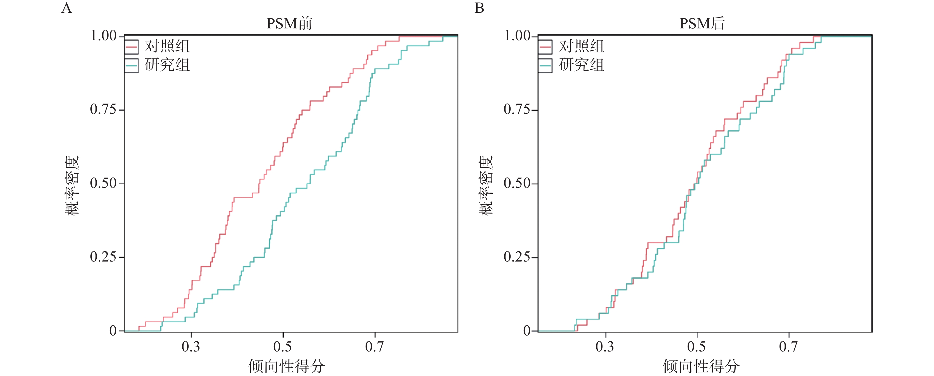
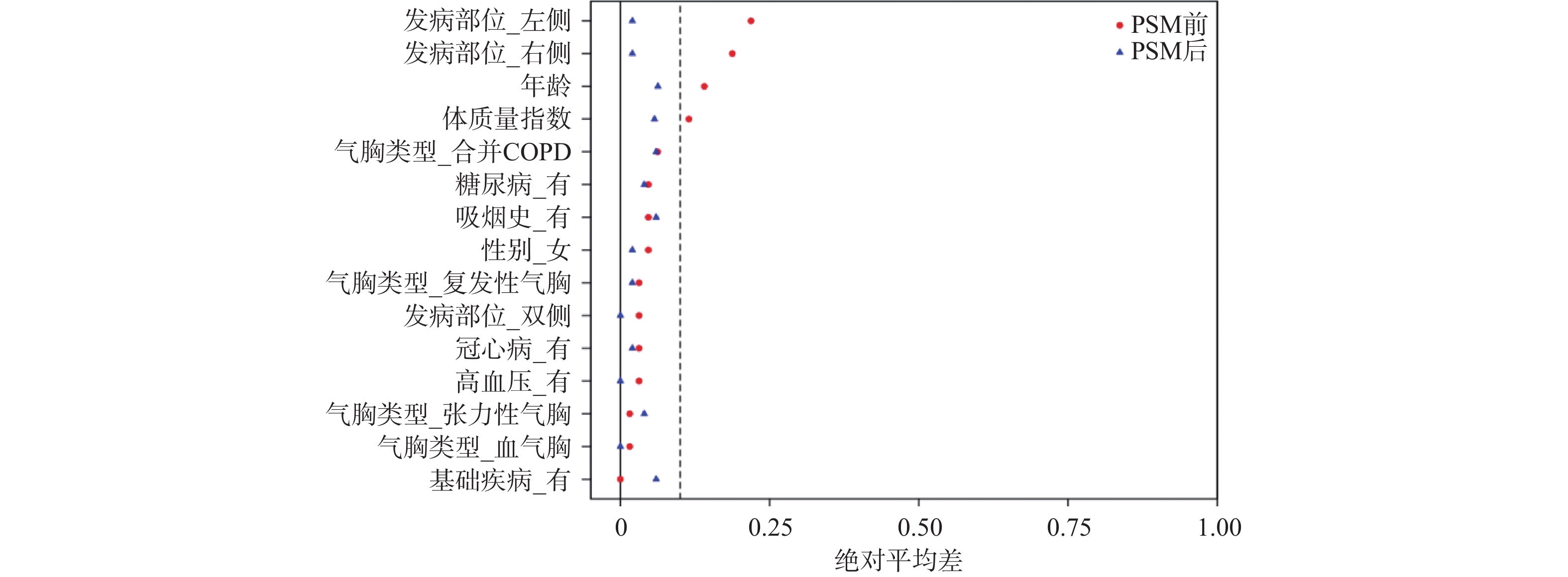
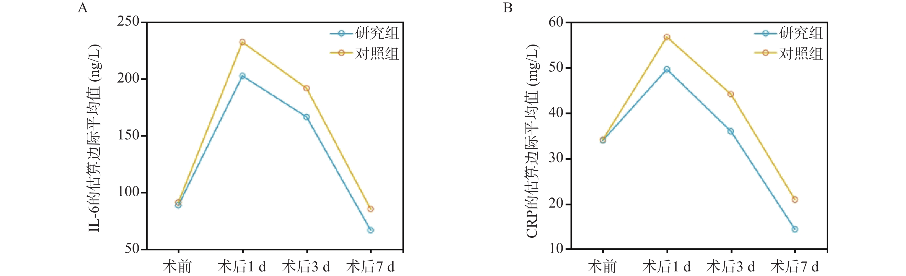
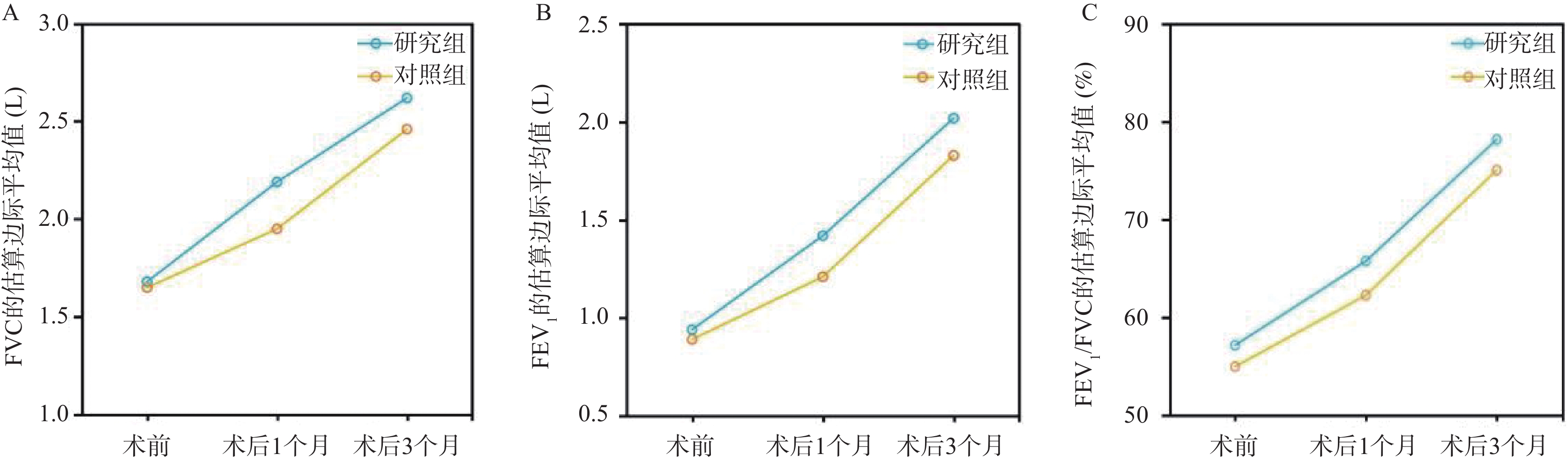
Objective To investigate the therapeutic effects of ultrasound-guided closed thoracic drainage versus conventional closed thoracic drainage in patients with complex and refractory pneumothorax, and to compare the effects of both approaches on inflammatory factor levels and pulmonary function indices in these patients. Methods A total of 128 patients with complex and refractory spontaneous pneumothorax admitted to Mianyang 404 Hospital between September 2021 and September 2024 were selected as the study subjects. Patients were non-randomly divided into a study group (n = 64, receiving ultrasound-guided closed thoracic drainage) and a control group (n = 64, receiving conventional closed thoracic drainage). To further balance potential differences between the two groups, propensity score matching (PSM) was adopted to match the baseline data at a 1∶1 ratio. Clinical data of matched patients were analyzed. Repeated measures analysis of variance was used to compare the levels of inflammatory factors [interleukin-6 (IL-6), C-reactive protein (CRP)] and pulmonary function indicators [forced vital capacity (FVC), forced expiratory volume in 1 second (FEV1), forced expiratory volume in 1 second/forced vital capacity (FEV1/FVC)] between the two groups. Pearson correlation analysis was performed to examine the correlation between preoperative inflammatory factor indicators and pulmonary function indicators. Logistic regression models were used to analyze the impact of ultrasound guidance on postoperative complications following closed thoracic drainage, with subgroup analyses performed. Results Following PSM, 50 patients were matched in each group, with comparable baseline characteristics(P > 0.05). Compared with the control group, the study group had shorter operation time, drainage time, and postoperative hospital stay (P < 0.05), higher drainage volume (P < 0.05), lower total complication rate (P < 0.05), and higher overall treatment efficacy rate (P < 0.05). Repeated measures analysis of variance showed: (1) IL-6 and CRP levels in both groups increased initially then decreased over time, while FVC, FEV1, and FEV1/FVC levels increased over time, with statistically significant time effects (P < 0.05); (2) IL-6 and CRP levels at postoperative day 1, 3, and 7 in the study group were significantly lower than in the control group, and FVC and FEV1 at postoperative 1 month and 3 months were significantly higher than in the control group (P < 0.05), with statistically significant group effects for all indicators (P < 0.05); (3) The effects of time on IL-6, CRP, FVC, and FEV1 differed between groups based on whether ultrasound guidance was used during closed thoracic drainage, with statistically significant interaction effects (P < 0.05). Pearson analysis revealed negative correlations between IL-6, CRP and FVC, FEV1, and FEV1/FVC (P < 0.05). Logistic regression analysis showed that after adjusting for covariates, ultrasound guidance during closed thoracic drainage remained an independent protective factor for reducing postoperative complications in patients with complex and refractory pneumothorax (OR = 0.196, 95%CI: 0.047~0.816, P = 0.025), with consistent results across subgroups (P for interaction > 0.05). Conclusion Ultrasound-guided closed thoracic drainage demonstrates superior efficacy in treating patients with complex and difficult spontaneous pneumothorax, more effectively regulating inflammatory factor levels, improving pulmonary function, and reducing the risk of postoperative complications.
2026,
47(6):
105-115.
doi: 10.12259/j.issn.2095-610X.S20260611
Abstract:
Objective To explore the predictive value of multimodal ultrasound combined with serum Amphiregulin (AREG) for sentinel lymph node (SLN) metastasis in patients with clinically negative axillary lymph node (cN0) invasive breast cancer (IBC). Methods A total of 218 IBC patients admitted to Xuzhou Central Hospital from July 2023 to January 2025 with preoperative clinical assessment of cN0 were randomly divided into a training set (153 cases) and validation set (65 cases) in a 7:3 ratio. Based on postoperative pathology, patients were classified into SLN-positive and SLN-negative groups. Multimodal ultrasound features and serum AREG levels were compared between the two groups. The predictive value of multimodal ultrasound alone and combined with serum AREG for SLN metastasis was analyzed and validated. Results Age, menstrual status, tumor size, differentiation degree, molecular subtype, Ki-67 index, TNM stage and family history of breast cancer were comparable between the training set and the validation set (P > 0.05). There were statistically significant differences in lymph hilum echogenicity, penetrating vessels, maximum elastic modulus (Emax), mean elastic modulus (Esd), peak intensity and peak time between the SLN positive group and the SLN negative group in the training set and validation set (P < 0.05). Serum AREG levels were significantly higher in the SLN-positive group compared to the SLN-negative group in both datasets (P < 0.05). Multivariate Logistic regression analysis showed that lymphatic echo (abnormal vs normal: OR = 3.758, 95%CI: 1.523~9.277), perforator vessels (with vs without: OR = 3.019, 95%CI: 1.323~6.891), Emax (every 1 kPa increase: OR = 1.046, 95%CI: 1.024~1.069), peak intensity (every 1 dB increase: OR = 1.037, 95%CI: 1.011~1.063), serum AREG (every 1 ng/mL increase: OR = 1.005, 95%CI: 1.001~1.009) were independent influencing factors of SLN metastasis in IBC patients (P < 0.05). The Logit(P) predictive model based on multivariate logistic regression achieved areas under the curve (AUC) of 0.890 (95%CI: 0.848~0.954) in the training set and 0.879 (95%CI: 0.816~0.926) in the validation set. Calibration analysis demonstrated good model fit with no significant difference between predicted and observed probabilities. Decision curve analysis showed positive net clinical benefit within the threshold probability range of 10%~80% in both datasets. Conclusion Multimodal ultrasound combined with serum AREG has high predictive value for SLN metastasis in cN0-stage IBC patients, enabling more accurate preoperative prediction of SLN metastasis status and providing evidence to guide surgical decision-making.
2026,
47(6):
116-122.
doi: 10.12259/j.issn.2095-610X.S20260612
Abstract:
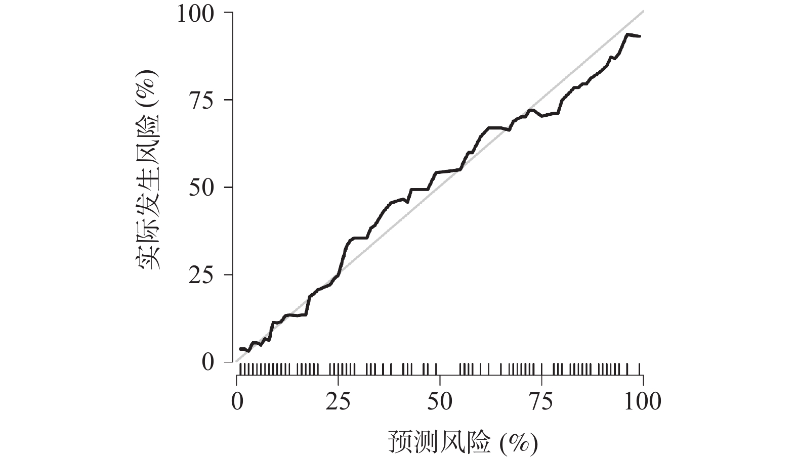
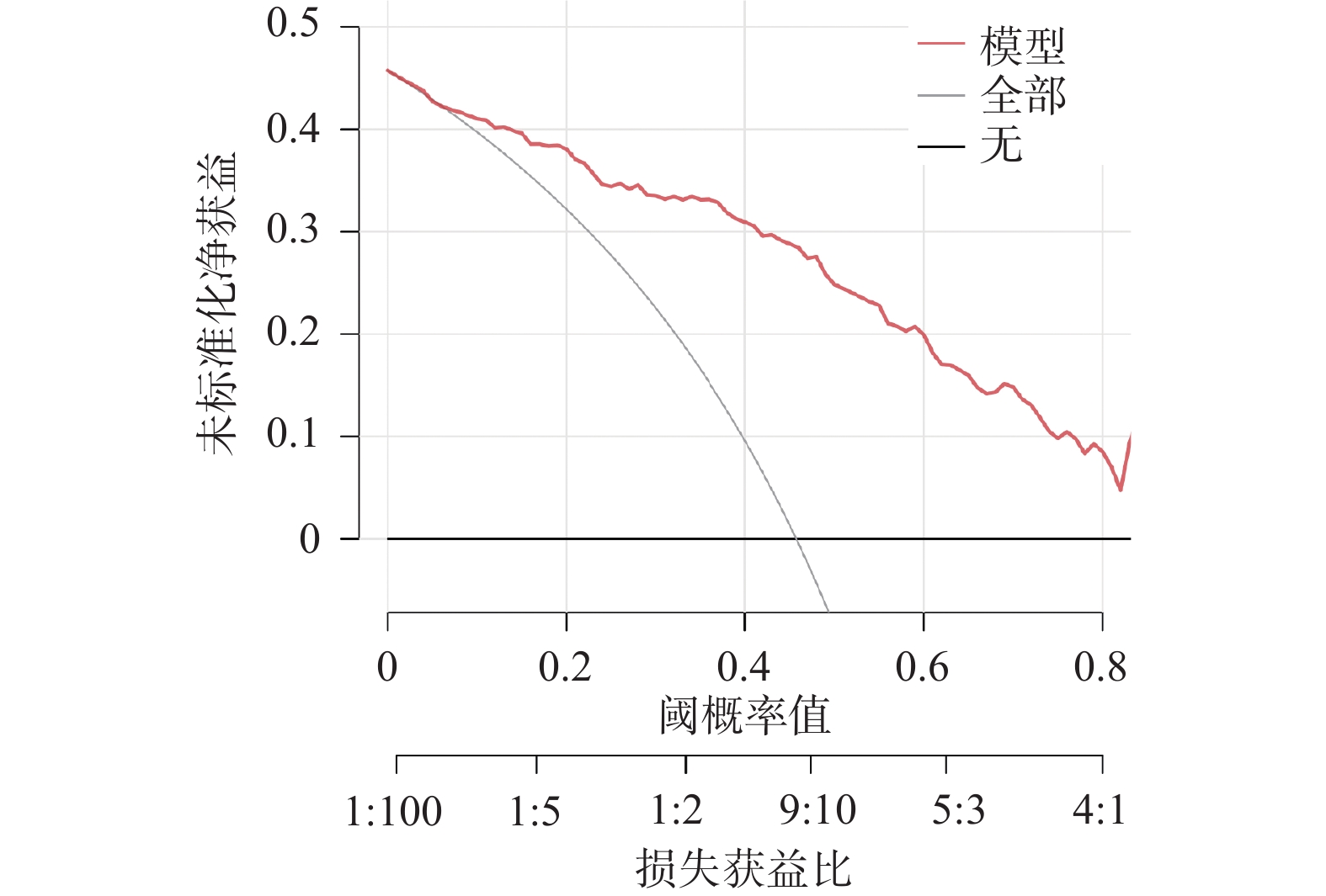
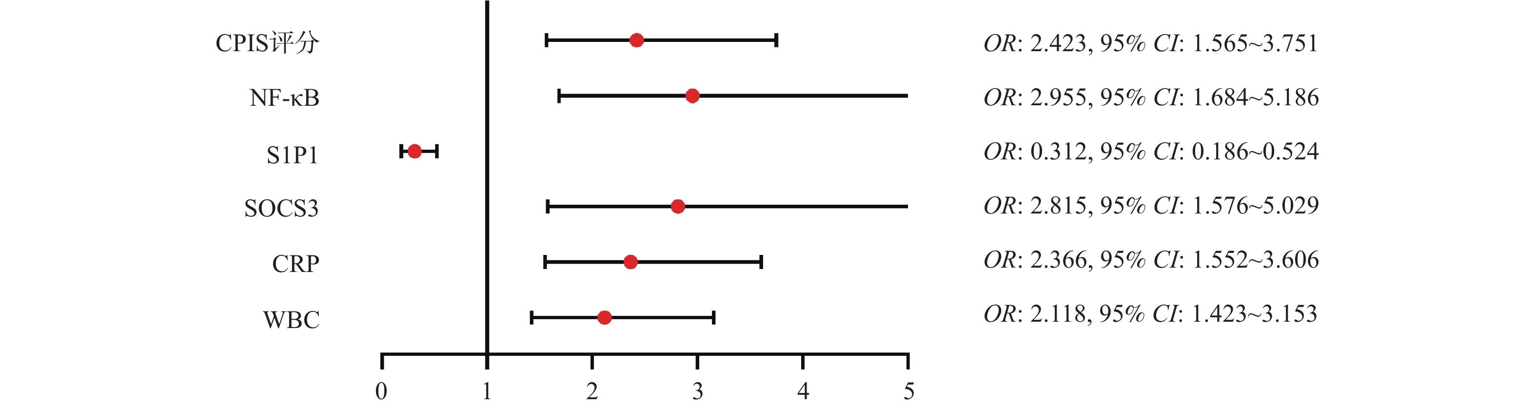
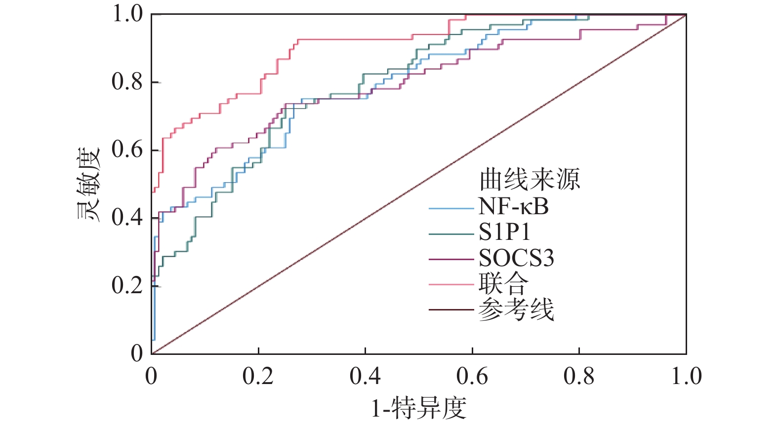
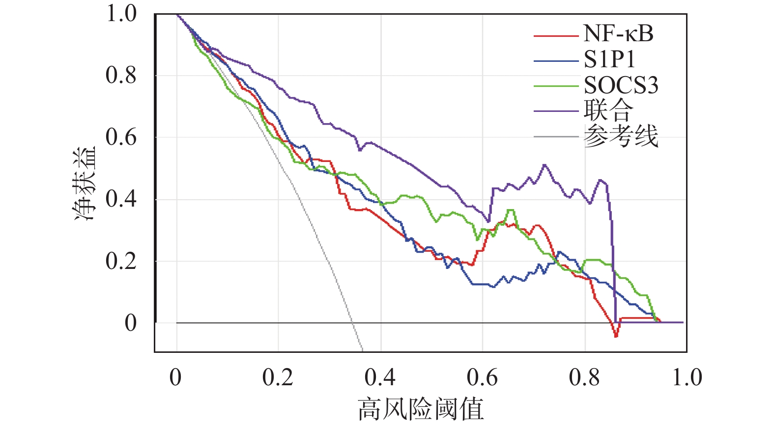
Objective To investigate the correlation between serum nuclear factor-kappa B (NF-κB), sphingosine-1-phosphate 1 (S1P1), suppressor of cytokine signaling 3 (SOCS3) levels and disease severity in children with bacterial infectious pneumonia, and to evaluate their predictive value for patient prognosis. Methods A total of 200 children with bacterial infectious pneumonia admitted to Xi'an International Medical Center Hospital from January 2022 to December 2024 were enrolled and divided into mild pneumonia group (n = 120) and severe pneumonia group (n = 80). Additionally, 120 healthy children from routine physical examinations served as the control group. Serum levels of NF-κB, S1P1, and SOCS3 in all three groups were detected using ELISA. Spearman and Pearson correlation analyses were performed to assess the correlations between these markers and disease indicators and clinical parameters. Children with bacterial infectious pneumonia were followed up for 6 months and categorized into favorable prognosis group (n = 131) and unfavorable prognosis group (n = 69). Logistic regression analysis was used to identify risk factors for poor prognosis. ROC curves were employed to evaluate the predictive value of serum NF-κB, S1P1, and SOCS3 levels for prognosis. Decision curve analysis (DCA) was used to assess the clinical net benefit of these biomarkers. Results Serum NF-κB, SOCS3, C-reactive protein (CRP), white blood cell count (WBC), and clinical pulmonary infection score (CPIS) in both mild and severe pneumonia groups were higher than in the control group, while S1P1, systolic blood pressure, and diastolic blood pressure were lower than in the control group (P < 0.05). Serum NF-κB, SOCS3, CRP, WBC, and CPIS scores in the severe group were higher than in the mild group, S1P1 was lower, and heart rate was higher than in both the control and mild groups (P < 0.05). Disease severity, CPIS score, CRP, and WBC showed positive correlations with serum NF-κB and SOCS3 levels, and negative correlations with S1P1 levels (P < 0.05). Serum NF-κB, SOCS3, CRP, WBC, and CPIS scores in the unfavorable prognosis group were higher than in the favorable prognosis group, while S1P1 was lower (P < 0.05). Elevated NF-κB, SOCS3, CRP, WBC, CPIS scores, as well as decreased S1P1, were identified as risk factors for poor prognosis in children with bacterial infectious pneumonia (P < 0.05). The AUC and net benefit of serum NF-κB, S1P1, and SOCS3 for poor prognosis were superior to individual predictors (P < 0.05). Conclusion Serum NF-κB and SOCS3 levels are elevated in children with bacterial pneumonia, while S1P1 level is decreased. The three factors are closely related to disease severity and prognosis, and have high predictive value and clinical net benefit for poor prognosis.
2026,
47(6):
123-133.
doi: 10.12259/j.issn.2095-610X.S20260613
Abstract:
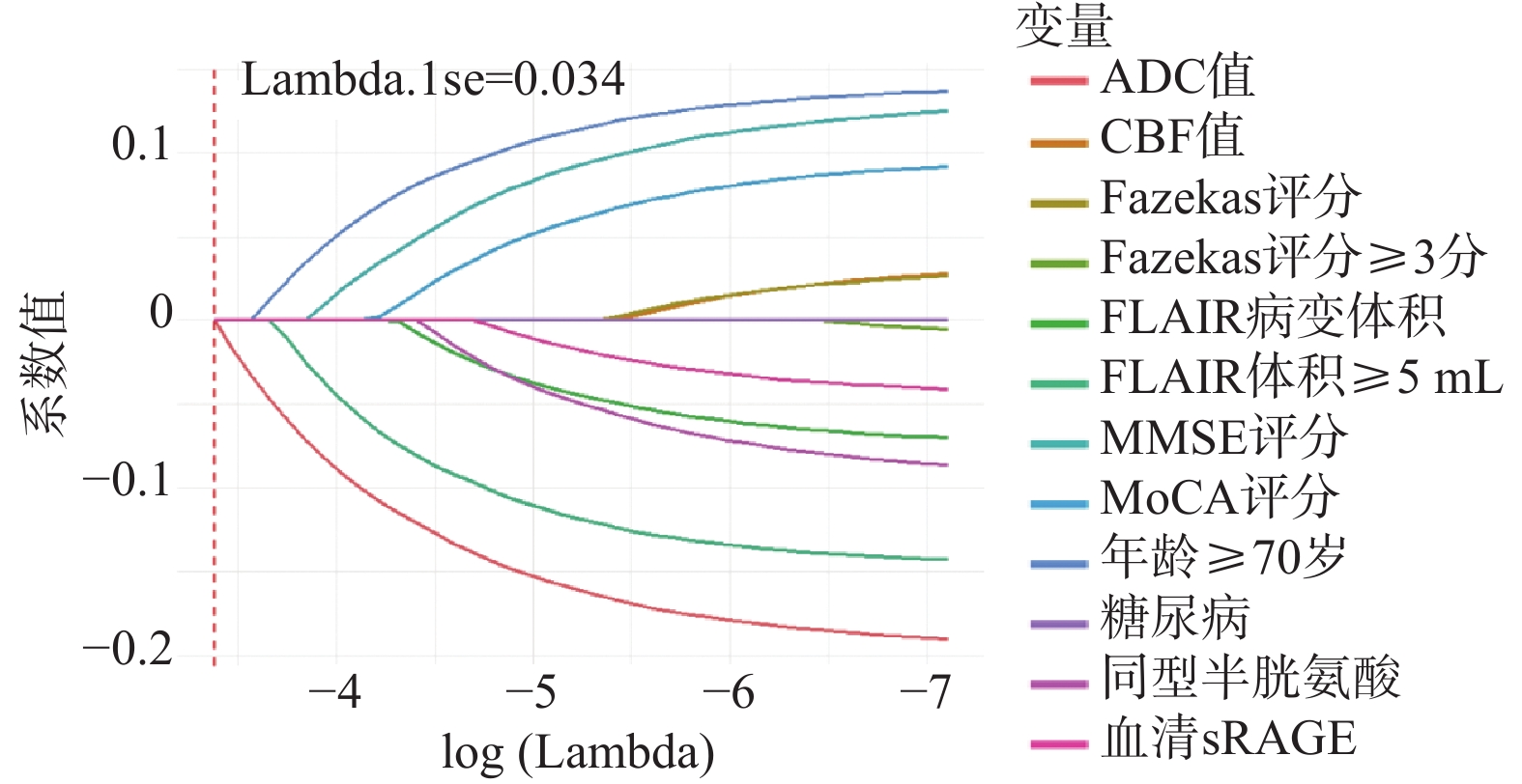
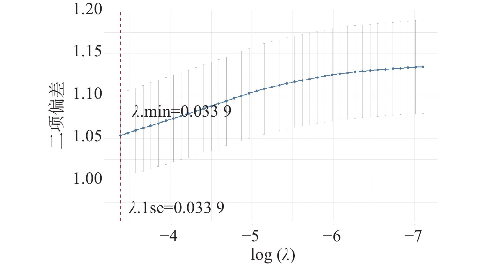
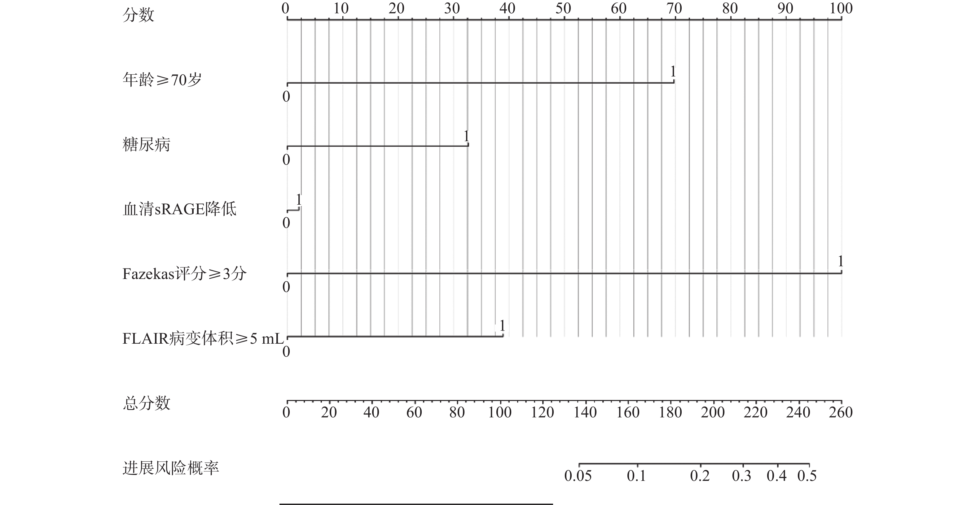
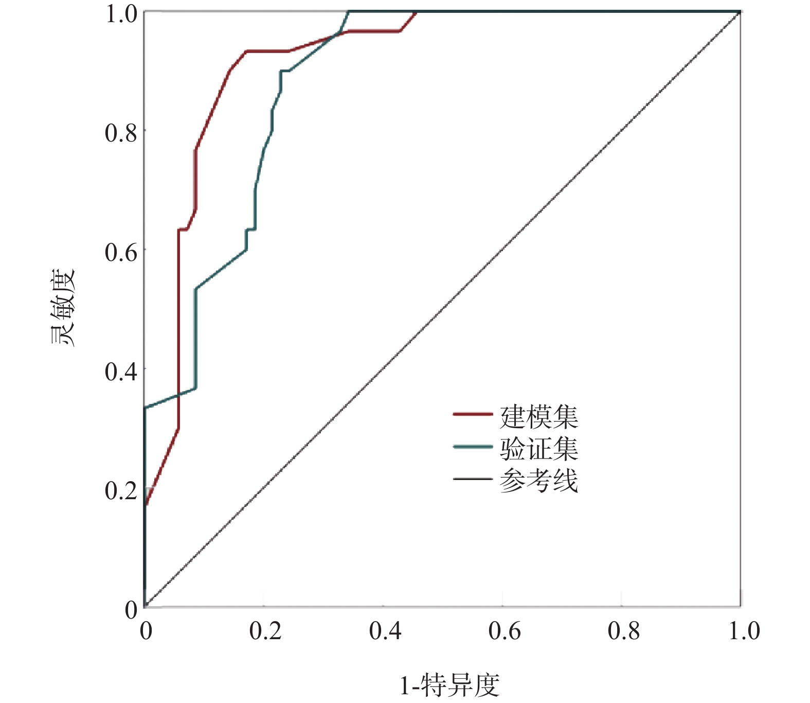
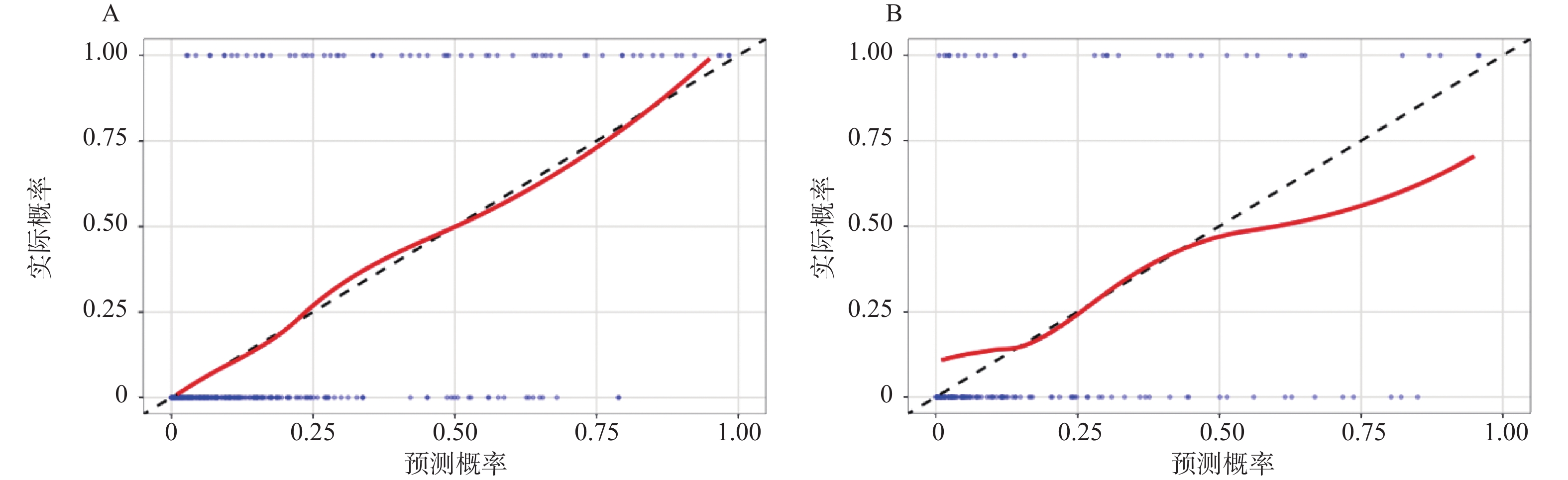
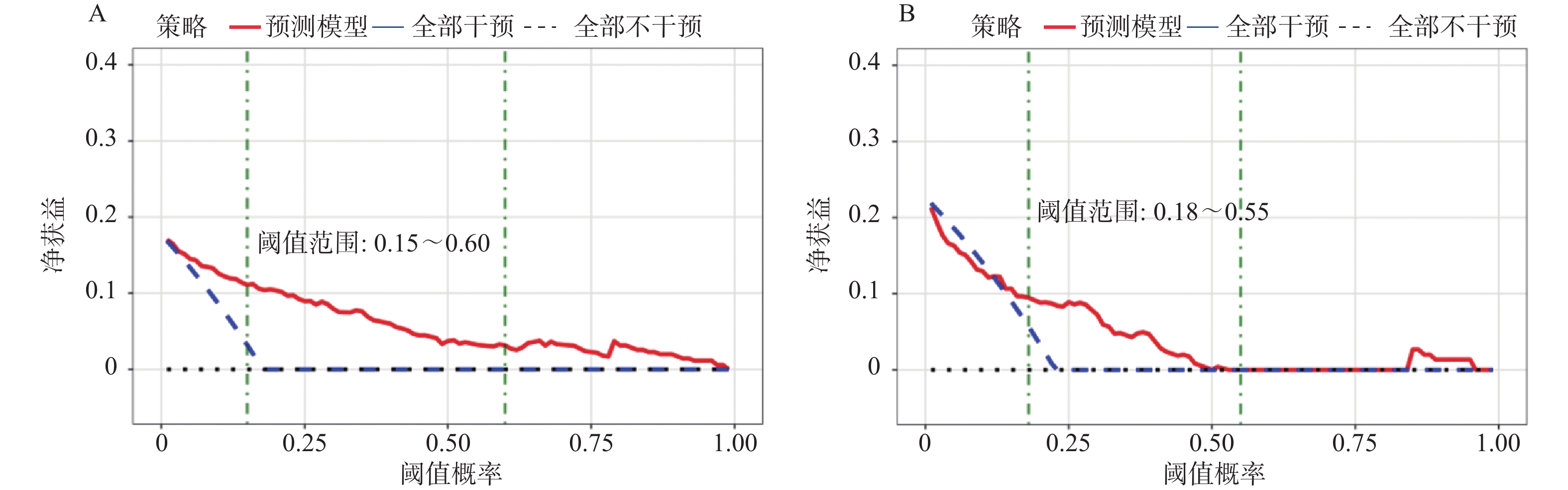
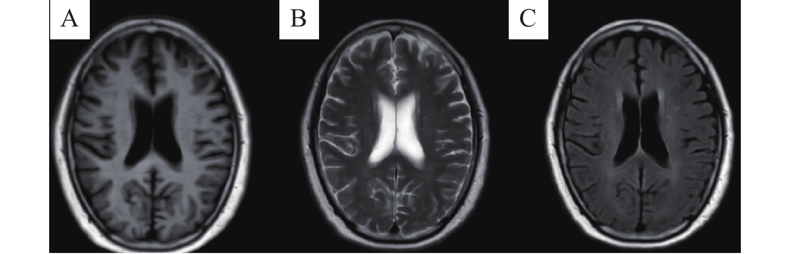
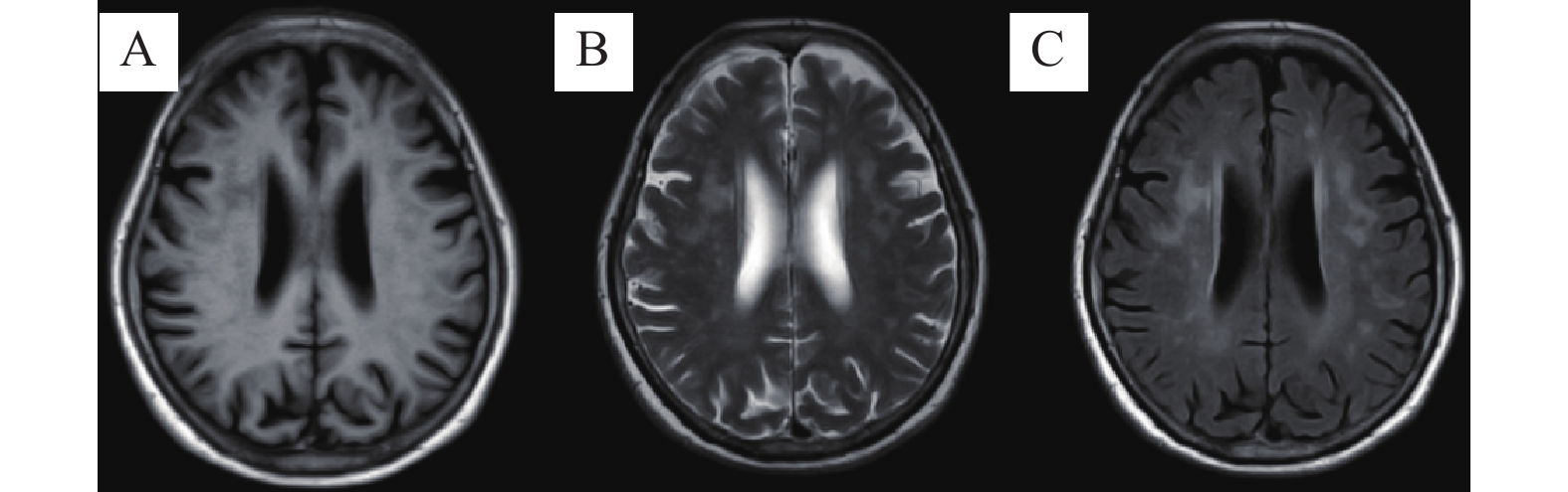
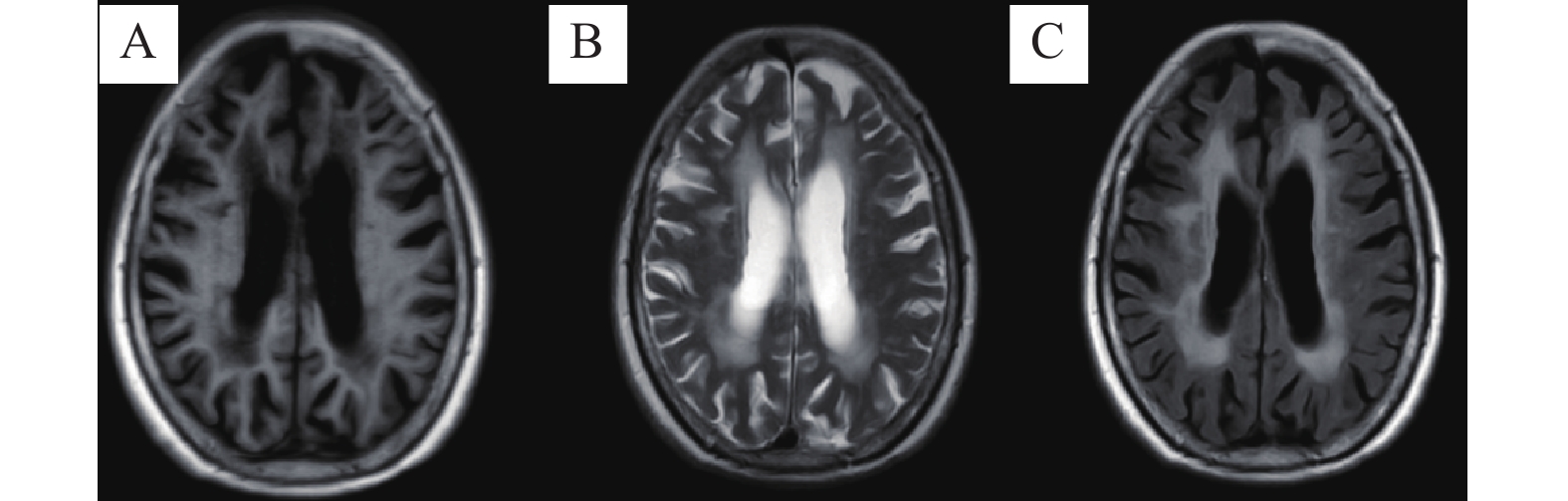
Objective To construct and validate a predictive model for the progression risk of vascular white matter lesions (WML) based on multimodal MRI combined with serum soluble receptor for advanced glycation end products (sRAGE). Methods A retrospective cohort study design was employed. A total of 330 patients diagnosed with WML diagnosed at the First Hospital of Handan from January 2020 to October 2023 were enrolled and randomly divided into a modeling set (n = 231) and a validation set (n = 99) in a 7∶3 ratio. Eighteen candidate predictive factors were collected, including general data, underlying diseases, laboratory indicators (including serum sRAGE), and multimodal MRI parameters (Fazekas score, FLAIR lesion volume, etc.). The outcome event was defined as lesion progression within 2 years of follow-up. Core factors were selected using LASSO regression, and a nomogram prediction model was constructed using multivariable logistic regression. The discriminative ability, calibration, and clinical applicability of the model were assessed using receiver operating characteristic (ROC) curves, Hosmer-Lemeshow test, and decision curve analysis (DCA), respectively. Results The lesion progression rates in the modeling and validation sets were 21.6% (50/231) and 22.2% (22/99), respectively. LASSO regression identified five core predictors: age ≥70 years, diabetes mellitus, decreased serum sRAGE level, Fazekas score ≥3, and FLAIR lesion volume ≥5 mL. Multivariate logistic regression showed that age ≥70 years, diabetes mellitus, Fazekas score ≥3, decreased serum sRAGE level, and FLAIR lesion volume ≥5 mL were independent risk factors for WML progression (all P < 0.05). In the modeling set, the area under the ROC curve (AUC) was 0.912 (95%CI: 0.875~0.949), with sensitivity of 0.840, specificity of 0.884, and Youden index of 0.724. In the validation set, the AUC was 0.885 (95%CI: 0.821~0.949), with sensitivity of 0.818, specificity of 0.869, and Youden index of 0.687. The Hosmer-Lemeshow test showed good calibration in both modeling set (χ2 = 8.762, P = 0.363) and validation set (χ2 = 9.541, P = 0.308). The DCA curve demonstrated that the model provides high net benefit within the clinical decision threshold range. Conclusion The prediction model constructed based on multimodal MRI combined with serum sRAGE can effectively identify patients at high risk for WML progression, exhibiting excellent predictive performance and clinical applicability. It provides an intuitive quantitative reference for individualized clinical management and early intervention decision-making.
2026,
47(6):
134-140.
doi: 10.12259/j.issn.2095-610X.S20260614
Abstract:
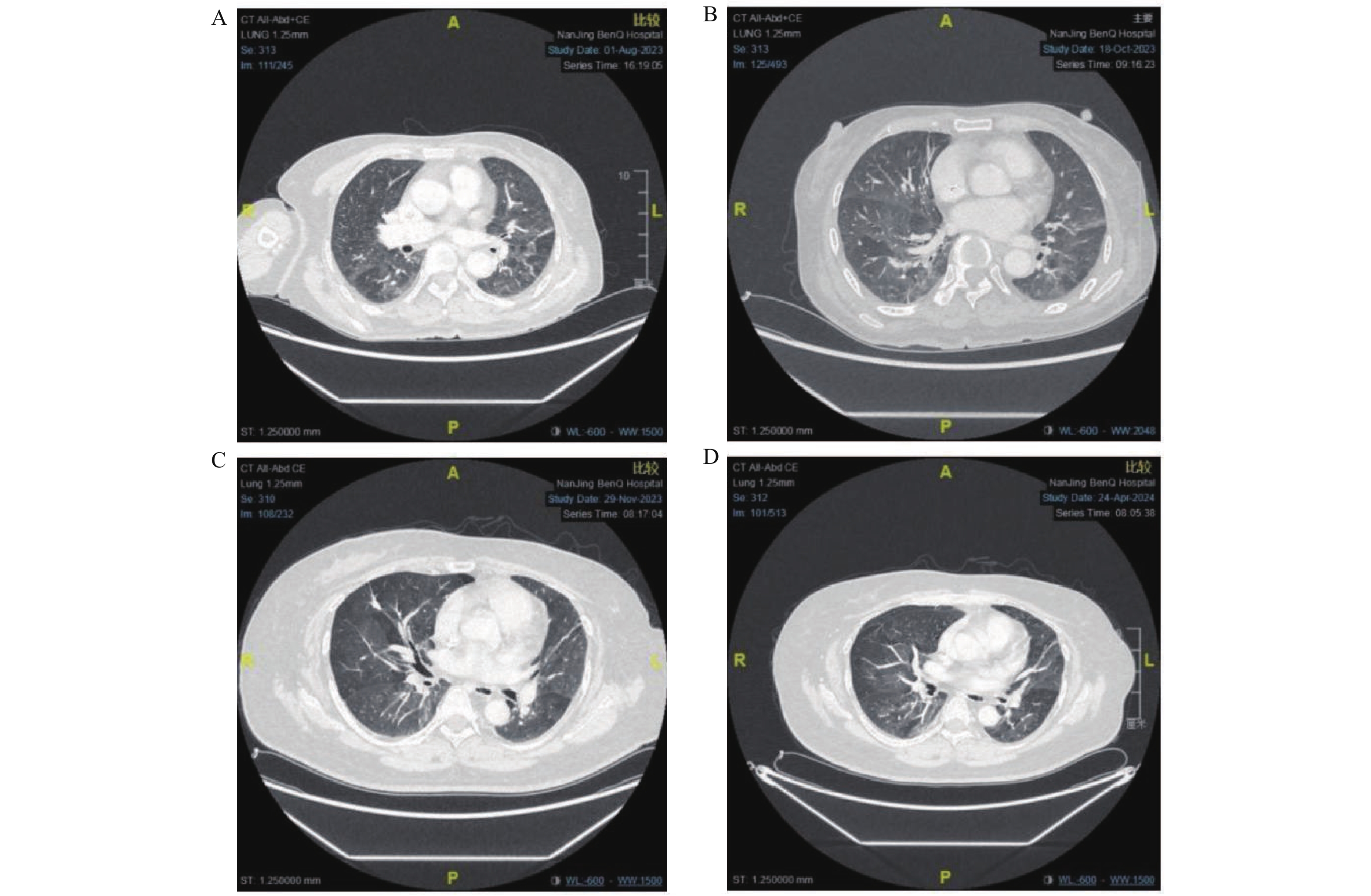
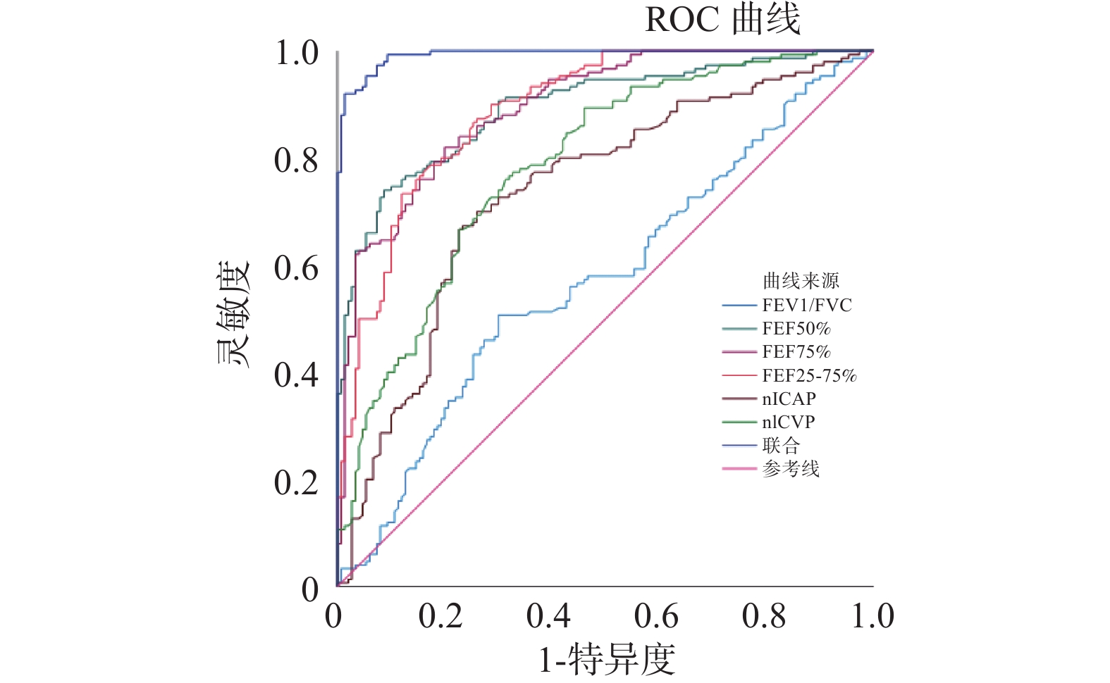
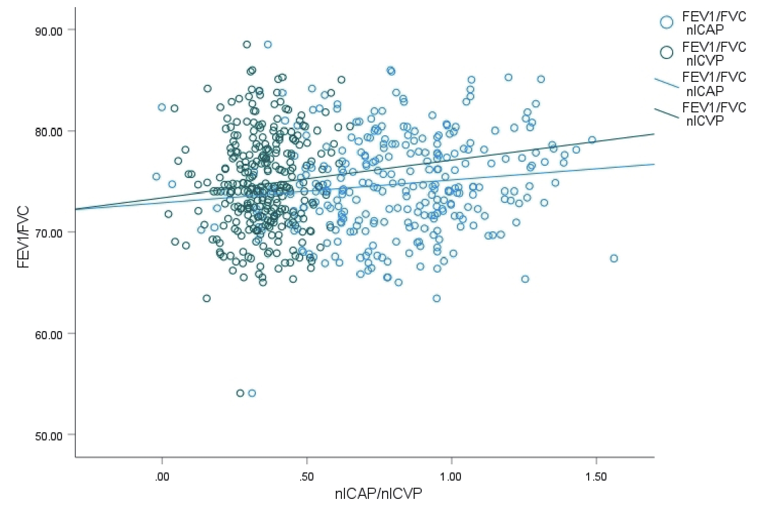
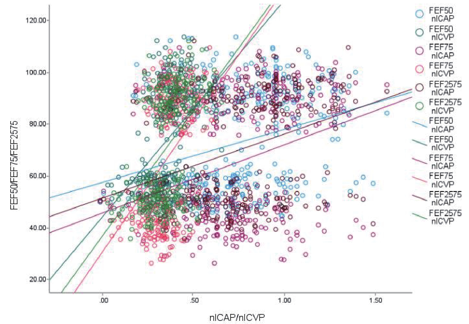
Objective To investigate the relationship between spectral computed tomography (CT) iodine-based values and pulmonary ventilation function parameters in patients with early-stage pulmonary small airway dysfunction. Methods A retrospective analysis was conducted on the clinical data of 152 patients with early-stage pulmonary small airway dysfunction admitted to Nanjing Mingji Hospital from August 2021 to December 2024, designated as the observation group. Additionally, 152 cases of clinical data from healthy individuals who underwent physical examination at the hospital during the same period were collected as the control group. All subjects had complete data including spectral CT imaging data, pulmonary function test data, and general information. Spectral CT parameters and pulmonary function parameters between the two groups were compared. The value of spectral CT iodine-based values in screening for early-stage pulmonary small airway dysfunction was analyzed, and the correlation between spectral CT iodine-based values and forced expiratory volume in one second (FEV1)/forced vital capacity (FVC) in patients with pulmonary small airway dysfunction was examined. Results FEV1/FVC, FEF50%, FEF75%, and FEF25%~75% in the observation group were significantly lower than those in the control group (P < 0.05). Normalized iodine concentration in arterial phase (nICAP) and normalized iodine concentration in venous phase (nICVP) values on spectral CT in the observation group were significantly lower than those in the control group (P < 0.05). Logistic regression analysis revealed that increases in FEV1/FVC, FEF50%, FEF75%, FEF25%~75%, nICAP, and nICVP were all protective factors against early-stage pulmonary small airway dysfunction (OR < 1, P < 0.05). ROC curve analysis demonstrated that the area under the curve (AUC) for spectral CT nICAP and nICVP values and FEV1/FVC, FEF50%, FEF75%, FEF25%~75% in screening for early-stage pulmonary small airway dysfunction were all greater than 0.50, with combined screening achieving an AUC greater than 0.90, indicating high screening value. Bivariate Pearson linear correlation analysis showed no correlation between FEV1/FVC and nICAP and nICVP in patients with early-stage pulmonary small airway dysfunction (r = 0.096, 0.076; P = 0.098, 0.189), while positive correlations were found with FEF50%, FEF75%, and FEF25%~75% (P < 0.05). Conclusion Spectral CT iodine-based values are correlated with pulmonary small airway dysfunction; however, no correlation exists between spectral CT iodine-based values and FEV1/FVC in patients with early-stage pulmonary small airway dysfunction.
2026,
47(6):
141-151.
doi: 10.12259/j.issn.2095-610X.S20260615
Abstract:
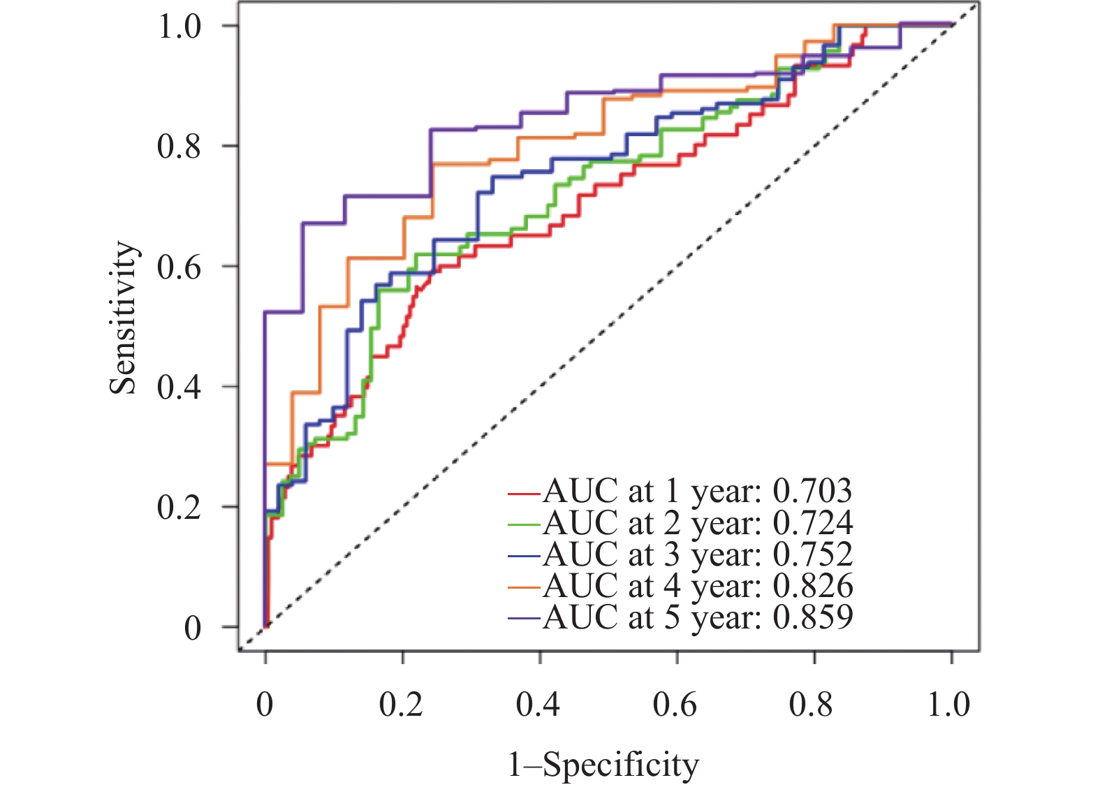
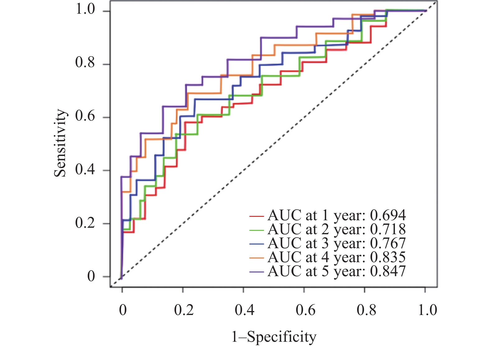
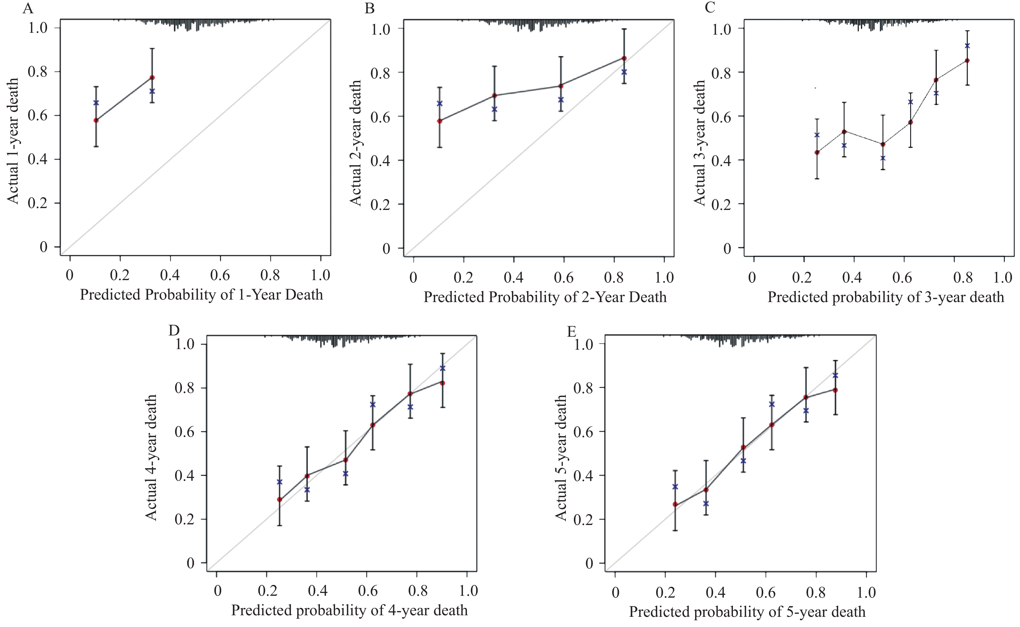
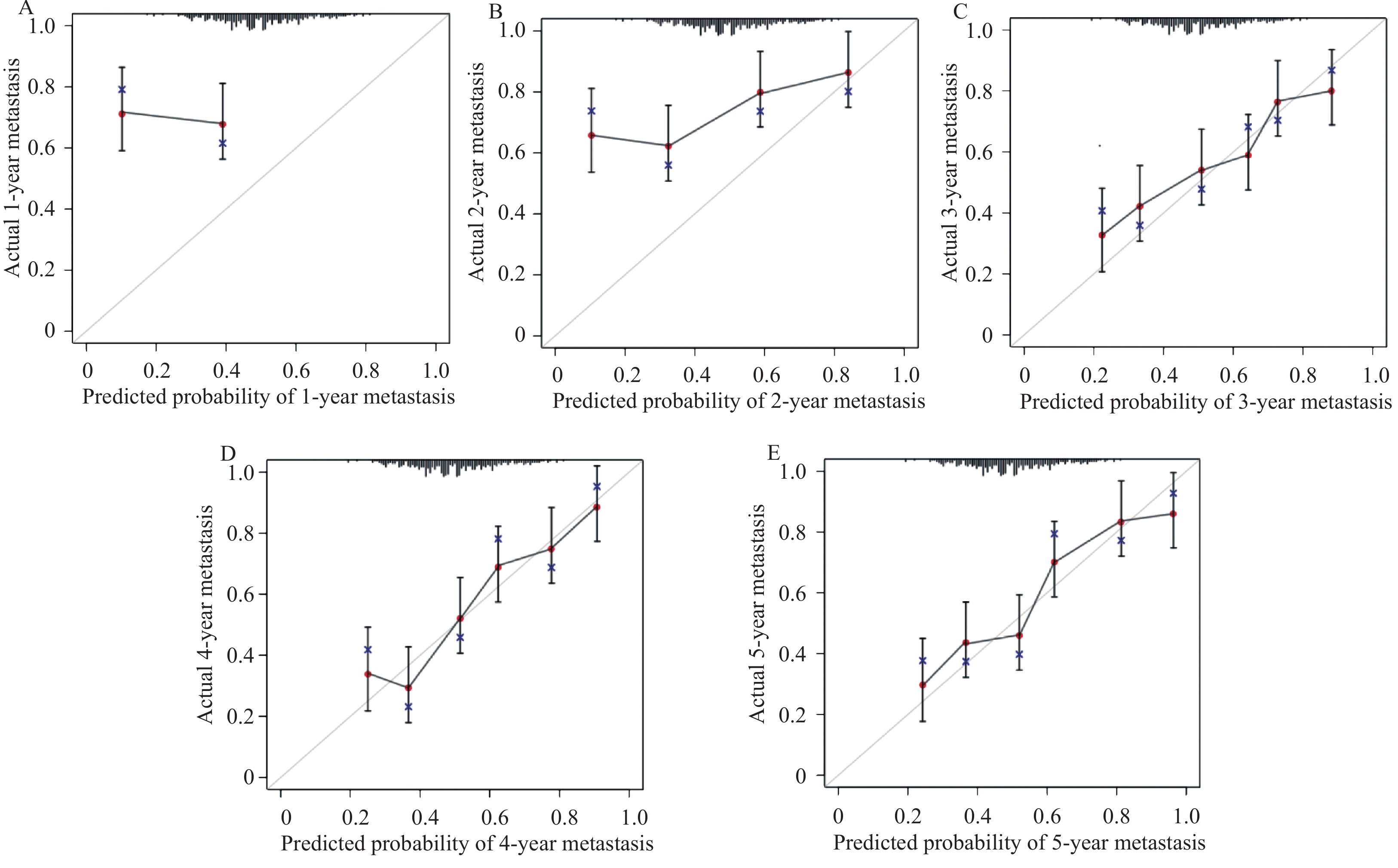
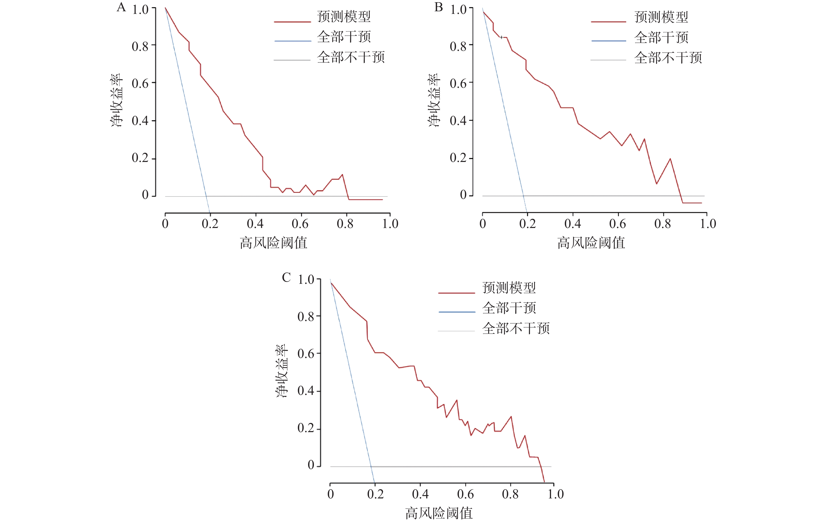
Objective To establish and internally validate a multi-indicator predictive model incorporating serum miR-504-3p, miR-151a-5p, and vascular endothelial growth factor (VEGF) to evaluate its predictive value for the 5-year survival rate and metastasis risk in patients with advanced non-small cell lung cancer (NSCLC). Methods A retrospective cohort study was conducted on 264 advanced NSCLC patients diagnosed between January 2016 and December 2019 at our institution, all of whom completed a 5-year follow-up. Patients were stratified into survival (n = 58) and mortality (n = 206) groups based on survival status. Multivariate Cox regression analysis was used to identify key factors affecting 5-year survival in advanced NSCLC patients, and a linear prognostic index (PI) model was constructed. Time-dependent receiver operating characteristic (ROC) curves were generated, and the area under the curve (AUC) was calculated to evaluate model discrimination. Internal validation was performed using the Bootstrap method (500 iterations), with calibration curves, intercepts, and slopes used to assess model calibration. Decision curve analysis (DCA) was employed to evaluate the clinical net benefit of the model. Results miR-151a-5p, VEGF, and clinical stage IV were independent risk factors for 5-year survival in patients with advanced NSCLC (P < 0.05), while miR-504-3p was a protective factor (P < 0.05). Time-dependent ROC curves demonstrated that the model's AUC for predicting mortality risk increased from 0.703 (95%CI: 0.637~0.769) at 1 year to 0.859 (95%CI: 0.814~0.904) at 5 years, and for predicting metastasis risk increased from 0.694 (95%CI: 0.624~0.764) at 1 year to 0.847 (95%CI: 0.802~0.892) at 5 years, with discriminatory ability progressively strengthening over time. Calibration curves and quantitative metrics demonstrated good calibration of the model for predicting 3-, 4-, and 5-year mortality and metastasis risk (calibration intercept approaching 0, calibration slope approaching 1), but suboptimal calibration at 1 and 2 years. DCA results demonstrated that for predicting 3-, 4-, and 5-year mortality and metastasis risk, the PI model provided net benefit superior to both "intervene all" and “intervene none” strategies across a wide range of threshold probabilities. Using the PI median, 264 patients were stratified into high-risk (n = 175) and low-risk (n = 89) groups. Kaplan-Meier survival analysis showed that 5-year survival and metastasis-free rates in the low-risk group were significantly superior to the high-risk group (P < 0.05). Conclusion Serum miR-504-3p, miR-151a-5p, and VEGF are closely associated with long-term survival and metastasis in advanced NSCLC patients. A multi-indicator model integrating these three markers and clinical staging demonstrates good predictive accuracy for long-term survival and metastasis risk at 3 years and beyond in advanced NSCLC patients and may facilitate clinical identification of patients at high risk for recurrence and metastasis.
2026,
47(6):
152-161.
doi: 10.12259/j.issn.2095-610X.S20260616
Abstract:
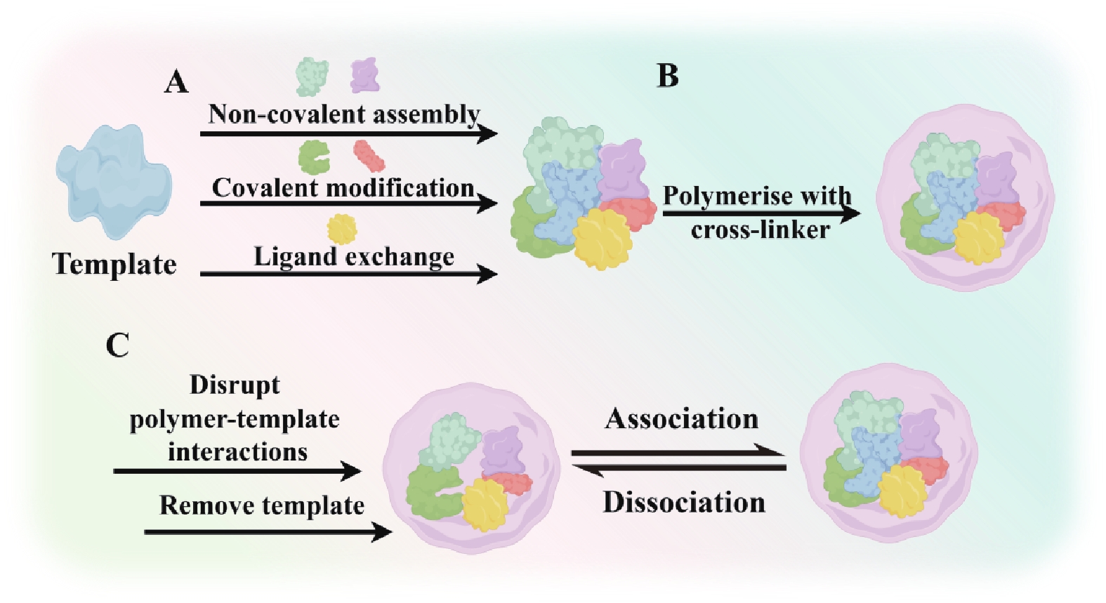
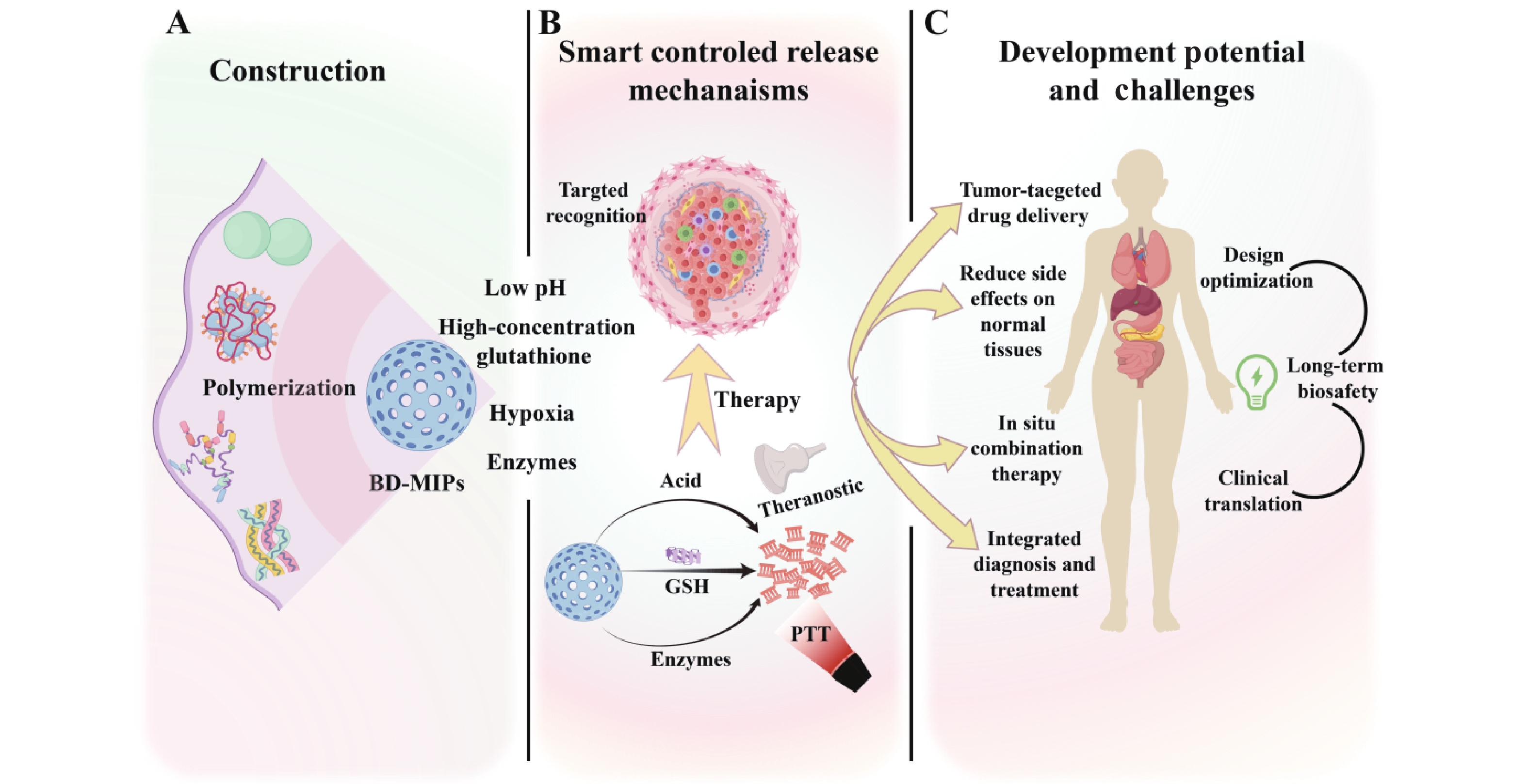
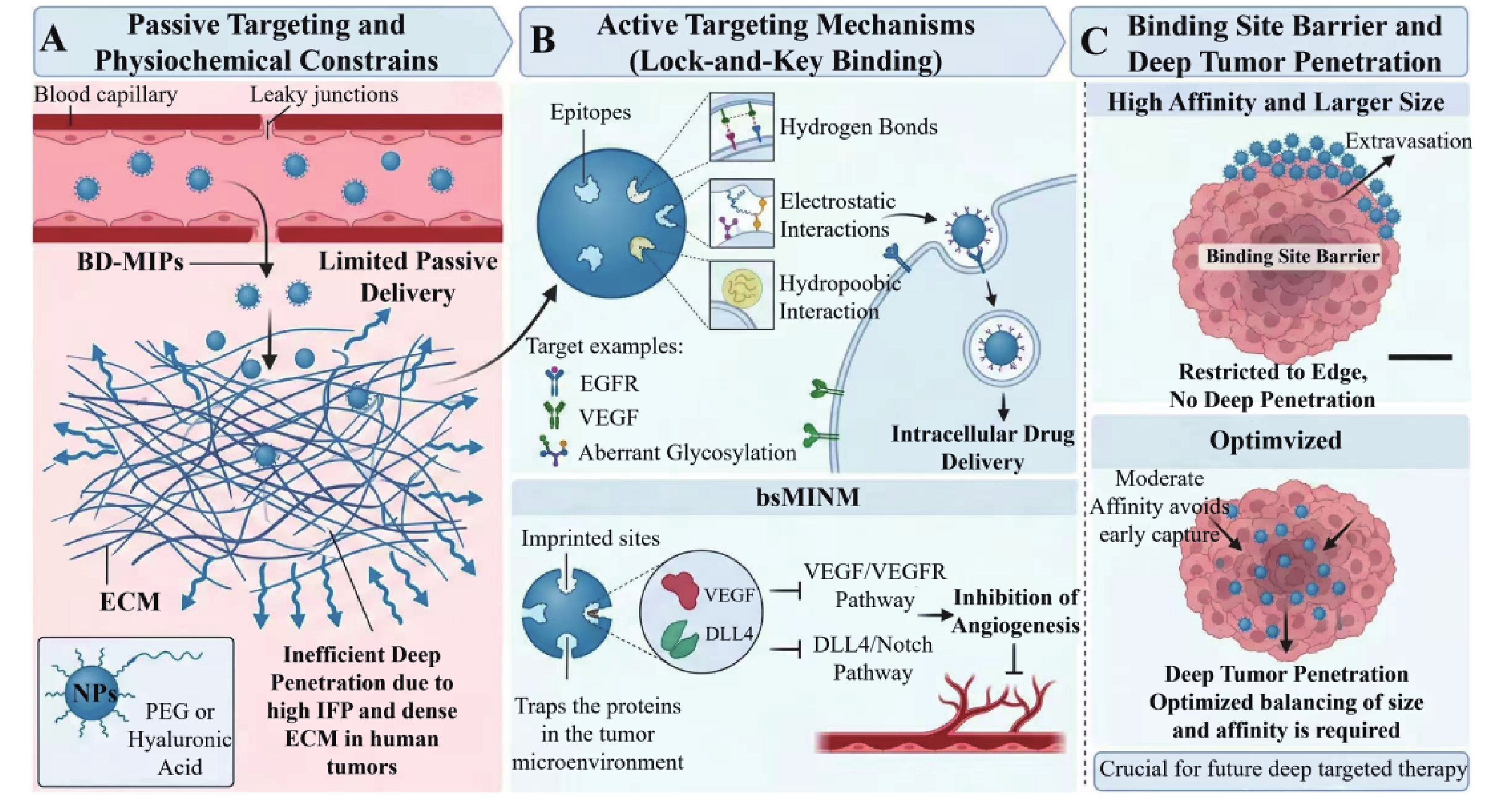
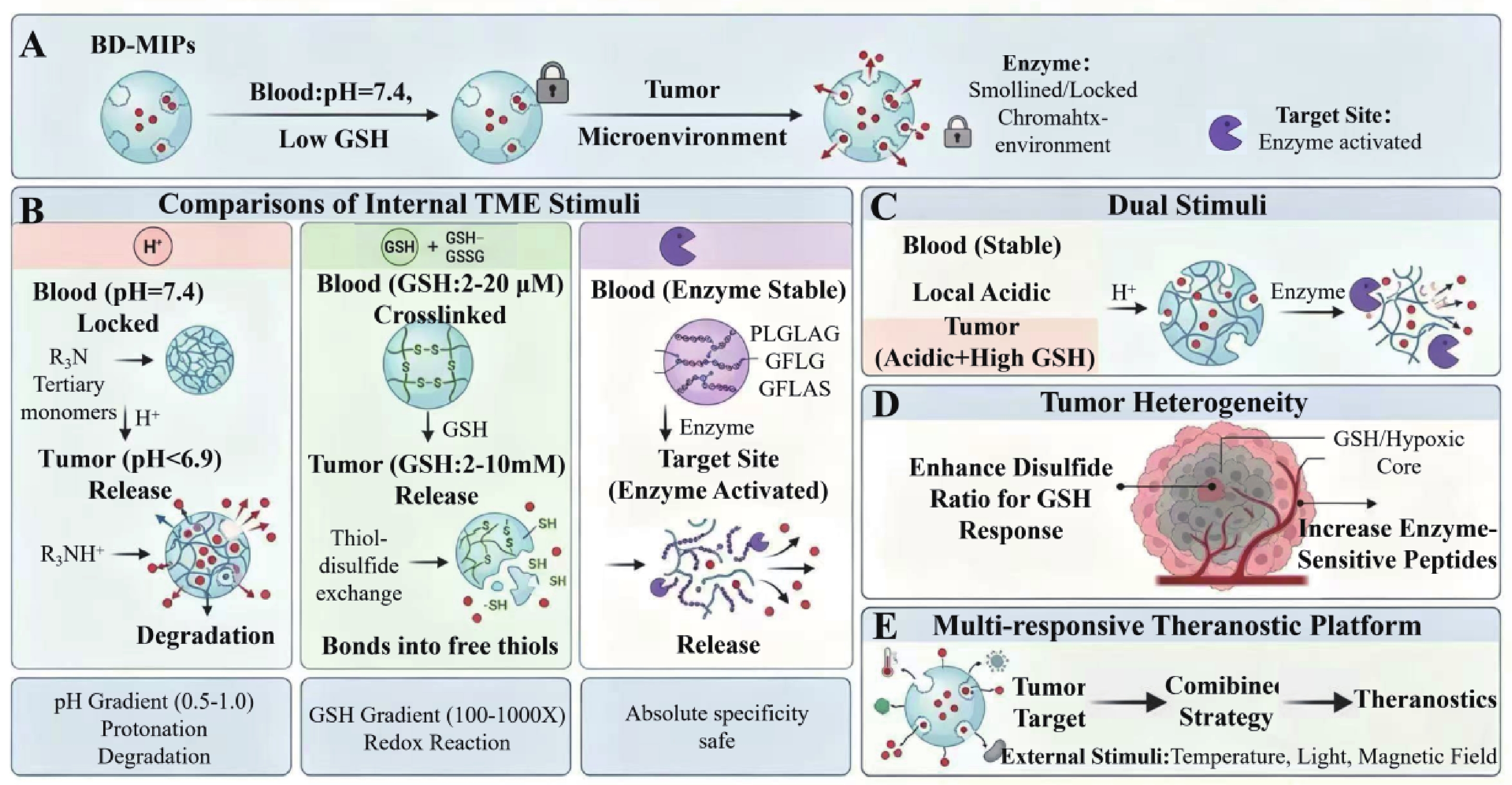
Tumor treatment remains a core challenge in global medical research. Conventional therapeutic modalities such as chemotherapy and radiotherapy suffer from lack of selectivity, propensity for drug resistance, and severe adverse reactions, which limit their therapeutic efficacy. Molecularly imprinted polymers (MIPs), as intelligent materials with customizable recognition cavities, can specifically bind tumor targets; however, their in vivo applications face limitations including poor stability and difficulties in metabolic clearance. To develop efficient, precise, and highly biocompatible novel delivery systems, biodegradable molecularly imprinted polymers (BD-MIPs) have emerged. This review systematically examines the targeted delivery mechanisms of BD-MIPs and their influencing factors, construction strategies, and design principles, with emphasis on their intelligent controlled-release mechanisms. It details endogenous strategies for drug release triggered by the unique characteristics of the tumor microenvironment (TME), as well as integrated theranostic approaches based on external triggers such as light and heat. Finally, it discusses the limitations of BD-MIPs in drug release kinetics and biocompatibility, analyzes challenges and prospects for future development, and highlights the tremendous potential of BD-MIPs as an innovative platform for precision cancer therapy. Furthermore, it provides strategic guidance for overcoming current research limitations and advancing their translation toward broad clinical application.
Tumor treatment remains a core challenge in global medical research. Conventional therapeutic modalities such as chemotherapy and radiotherapy suffer from lack of selectivity, propensity for drug resistance, and severe adverse reactions, which limit their therapeutic efficacy. Molecularly imprinted polymers (MIPs), as intelligent materials with customizable recognition cavities, can specifically bind tumor targets; however, their in vivo applications face limitations including poor stability and difficulties in metabolic clearance. To develop efficient, precise, and highly biocompatible novel delivery systems, biodegradable molecularly imprinted polymers (BD-MIPs) have emerged. This review systematically examines the targeted delivery mechanisms of BD-MIPs and their influencing factors, construction strategies, and design principles, with emphasis on their intelligent controlled-release mechanisms. It details endogenous strategies for drug release triggered by the unique characteristics of the tumor microenvironment (TME), as well as integrated theranostic approaches based on external triggers such as light and heat. Finally, it discusses the limitations of BD-MIPs in drug release kinetics and biocompatibility, analyzes challenges and prospects for future development, and highlights the tremendous potential of BD-MIPs as an innovative platform for precision cancer therapy. Furthermore, it provides strategic guidance for overcoming current research limitations and advancing their translation toward broad clinical application.
2026,
47(6):
162-172.
doi: 10.12259/j.issn.2095-610X.S20260617
Abstract:
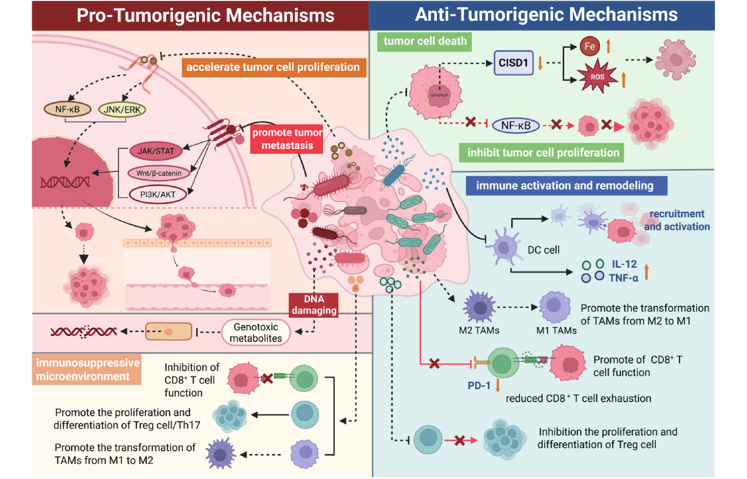
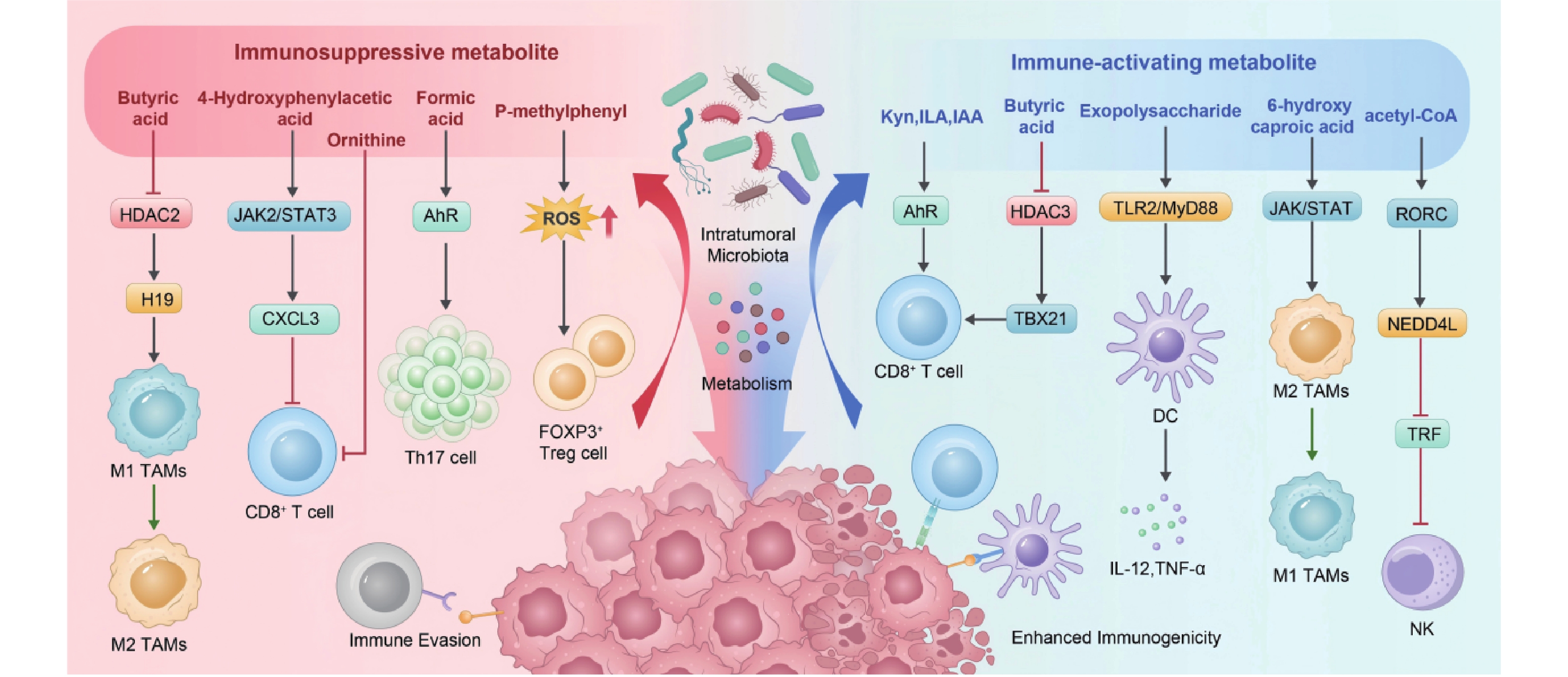
Research has confirmed that tumor tissues are not in a sterile state, but rather host microbial communities with tumor-specific characteristics. These intratumoral microorganisms not only directly promote or inhibit tumorigenesis themselves, but can also influence tumor development and therapeutic responses through their active metabolic activities. This review examines the effects and mechanisms of intratumoral microbial metabolism on tumors from both pro-tumoral and anti-tumoral perspectives, and summarizes novel strategies for tumor prevention and treatment based on intratumoral microbial metabolism. The precise regulatory network of intratumoral microbial metabolism on tumors requires further investigation. Elucidating its dual effects and molecular mechanisms on tumors is expected to provide new directions for tumor prevention and treatment.
Research has confirmed that tumor tissues are not in a sterile state, but rather host microbial communities with tumor-specific characteristics. These intratumoral microorganisms not only directly promote or inhibit tumorigenesis themselves, but can also influence tumor development and therapeutic responses through their active metabolic activities. This review examines the effects and mechanisms of intratumoral microbial metabolism on tumors from both pro-tumoral and anti-tumoral perspectives, and summarizes novel strategies for tumor prevention and treatment based on intratumoral microbial metabolism. The precise regulatory network of intratumoral microbial metabolism on tumors requires further investigation. Elucidating its dual effects and molecular mechanisms on tumors is expected to provide new directions for tumor prevention and treatment.
2026,
47(6):
173-180.
doi: 10.12259/j.issn.2095-610X.S20260618
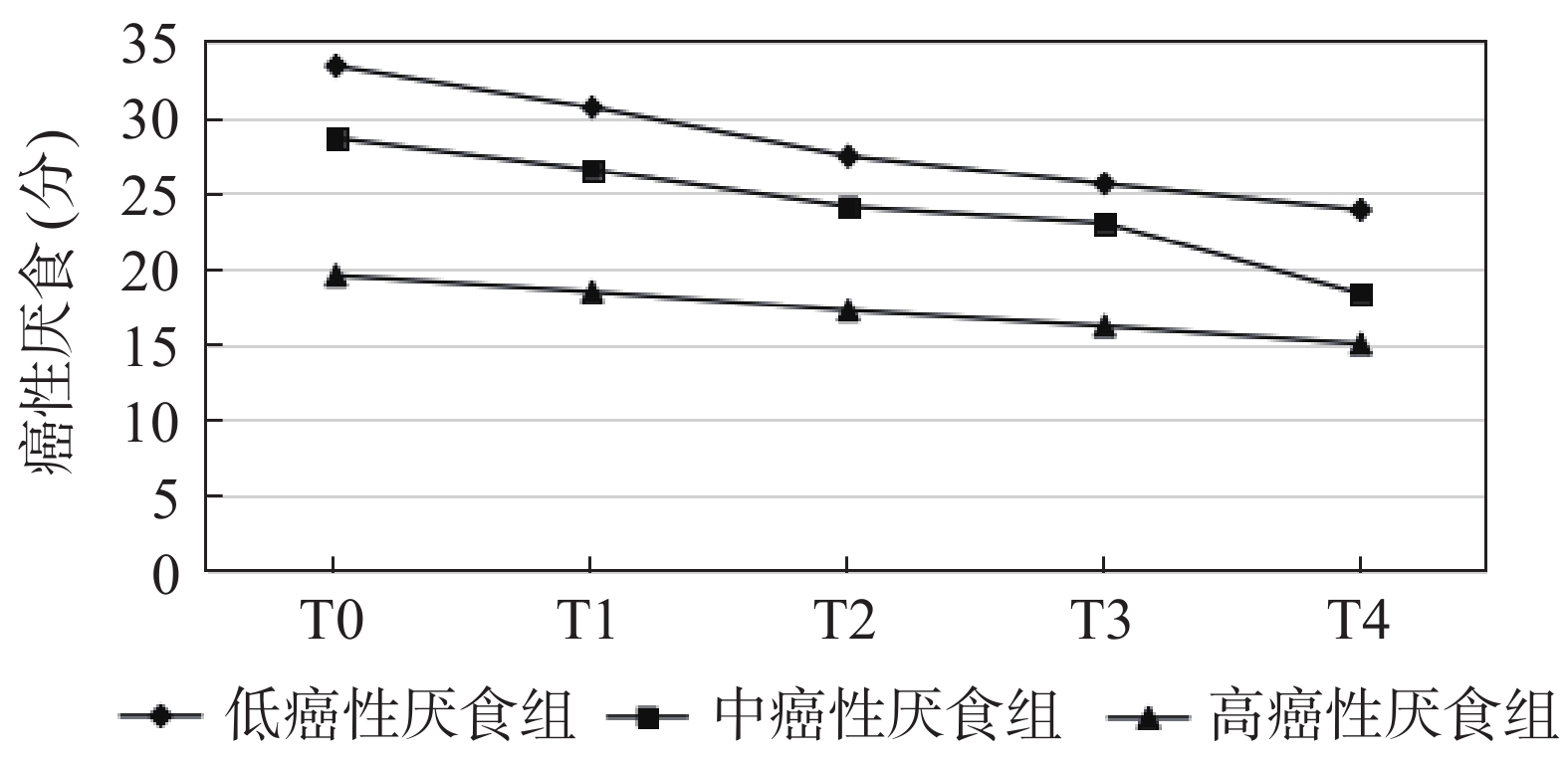
Abstract:
Objective To explore the trajectories of cancer-related anorexia and its influencing factors in nasopharyngeal carcinoma patients receiving local hypothermia care combined with nutritional intervention for radiation-induced oral mucositis. Methods A total of 120 patients with radiation-induced oral mucositis from nasopharyngeal carcinoma admitted to Nanfang Hospital, Southern Medical University from January 2024 to December 2024 were enrolled and received local hypothermia care combined with nutritional intervention. Cancer-related anorexia was assessed using the Functional Assessment of Anorexia/Cachexia Therapy (FAACT) questionnaire at week 1 (T0), week 3 (T1), week 5 (T2), week 7 (T3), and upon completion of radiotherapy (T4). Patients were stratified into three groups: low cancer-related anorexia group (n = 48), moderate cancer-related anorexia group (n = 42), and high cancer-related anorexia group (n = 30). The dynamic trajectory of anorexia nervosa scores in three groups was analyzed using the latent class growth model. Concurrently, the World Health Organization (WHO) classification of radiation-induced oral mucositis was employed to assess mucosal injury severity, serum albumin levels were measured, and nutritional status was evaluated using the PGS-GA scale. Univariate analysis and multivariate logistic regression were conducted to identify influencing factors of the anorexia nervosa trajectory. The classification of oral mucositis was performed according to the WHO classification of radiation-induced oral mucositis. Results The latent category growth model demonstrated that three models provided the best fit. In the low cancer-related anorexia group (25.00%), FAACT scores ≥24 were observed at T0~T2; in the moderate cancer-related anorexia group (35.00%), scores at all timepoints were lower than the low group but higher than the high group (P < 0.05); in the high cancer-related anorexia group (40.00%), T0~T4 scores were < 24 points, with 83.33% reporting eating-related pain and 66.67% reporting aversion to fatty foods, compared to only 12.50% with mild discomfort in the low group. Univariate analysis revealed that age, radiotherapy dose, oral mucositis grading, serum albumin level, FAACT score, and the Patient-Generated Subjective Global Assessment (PGS-GA) score were significantly correlated with anorexia trajectory (P < 0.05), whereas gender and other factors showed no significant correlation (P > 0.05). Multivariate logistic regression analysis identified these six factors as independent influencing factors (P < 0.05). Conclusion Cancer-related anorexia presents three distinct trajectories: low, moderate, and high. Age, radiotherapy dose, oral mucositis grade, serum albumin level, FAACT score, and PG-SGA score are independent influencing factors that may provide insights for clinical nursing intervention planning.
