

Effects of Decursin on Myocardial Fibrosis Following Myocardial Infarction
-
摘要:
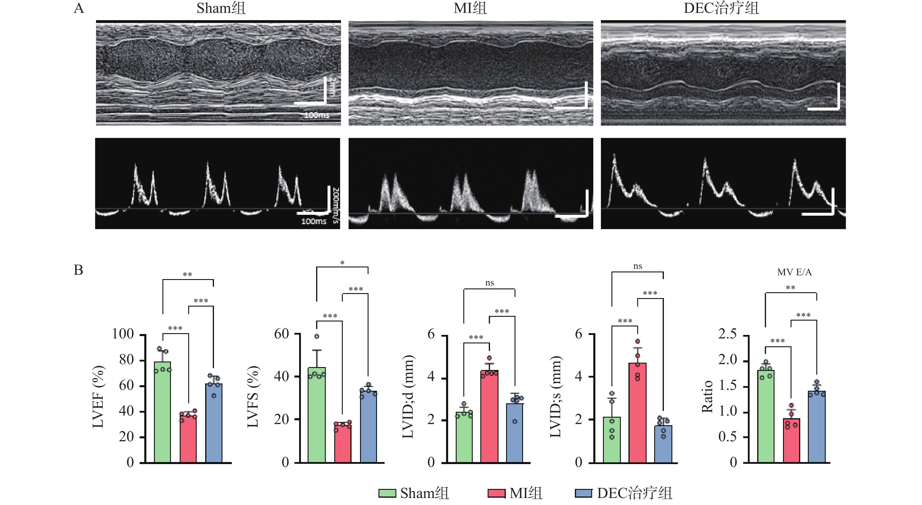
目的 探讨紫花前胡素(decursin,DEC)对小鼠心肌梗死后心肌纤维化的作用,及其对心肌成纤维细胞(cardiac fibroblasts,CFs)活化的调控机制。 方法 选用C57BL/6J小鼠,行冠状动脉左前降支结扎术,构建小鼠心肌梗死模型,随机分组(每组n = 5只),假手术组(Sham组)、心肌梗死模型组(MI组)、紫花前胡素治疗组(DEC组),通过灌胃给药的方式,给予DEC治疗,术后28 d,通过超声心动图评估心功能,免疫组织化学法检测心肌组织的病理形态变化,观察心肌纤维化程度;细胞划痕实验检测心肌成纤维细胞迁移能力;EdU技术检测心肌成纤维细胞增殖;Western blot检测心肌纤维化相关信号通路及关键蛋白表达水平。 结果 超声心动图评估结果发现,DEC治疗组与MI组相比,左心室射血分数(LVEF,升高25%,P < 0.001)、左心室短轴缩短率(left ventricular ejection fraction,LVFS)升高16%,P < 0.001,二尖瓣E/A比值显著升高(MV E/A ,P < 0.001)。免疫组化结果显示,MI组心肌间质胶原沉积增多、纤维化面积扩大(P < 0.001),而DEC干预可明显抑制纤维化进程(P < 0.01)。体外实验表明,DEC能显著抑制心肌成纤维细胞增殖(P < 0.01)与迁移(P < 0.05),使Fibronectin(P < 0.05)和α-SMA、Collagen I等蛋白表达下调(P < 0.01),并抑制心肌成纤维细胞的活化。 结论 紫花前胡素具有改善心肌梗死后的心功能及抑制心肌纤维化的作用,其机制可能与PI3K/AKT信号通路相关。 -
关键词:
- 紫花前胡素 /
- 心肌梗死 /
- 心肌纤维化 /
- 成纤维细胞 /
- PI3K/AKT信号通路
Abstract:Objective To investigate the effects of Decursin (DEC) on myocardial fibrosis after myocardial infarction in mice and its regulatory mechanism underlying the activation of cardiac fibroblasts (CFs). Methods C57BL/6J mice were subjected to ligation of the left anterior descending coronary artery to establish a mouse myocardial infarction model. The mice were randomly divided into three groups (n = 5 per group): sham operation group (Sham), myocardial infarction model group (MI), and decursin treatment group (DEC). DEC was administered by intragastric gavage. For 28 days post-surgery, cardiac function was evaluated by echocardiography; histopathological changes and the degree of myocardial fibrosis were assessed by immunohistochemistry. The migratory capacity of cardiac fibroblasts was determined by wound-healing assay; the proliferation of cardiac fibroblasts was detected by EdU assay. The expression levels of key proteins related to myocardial fibrosis signaling pathways were measured by Western blot (WB). Results Echocardiographic assessment revealed that compared with the MI group, the DEC treatment group exhibited significantly increased left ventricular ejection fraction (LVEF, increased by 25%, P < 0.001), left ventricular fractional shortening (LVFS, increased by 16%, P < 0.001), and mitral valve E/A ratio (MV E/A, P < 0.001). Immunohistochemical results showed that the MI group presented increased myocardial interstitial collagen deposition and enlarged fibrotic area (P < 0.001), whereas DEC intervention markedly attenuated the fibrotic process (P < 0.01). In vitro experiments demonstrated that DEC significantly inhibited the proliferation (P < 0.01) and migration (P < 0.05) of cardiac fibroblasts, downregulated the expression of fibronectin (P < 0.05), α-SMA, collagen I and other proteins (P < 0.01), and suppressed the activation of cardiac fibroblasts. Conclusion Decursin can improve cardiac function and attenuate myocardial fibrosis after myocardial infarction, and its mechanism may be associated with the PI3K/AKT signaling pathway. -
Key words:
- Decursin /
- Myocardial infarction /
- Myocardial fibrosis /
- Fibroblasts /
- PI3K/AKT signaling pathway
-
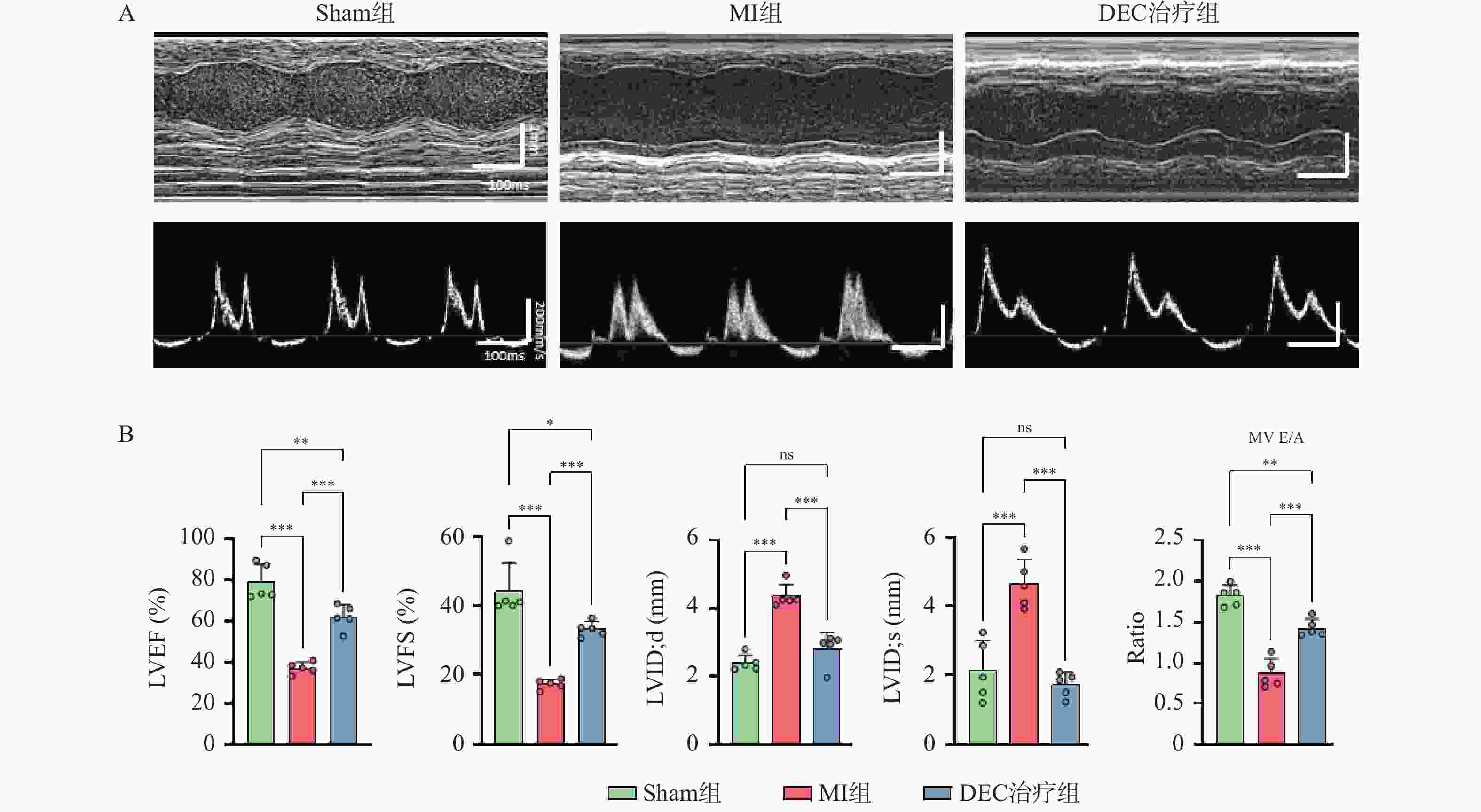
图 1 紫花前胡素对心肌梗死小鼠心功能的影响($\bar x \pm s $,n = 5)
A:不同组小鼠超声心动图;B:不同组小鼠心功能指标;LVEF(%):左室射血分数,LVFS(%):左室短轴缩短率,LVID'd (mm):左室舒张末期内径,LVID's(mm):左室收缩末期内径,MV E/A:二尖瓣E/A比值。*P < 0.05;**P < 0.01;***P < 0.001。
Figure 1. Effects of decursin on cardiac function in mice with myocardial infarction ($\bar x \pm s $,n = 5)
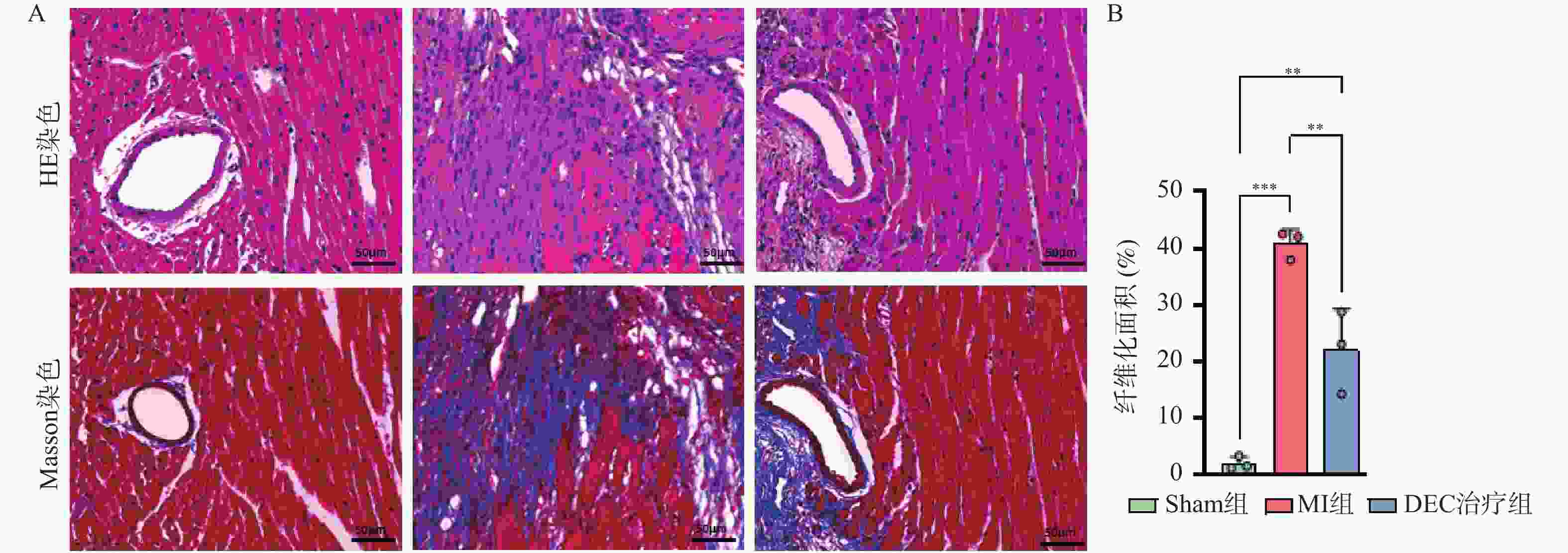
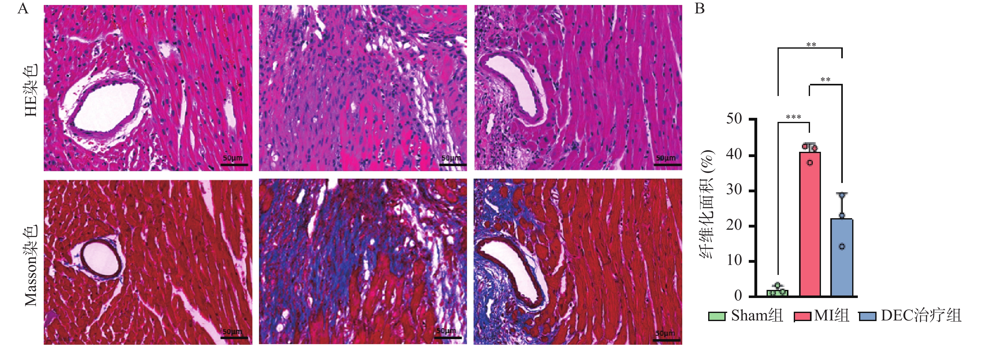
图 2 紫花前胡素对心肌梗死小鼠心肌纤维化的影响($\bar x \pm s $,n = 3)
A:HE和Masson染色结果(×200);B:Masson染色心肌相对纤维化面积统计(n = 3);**P < 0.01;***P < 0.001。
Figure 2. Effects of decursin on myocardial fibrosis in mice with myocardial infarction($\bar x \pm s $,n = 3)
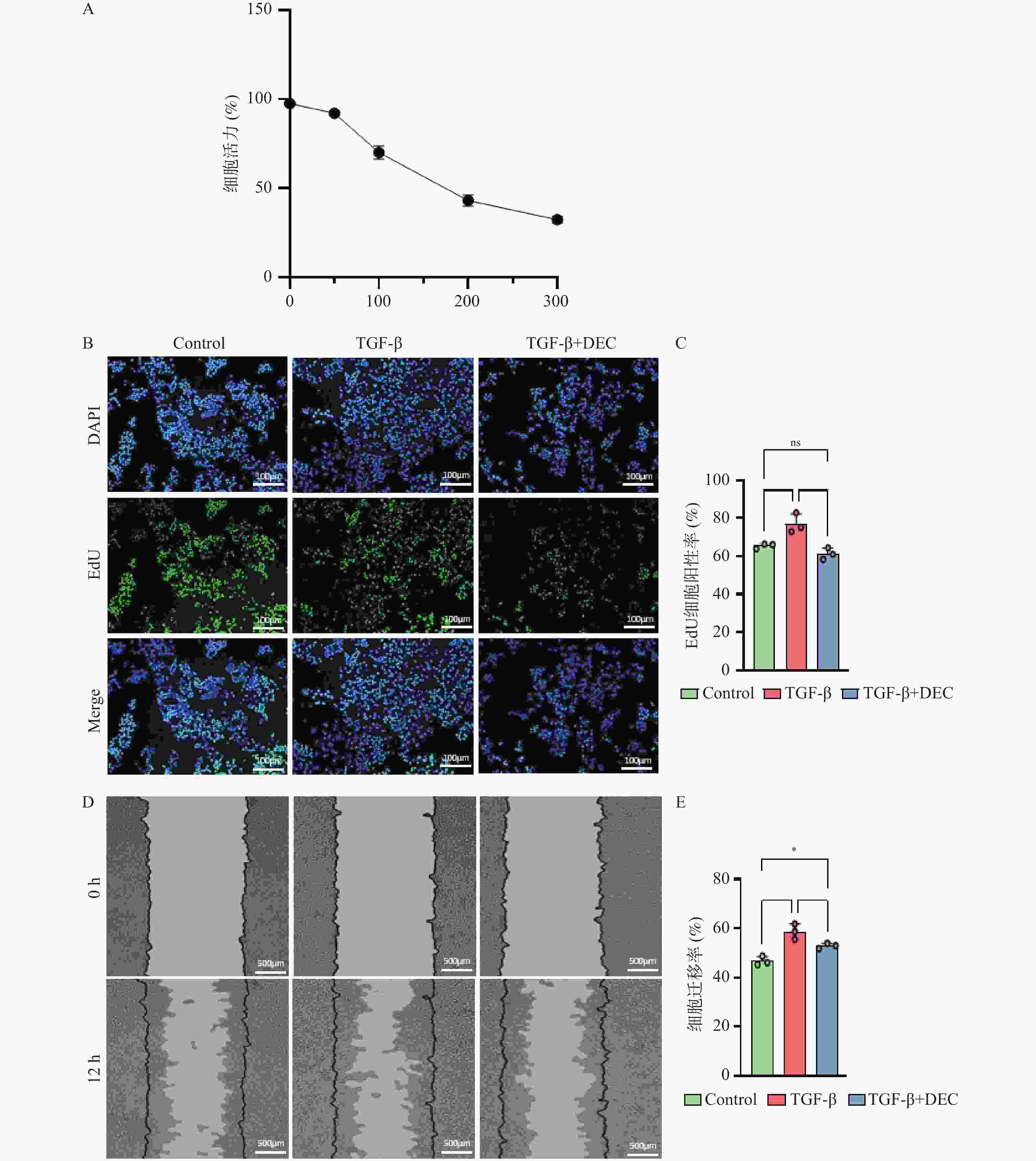
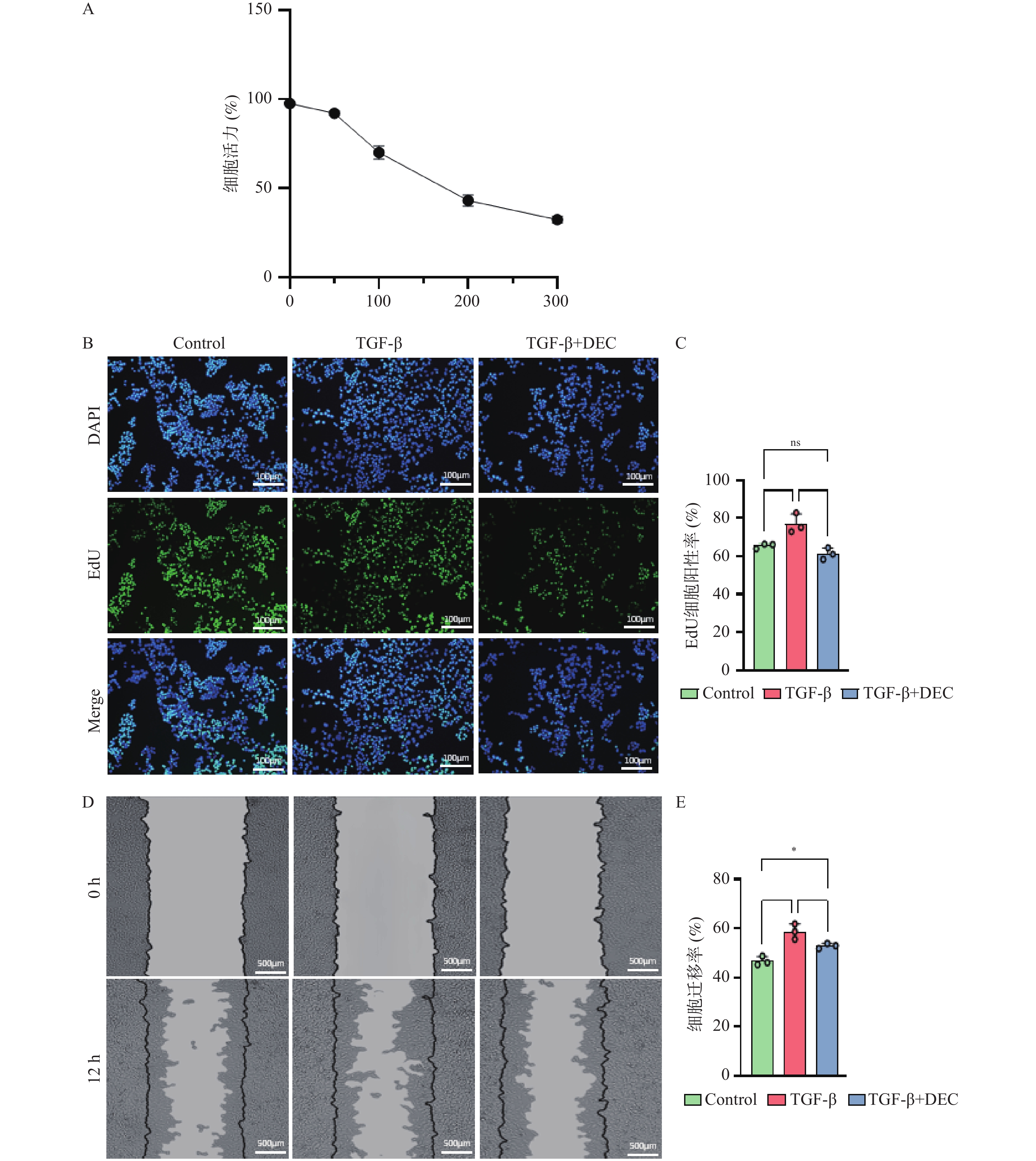
图 3 紫花前胡素对抗TGF-β刺激的MCFs的增殖和迁移($\bar x \pm s $,n = 3)
A:CCK8检测结果;B~C:EdU细胞增殖实验结果;D~E:划痕实验结果;*P < 0.05;**P < 0.01。
Figure 3. Decursin antagonizes TGF‑β‑induced proliferation and migration of cardiac MCFs($\bar x \pm s $,n = 3)
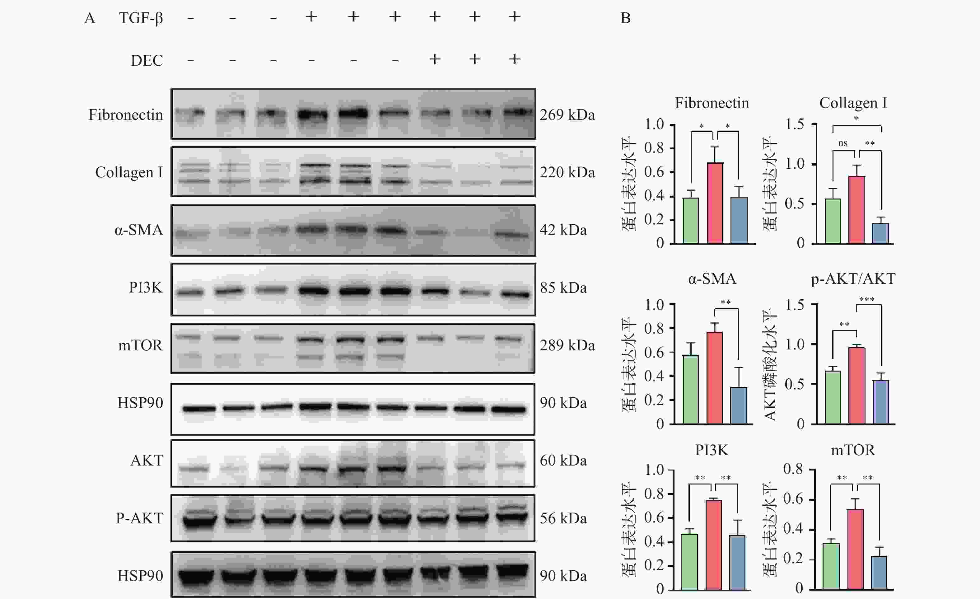
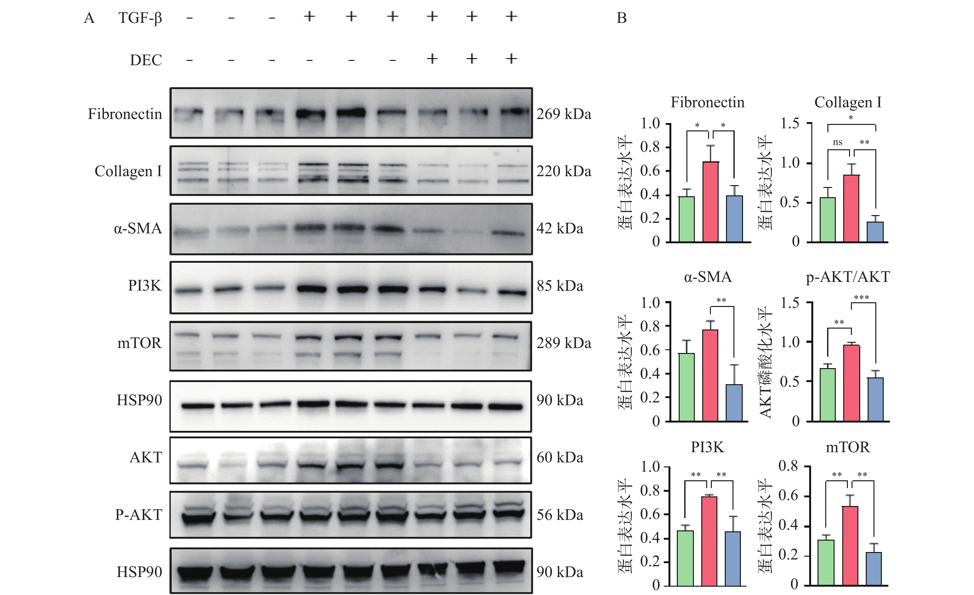
图 5 紫花前胡素可抑制TGF-β诱导的纤维化相关蛋白的表达($\bar x \pm s $,n = 3)
A:Western blot检测纤维化相关蛋白表达水平,内参为HSP90;B:Fibronectin,Collagen Ⅰ,α-SMA,PI3K,mTOR的蛋白表达水平和AKT磷酸化水平统计;*P < 0.05,**P < 0.01,***P < 0.001。
Figure 5. Decursin inhibits the expression of fibrosis-related proteins induced by TGF-β($\bar x \pm s $,n = 3)
-
[1] Mensah G A, Fuster V, Murray C J L, et al. Global burden of cardiovascular diseases and risks, 1990-2022[J]. J Am Coll Cardiol, 2023, 82(25): 2350-2473. doi: 10.1016/j.jacc.2023.11.007 [2] Sharma A, Vidusha K, Suresh H, et al. Global awareness of myocardial infarction symptoms in general population: A systematic review and meta-analysis[J]. Korean Circ J, 2021, 51(12): 983. doi: 10.4070/kcj.2021.0100 [3] Frangogiannis N G. Cardiac fibrosis[J]. Cardiovasc Res, 2021, 117(6): 1450-1488. doi: 10.1093/cvr/cvaa324 [4] Pesce M, Duda G N, Forte G, et al. Cardiac fibroblasts and mechanosensation in heart development, health and disease[J]. Nat Rev Cardiol, 2023, 20(5): 309-324. doi: 10.1038/s41569-022-00799-2 [5] Maruyama K, Imanaka-Yoshida K. The pathogenesis of cardiac fibrosis: A review of recent progress[J]. Int J Mol Sci, 2022, 23(5): 2617. doi: 10.3390/ijms23052617 [6] Plikus M V, Wang X, Sinha S, et al. Fibroblasts: Origins, definitions, and functions in health and disease[J]. Cell, 2021, 184(15): 3852-3872. doi: 10.1016/j.cell.2021.06.024 [7] Ghazal R, Wang M, Liu D, et al. Cardiac fibrosis in the multi-omics era: Implications for heart failure[J]. Circ Res, 2025, 136(7): 773-802. doi: 10.1161/CIRCRESAHA.124.325402 [8] Muralikrishnan A, Sekar M, Kumarasamy V, et al. Chemistry, pharmacology and therapeutic potential of decursin: A promising natural lead for new drug discovery and development[J]. Drug Des Dev Ther, 2024, 18: 3741-3763. doi: 10.2147/DDDT.S476279 [9] Ge Y, Yoon S H, Jang H, et al. Decursin promotes HIF-1α proteasomal degradation and immune responses in hypoxic tumour microenvironment[J]. Phytomedicine, 2020, 78: 153318. doi: 10.1016/j.phymed.2020.153318 [10] Son D B, Choi W, Kim M, et al. Decursin alleviates mechanical allodynia in a paclitaxel-induced neuropathic pain mouse model[J]. Cells, 2021, 10(3): 547. doi: 10.3390/cells10030547 [11] Choi Y J, Kim D H, Kim S J, et al. Decursin attenuates hepatic fibrogenesis through interrupting TGF-beta-mediated NAD(P)H oxidase activation and Smad signaling in vivo and in vitro[J]. Life Sci, 2014, 108(2): 94-103. doi: 10.1016/j.lfs.2014.05.012 [12] Que R, Cao M, Dai Y, et al. Decursin ameliorates carbon-tetrachloride-induced liver fibrosis by facilitating ferroptosis of hepatic stellate cells[J]. Biochem Cell Biol, 2022, 100(5): 378-386. doi: 10.1139/bcb-2022-0027 [13] 刘建, 范慧敏, 汪进益, 等. 小鼠心梗模型的建立与无创评价[J]. 中国实验动物学报, 2010, 18(3): 196-198. [14] Wang J, Ge S, Wang Y, et al. Puerarin alleviates UUO-induced inflammation and fibrosis by regulating the NF-κB P65/STAT3 and TGFβ1/smads signaling pathways[J]. Drug Des Dev Ther, 2021, 15: 3697-3708. doi: 10.2147/DDDT.S321879 [15] 贾迎起, 张敏, 李彪. 放射性核素标记的成纤维细胞活化蛋白抑制剂正电子发射断层扫描在心脏疾病诊断中的研究进展[J]. 诊断学理论与实践, 2025, 24(2): 220-225. doi: 10.16150/j.1671-2870.2025.02.014 [16] Chen G, Xu H, Xu T, et al. Calycosin reduces myocardial fibrosis and improves cardiac function in post-myocardial infarction mice by suppressing TGFBR1 signaling pathways[J]. Phytomedicine, 2022, 104: 154277. doi: 10.1016/j.phymed.2022.154277 [17] Qin W, Cao L, Massey I Y. Role of PI3K/Akt signaling pathway in cardiac fibrosis[J]. Mol Cell Biochem, 2021, 476(11): 4045-4059. doi: 10.1007/s11010-021-04219-w [18] Yang W, Wu Z, Yang K, et al. BMI1 promotes cardiac fibrosis in ischemia-induced heart failure via the PTEN-PI3K/Akt-mTOR signaling pathway[J]. Am J Physiol Heart Circ Physiol, 2019, 316(1): H61-H69. doi: 10.1152/ajpheart.00487.2018 [19] Stempien-Otero A, Kim D H, Davis J. Molecular networks underlying myofibroblast fate and fibrosis[J]. J Mol Cell Cardiol, 2016, 97: 153-161. doi: 10.1016/j.yjmcc.2016.05.002 [20] Tang Q, Markby G R, MacNair A J, et al. TGF-β-induced PI3K/AKT/mTOR pathway controls myofibroblast differentiation and secretory phenotype of valvular interstitial cells through the modulation of cellular senescence in a naturally occurring in vitro canine model of myxomatous mitral valve disease[J]. Cell Prolif, 2023, 56(6): e13435. doi: 10.1111/cpr.13435 -

 下载:
下载:
 点击查看大图
计量
- 文章访问数: 80
- HTML全文浏览量: 55
- PDF下载量: 66
- 被引次数: 0
