A Comparative Study on the Establishment of Animal Models of Chronic Renal Failure by Two Methods
-
摘要:
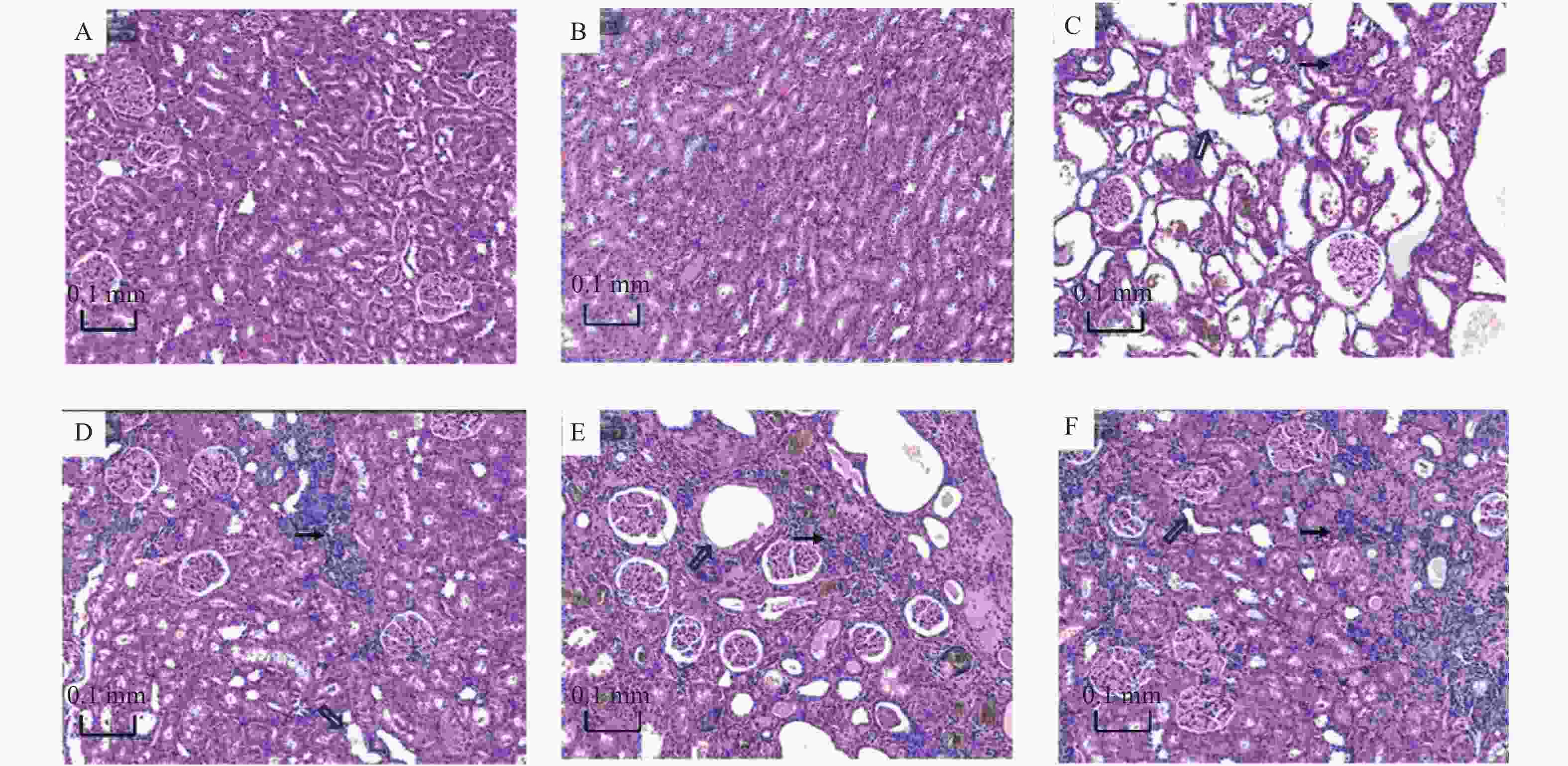
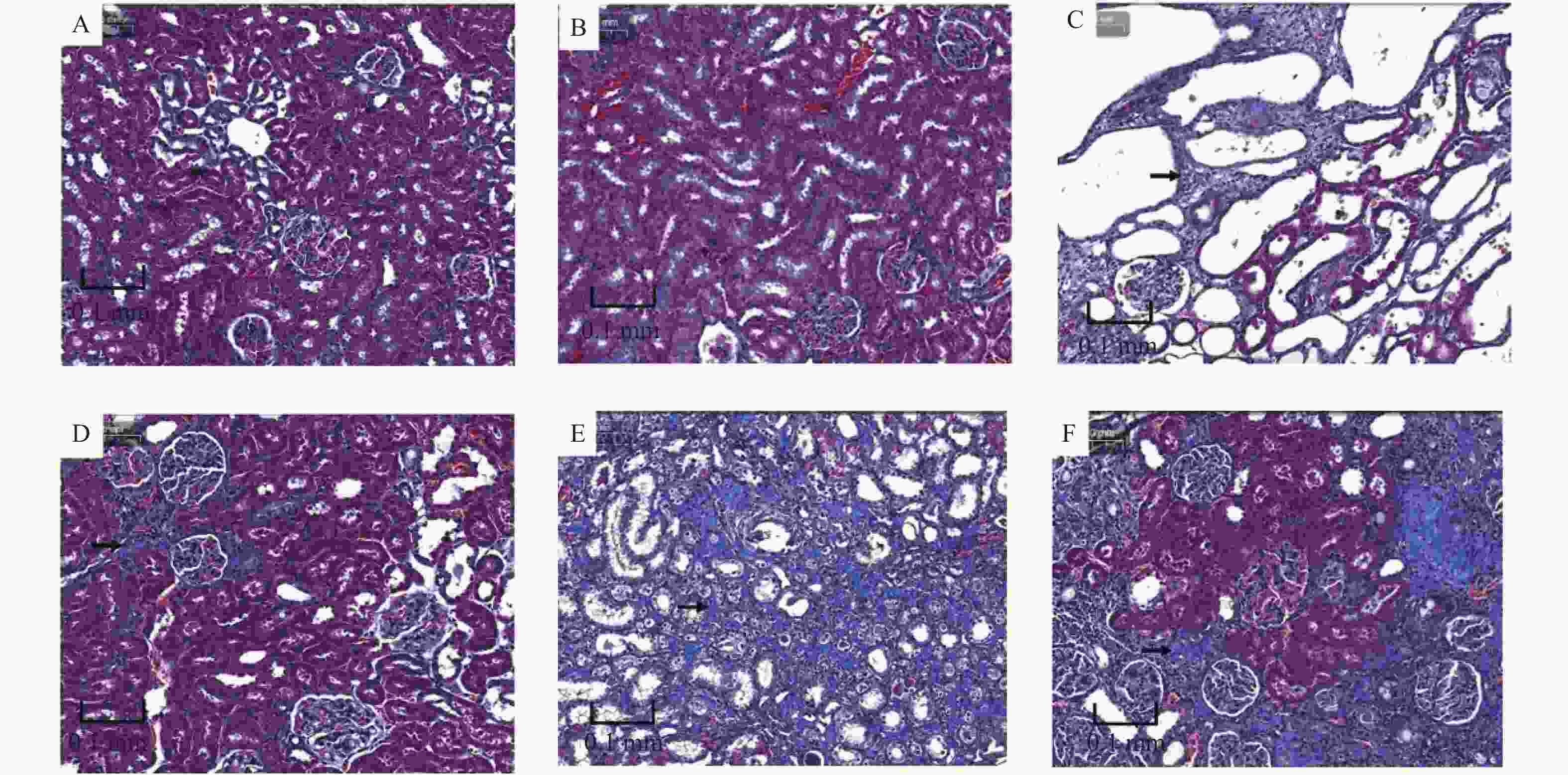
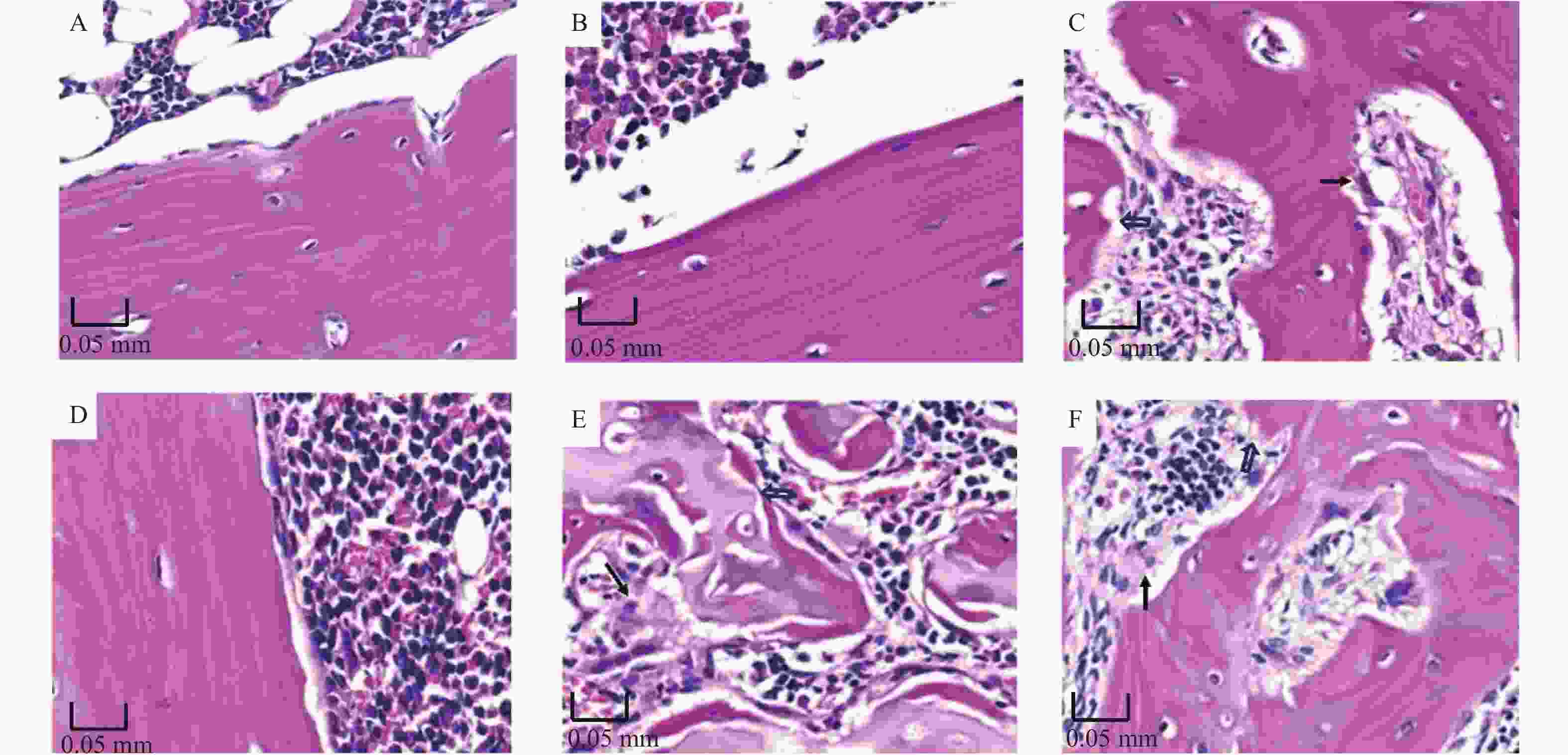
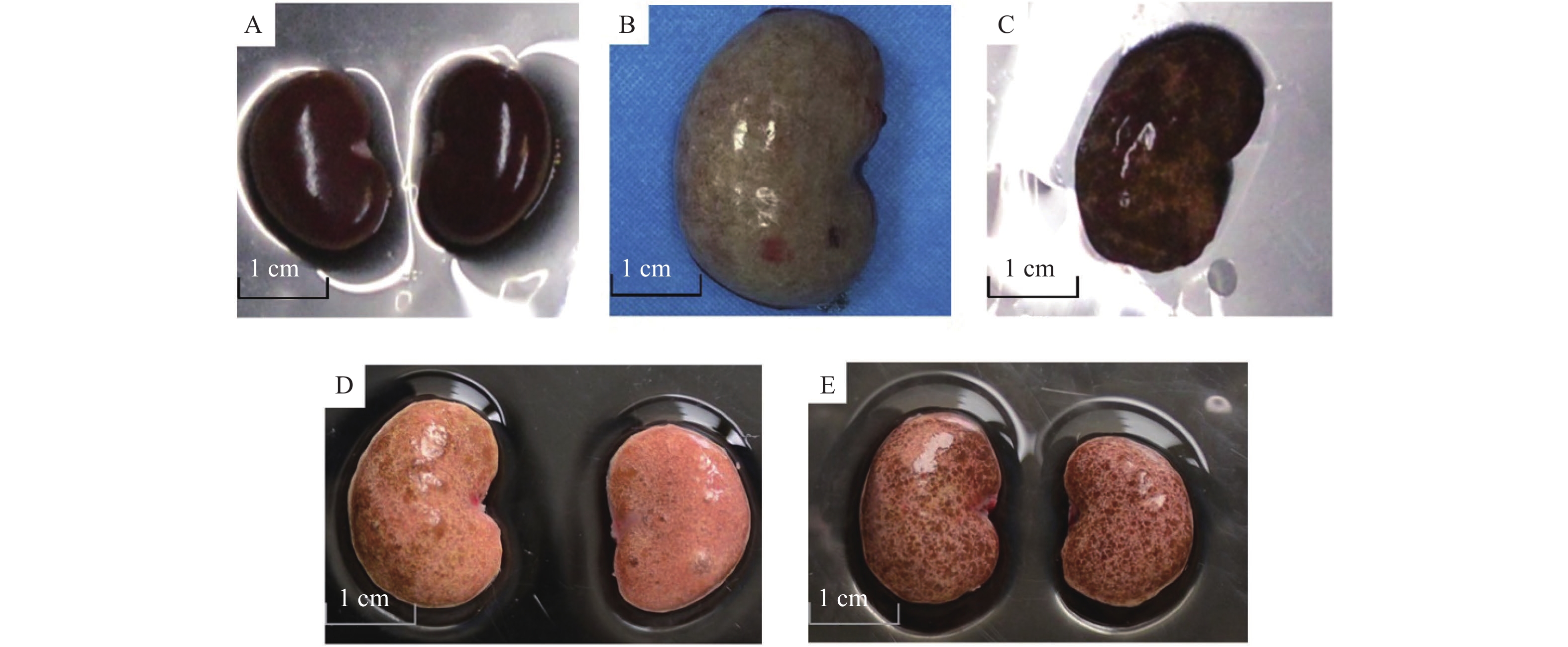
目的 分别使用2种方法建立慢性肾衰竭(chronic renal failure,CRF)大鼠模型,对比2种模型的稳定性,旨在为建立一种稳定性高的CRF动物模型提供依据。 方法 分别使用单侧肾切除+腺嘌呤灌胃+高磷饲料喂养法、高磷+腺嘌呤饲料喂养法建立CRF大鼠模型。造模结束及造模后6周检测模型动物的红细胞、血红蛋白,血清尿素、肌酐,并行肾脏、骨组织病理学检查,计算肾小管间质纤维化指数(tubulointerstitial fibrosis index,TBI)、肾组织胶原纤维面积百分比。 结果 模型一组(单侧肾切除+腺嘌呤灌胃+高磷饲料喂养法)造模结束时,与正常对照组比,红细胞、血红蛋白明显下降(P < 0.05),血清尿素、肌酐明显升高(P < 0.05),TBI、肾组织胶原纤维面积百分比轻度升高(P < 0.05),股骨病变明显。造模结束后6周,与正常对照组比,模型一组红细胞、血红蛋白轻度下降(P < 0.05),血清尿素、肌酐水平差异无统计学意义,TBI、肾组织胶原纤维面积百分比轻度升高(P < 0.05),股骨病变不明显。模型二组(高磷+腺嘌呤饲料喂养法)造模结束时,与正常组比,红细胞、血红蛋白明显下降(P < 0.05),血清尿素、肌酐、TBI、肾组织胶原纤维面积百分比明显升高(P < 0.05),股骨病变明显。造模后6周,与正常组比,模型二组红细胞、血红蛋白较正常组明显下降(P < 0.05),血清尿素、肌酐、TBI、肾组织胶原纤维面积百分比较正常组明显升高(P < 0.05),股骨病变明显。造模后6周,与模型一组比,模型二组大鼠红细胞、血红蛋白明显降低(P < 0.05),血清尿素、肌酐、TBI、肾组织胶原纤维面积百分比明显升高(P < 0.05),骨病变程度明显。 结论 高磷+腺嘌呤饲料喂养法可建立稳定性较高的CRF大鼠模型。 Abstract:Objective To investigate the stability of the two kinds of rat models of chronic renal failure, aiming to provide a basis for establishing a feasible and stable animal model of chronic renal failure. Methods The rat model of chronic renal failure was established by unilateral nephrectomy + adenine gavage + high-phosphorus diet and high-phosphorus + adenine diet, respectively. Erythrocyte and hemoglobin levels, serum urea and creatinine levels were detected at the end of modeling and 6 weeks after modeling, and histopathological examinations of kidney and bone were performed. The tubulointerstitial fibrosis index (TBI) and the percentage of collagen fiber area in renal tissue were calculated. Results At the end of modeling, compared with the normal group, the erythrocyte and hemoglobin in the model group 1(unilateral nephrectomy + adenine gavage + high-phosphorus) were significantly decreased(P < 0.05), the serum urea and creatinine were significantly increased(P < 0.05), TBI and the percentage of collagen fiber area in kidney tissue were slightly increased(P < 0.05), and the femur lesions were significantly increased. 6 weeks after modeling, compared with the normal group, the erythrocyte and hemoglobin in model group 1 showed a slight decrease (P < 0.05), while there was no significant difference in serum urea and creatinine levels. The TBI and collagen fiber area percentage in renal tissue showed a slight increase (P < 0.05), and there was no obvious change in femoral lesions. At the end of the modeling, the erythrocyte and hemoglobin in the model group 2 (high-phosphorus + adenine diet) were significantly decreased compared with the normal group(P < 0.05), serum urea, creatinine, TBI and the percentage of collagen fiber area in kidney tissue were significantly increased(P < 0.05), and the lesions of femur were obvious. 6 weeks after the end of modeling, compared with the normal group, the erythrocyte and hemoglobin in the model group 2 were significantly decreased(P < 0.05), serum urea, creatinine, TBI and the percentage of collagen fiber area in kidney tissue were significantly increased(P < 0.05), and the lesions of femur were obvious. 6 weeks after the end of modeling, compared with the model group 1, the erythrocyte and hemoglobin in the model group 2 were significantly decreased(P < 0.05), while the serum urea, creatinine, TBI, the collagen fiber area percentage of kidney tissue and femur lesions were significantly increased(P < 0.05). Conclusions A stable rat model of chronic renal failure can be established by high phosphorus + adenine diet. -
Key words:
- Rat /
- Chronic renal failure /
- Adenine /
- Stability
-
表 1 肾组织TBI评分和胶原纤维百分比($ \bar x \pm s $)
Table 1. TBI scores and percentage of collagen fibers of renal tissue ($ \bar x \pm s $)
时间 正常对照组
(n = 10)模型一组
(n = 10)模型二组
(n = 13)F P TBI评分 造模结束时 0.00 ± 0.00 1.69 ± 0.12# 2.58 ± 0.06#* 2876.793 < 0.001 造模结束后6周 0.00 ± 0.00 0.39 ± 0.19#¥ 2.04 ± 0.14#*¥ 743.100 < 0.001 胶原纤维百分比(%) 造模结束时 0.53 ± 0.13 5.77 ± 1.03# 35.20 ± 4.49#* 492.517 < 0.001 造模结束后6周 0.85 ± 0.09 7.33 ± 0.94#¥ 37.21 ± 4.98#* 433.534 < 0.001 注:与正常对照组比较,#P < 0.05;与模型一组比较,*P< 0.05;与造模结束时比较,¥P < 0.05。 表 2 2种实验方法大鼠红细胞和血红蛋白水平比较($ \bar x \pm s $)
Table 2. Comparison of erythrocyte and hemoglobin levels between the two methods
时间 正常对照组
(n = 10)模型一组
(n = 10)模型二组
(n = 13)F P 红细胞 (×1012/L) 造模结束时 7.55 ± 0.45 5.21 ± 1.27# 4.57 ± 0.71# 32.128 < 0.001 造模结束后6周 7.67 ± 0.29 6.34 ± 0.65#¥ 4.88 ± 1.11*# 34.416 < 0.001 血红蛋白 (g/L) 造模结束时 171.40 ± 9.51 109.90 ± 27.20# 83.40 ± 10.34*# 65.217 < 0.001 造模结束后6周 168.60 ± 9.25 144.60 ± 11.29#¥ 98.85 ± 16.50*#¥ 84.366 < 0.001 注:与正常对照组比较,#P < 0.05;与模型一组比较,*P < 0.05;与造模结束时比较,¥P < 0.05。 表 3 2种实验方法大鼠血清肌酐和尿素值($ \bar x \pm s $)
Table 3. Comparison of serum creatinine and urea values between the two methods ($ \bar x \pm s $)
时间 正常对照组
(n = 10)模型一组
(n = 10)模型二组
(n = 13)F P 肌酐(μmol/L) 造模结束时 53.95 ± 7.82 381.33 ± 112.45# 390.50 ± 40.30# 76.945 < 0.001 造模结束后6周 58.73 ± 4.05 71.55 ± 13.86¥ 213.53 ± 51.14*#¥ 78.604 < 0.001 尿素(mmol/L) 造模结束时 5.85 ± 1.52 58.33 ± 5.65# 45.23 ± 9.35*# 184.134 < 0.001 造模结束后6周 5.83 ± 1.34 8.35 ± 1.78¥ 35.25 ± 15.65*# 31.580 < 0.001 注:与正常对照组比较,#P < 0.05;与模型一组比较,*P < 0.05;与造模结束时比较,¥P < 0.05。 -
[1] Ku E, Del Vecchio L, Eckardt K U, et al. Novel anemia therapies in chronic kidney disease: Conclusions from a Kidney Disease: Improving Global Outcomes (KDIGO) Controversies Conference[J]. Kidney Int,2023,104(4):655-680. [2] Ku E,Del Vecchio L,Eckardt K U,et al. Novel anemia therapies in chronic kidney disease: conclusions from a Kidney Disease: Improving Global Outcomes (KDIGO) Controversies Conference[J]. Kidney Int,2023,104(4):655-680. doi: 10.1016/j.kint.2023.05.009 [3] Kaur R,Singh R. Mechanistic insights into CKD-MBD-related vascular calcification and its clinical implications[J]. Life Sci,2022,311(Pt B): 121148. [4] GBD Chronic Kidney Disease Collaboration. Global,regional,and national burden of chronic kidney disease,1990-2017: a systematic analysis for the global burden of disease study 2017[J]. Lancet,2020,395(10225):709-733. doi: 10.1016/S0140-6736(20)30045-3 [5] Ruiz-Ortega M,Rayego-Mateos S,Lamas S,et al. Targeting the progression of chronic kidney disease[J]. Nat Rev Nephrol,2020,16(5):269-288. doi: 10.1038/s41581-019-0248-y [6] Adam R J,Williams A C,Kriegel A J. Comparison of the surgical resection and infarct 5/6 nephrectomy rat models of chronic kidney disease[J]. Am J Physiol Renal Physiol,2022,322(6):F639-f654. doi: 10.1152/ajprenal.00398.2021 [7] Atteia H H,Alamri E S,Sirag N,et al. Soluble guanylate cyclase agonist,isoliquiritigenin attenuates renal damage and aortic calcification in a rat model of chronic kidney failure[J]. Life Sci,2023,317:121460. doi: 10.1016/j.lfs.2023.121460 [8] Diwan V,Mistry A,Gobe G,et al. Adenine-induced chronic kidney and cardiovascular damage in rats[J]. J Pharmacol Toxicol Methods,2013,68(2):197-207. doi: 10.1016/j.vascn.2013.05.006 [9] Yazgan B,Avcı F,Memi G,et al. Inflammatory response and matrix metalloproteinases in chronic kidney failure: Modulation by adropin and spexin[J]. Exp Biol Med (Maywood),2021,246(17):1917-1927. [10] Radford M G,Jr, Donadio J V,Jr, Bergstralh E J,et al. Predicting renal outcome in IgA nephropathy[J]. J Am Soc Nephrol,1997,8(2): 199-207. [11] Zhu J,Chen A,Gao J,et al. Diffusion-weighted,intravoxel incoherent motion,and diffusion kurtosis tensor MR imaging in chronic kidney diseases: Correlations with histology[J]. Magn Reson Imaging,2024,106:1-7. doi: 10.1016/j.mri.2023.07.002 [12] Yokozawa T,Zheng P D,Oura H,et al. Animal model of adenine-induced chronic renal failure in rats[J]. Nephron,1986,44(3):230-234. [13] Diwan V,Brown L,Gobe G C. Adenine-induced chronic kidney disease in rats[J]. Nephrology (Carlton),2018,23(1):5-11. doi: 10.1111/nep.13180 [14] Levey A S,Stevens L A,Coresh J. Conceptual model of CKD: Applications and implications[J]. Am J Kidney Dis,2009,53(3 Suppl 3): S4-16. [15] López-Novoa J M,Rodríguez-Peña A B,Ortiz A,et al. Etiopathology of chronic tubular,glomerular and renovascular nephropathies: clinical implications[J]. J Transl Med,2011,9:13. [16] Kim K,Anderson E M,Thome T,et al. Skeletal myopathy in CKD: A comparison of adenine-induced nephropathy and 5/6 nephrectomy models in mice[J]. Am J Physiol Renal Physiol,2021,321(1):F106-f119. doi: 10.1152/ajprenal.00117.2021 [17] Barratt J,Andric B,Tataradze A,et al. Roxadustat for the treatment of anaemia in chronic kidney disease patients not on dialysis: A Phase 3,randomized,open-label,active-controlled study (DOLOMITES)[J]. Nephrol Dial Transplant,2021,36(9):1616-1628. doi: 10.1093/ndt/gfab191 -


 下载:
下载:
