




Effects of Losartan on Transwall Repolarization Dispersion and Delayed Potassium Rectification Currents in Hypertensive Left Ventricular Hypertrophy
-
摘要:
目的 探讨正常犬左心室心肌跨室壁复极异质性(transmural dispersion of repolarization,TDR)的电生理特征,并观察肾性高血压诱导的压力超负荷性左心室肥厚(left ventricular hypertrophy,LVH)对TDR及延迟整流钾电流的影响。 方法 15只成年比格犬随机分为正常组(Control)、LVH组和Losartan组,每组5只。成年比格犬采用“一肾一夹”法建立肾性高血压性LVH模型。超声心动图及HE、Masson染色评价模型建立情况。采用全细胞膜片钳技术记录左心室心内膜下层(Endo)、M层及心外膜下层(Epi)心肌细胞动作电位90%复极时程(action potential duration at 90% repolarization,APD90)及快速、缓慢延迟整流钾电流(IKr、IKs)密度。 结果 正常犬左心室心肌存在跨室壁复极异质性,M层心肌细胞APD90较Endo层及Epi层明显延长(P < 0.05)。与Control比较,LVH组M层及Epi层心肌细胞APD90进一步延长,其中M层延长最为显著(P < 0.01);同时M层心肌细胞IKr和IKs电流密度均明显降低(P < 0.05),提示压力超负荷性LVH进一步加重心肌复极异质性。Losartan干预后,M层APD90较LVH组明显缩短,IKr及IKs电流密度均有所恢复(P < 0.05),跨室壁复极离散度较LVH组减小。 结论 肾性高血压所致左心室肥厚可加重犬心肌跨室壁复极离散度,可能与M层心肌细胞IKr、IKs电流密度降低及复极延迟有关,氯沙坦可部分改善上述电生理异常。 -
关键词:
- 犬 /
- 左心室肥厚 /
- 复极异质性 /
- 肾素-血管紧张素系统
Abstract:Objective To investigate the electrophysiological characteristics of transmural dispersion of repolarization (TDR) in the normal canine left ventricular myocardium and to evaluate the effects of renal hypertension-induced pressure overload left ventricular hypertrophy (LVH) on TDR and delayed rectifier potassium currents. Methods Fifteen adult beagle dogs were randomly assigned to three groups: the control group, LVH group, and losartan group (n = 5 per group). Renal hypertension-induced LVH was established using the one-kidney, one-clip (1K1C) model. Echocardiography, hematoxylin and eosin (HE) staining, and Masson's trichrome staining were performed to assess the establishment of the LVH model. The whole-cell patch-clamp technique was used to record action potential duration at 90% repolarization (APD90) and the densities of the rapid (IKr) and slow (IKs) components of the delayed rectifier potassium current in cardiomyocytes isolated from the subendocardial (Endo), midmyocardial (M), and subepicardial (Epi) layers of the left ventricular wall. Results Transmural heterogeneity of repolarization was observed in the normal canine left ventricular myocardium, with significantly longer APD90 in M-layer cardiomyocytes than in Endo- and Epi-layer cardiomyocytes (P < 0.05). Compared with the control group, the LVH group exhibited further prolongation of APD90 in M- and Epi-layer cardiomyocytes, with the most pronounced prolongation observed in the M layer (P < 0.01). Meanwhile, both IKr and IKs current densities were significantly reduced in M-layer cardiomyocytes (P < 0.05), suggesting that pressure overload-induced LVH exacerbated transmural repolarization heterogeneity. Following losartan treatment, APD90 in M-layer cardiomyocytes was significantly shortened, while IKr and IKs current densities were partially restored compared with those in the LVH group (P < 0.05). Consequently, transmural dispersion of repolarization was attenuated in the losartan group compared with the LVH group. Conclusions Renal hypertension-induced LVH exacerbates transmural dispersion of repolarization in the canine left ventricular myocardium, potentially through reductions in IKr and IKs current densities and delayed repolarization in M-layer cardiomyocytes. Losartan partially ameliorates these electrophysiological abnormalities. -
Key words:
- Canine /
- Left ventricular hypertrophy /
- Repolarization heterogeneity /
- RAS
-
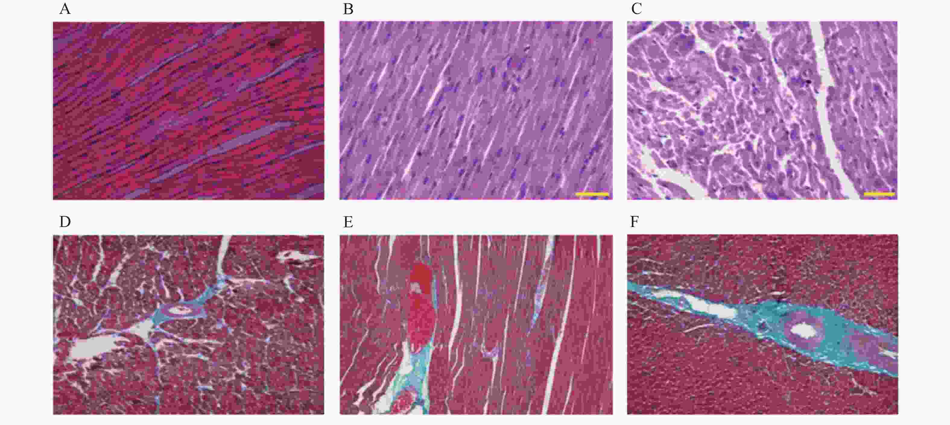
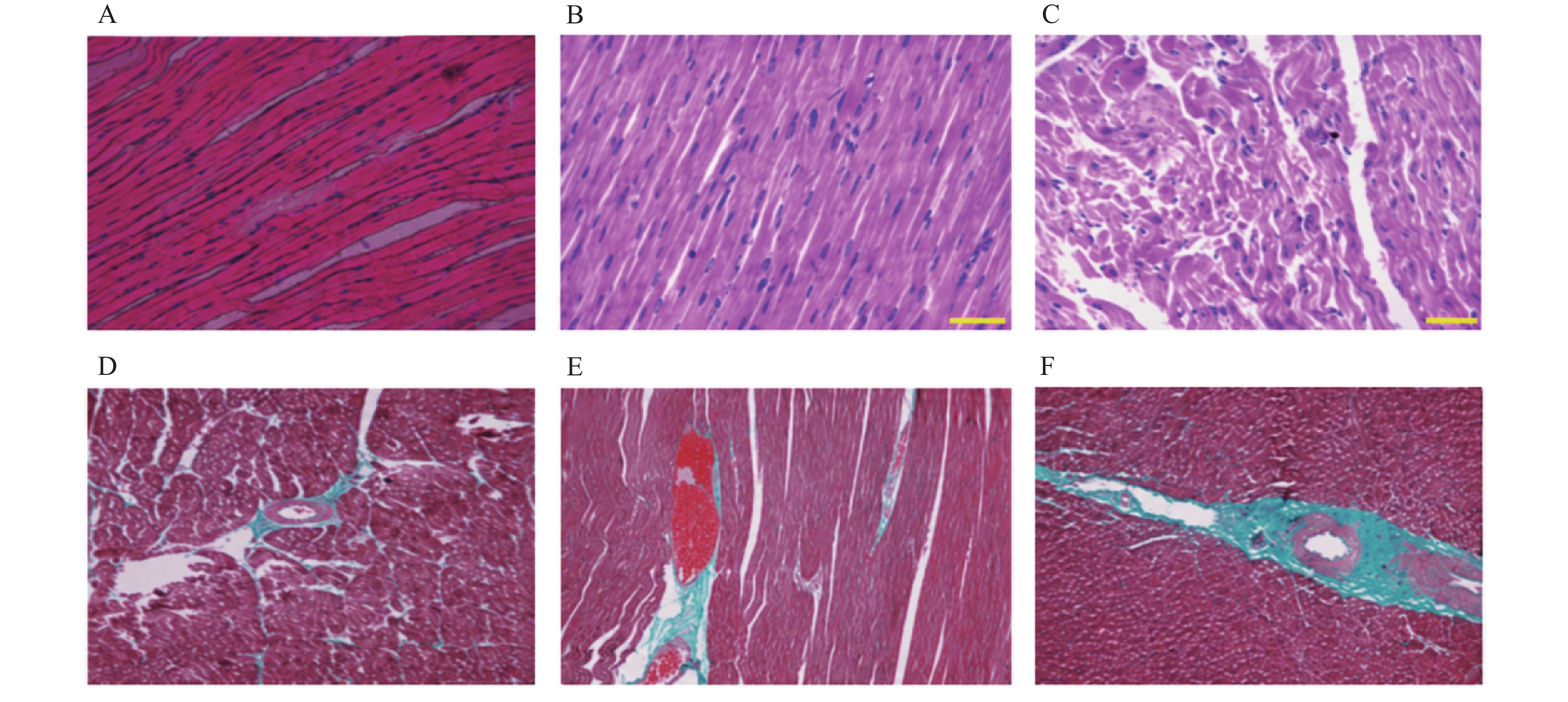
图 1 HE和Masson染色普通光学显微镜(×20)
A:正常组左室心肌组织HE染色,B:Losartan组左室心肌组织HE染色,C:LVH组左室心肌组织HE染色; D:正常组左室心肌组织Masson染色,E:Losartan组左室心肌组织Masson染色,F:LVH组左室心肌组织Masson染色。
Figure 1. HE and Masson staining under a conventional light microscope (×20)
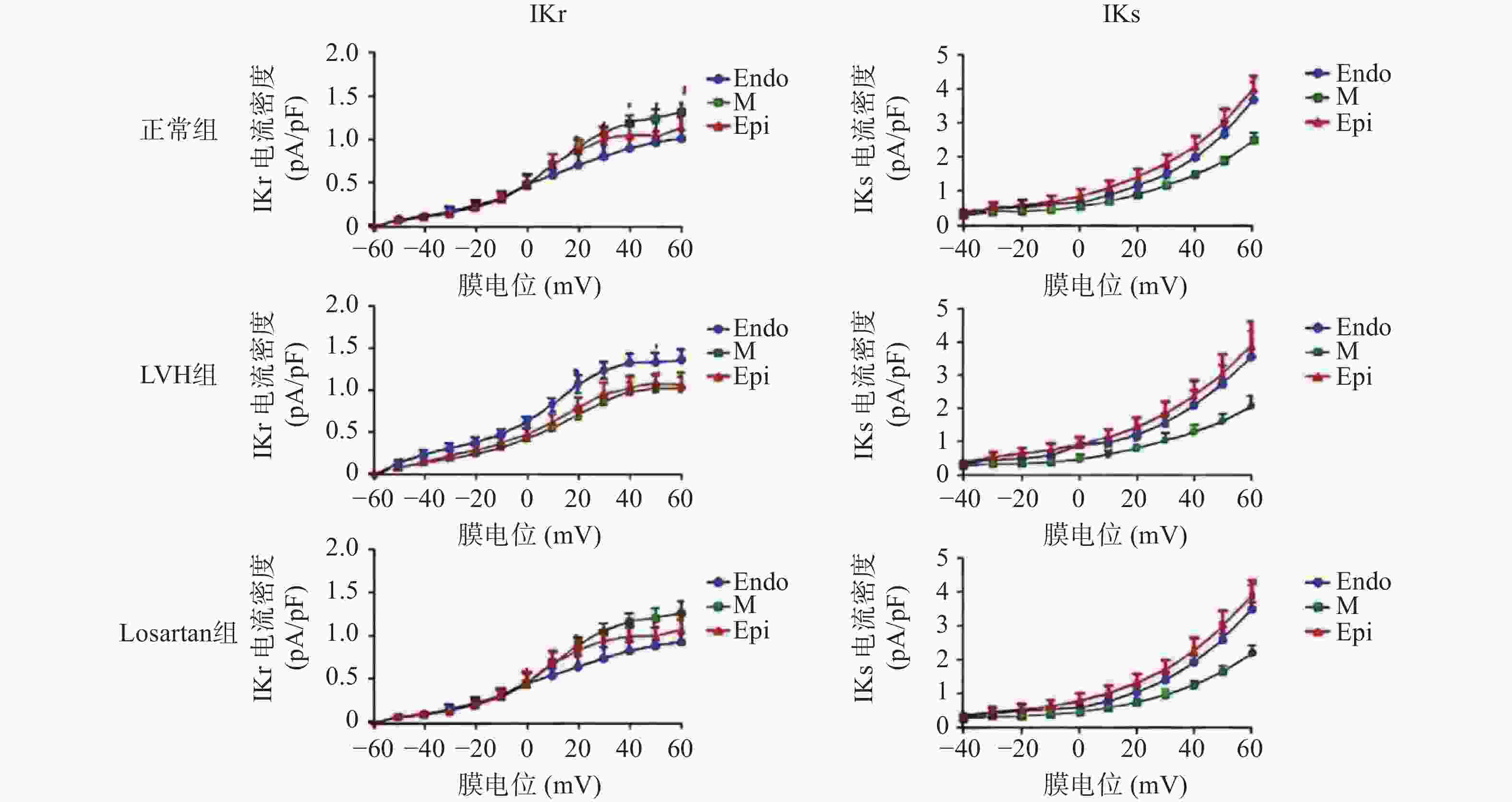
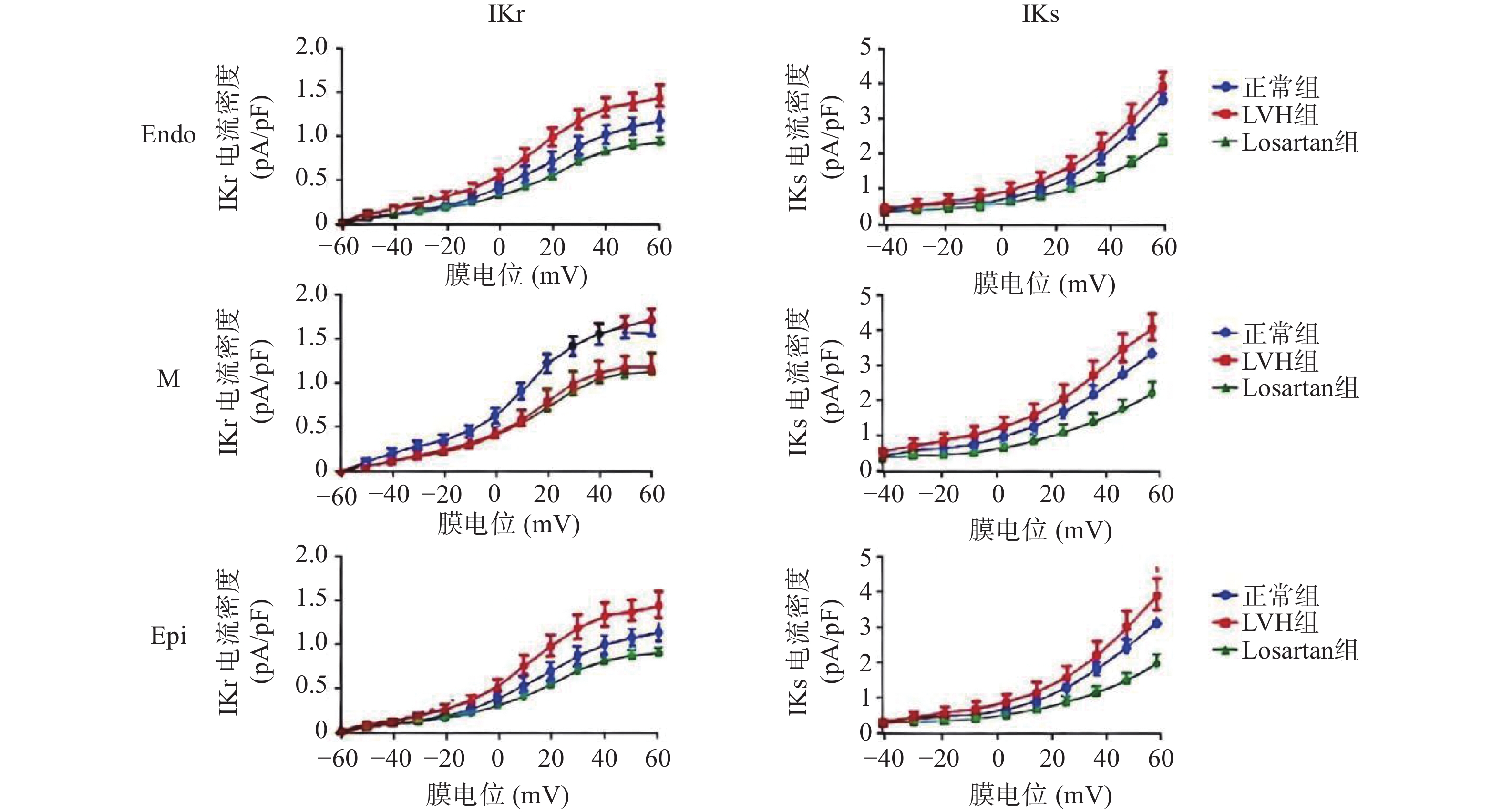
图 2 正常组、LVH组、Losartan组犬三层心肌细胞之间IKs、Ikr的I-V曲线
Figure 2. I-V curves of IKs and Ikr among three layers of cardiomyocytes in the normal group,LVH group,and losartan-treated group in dogs
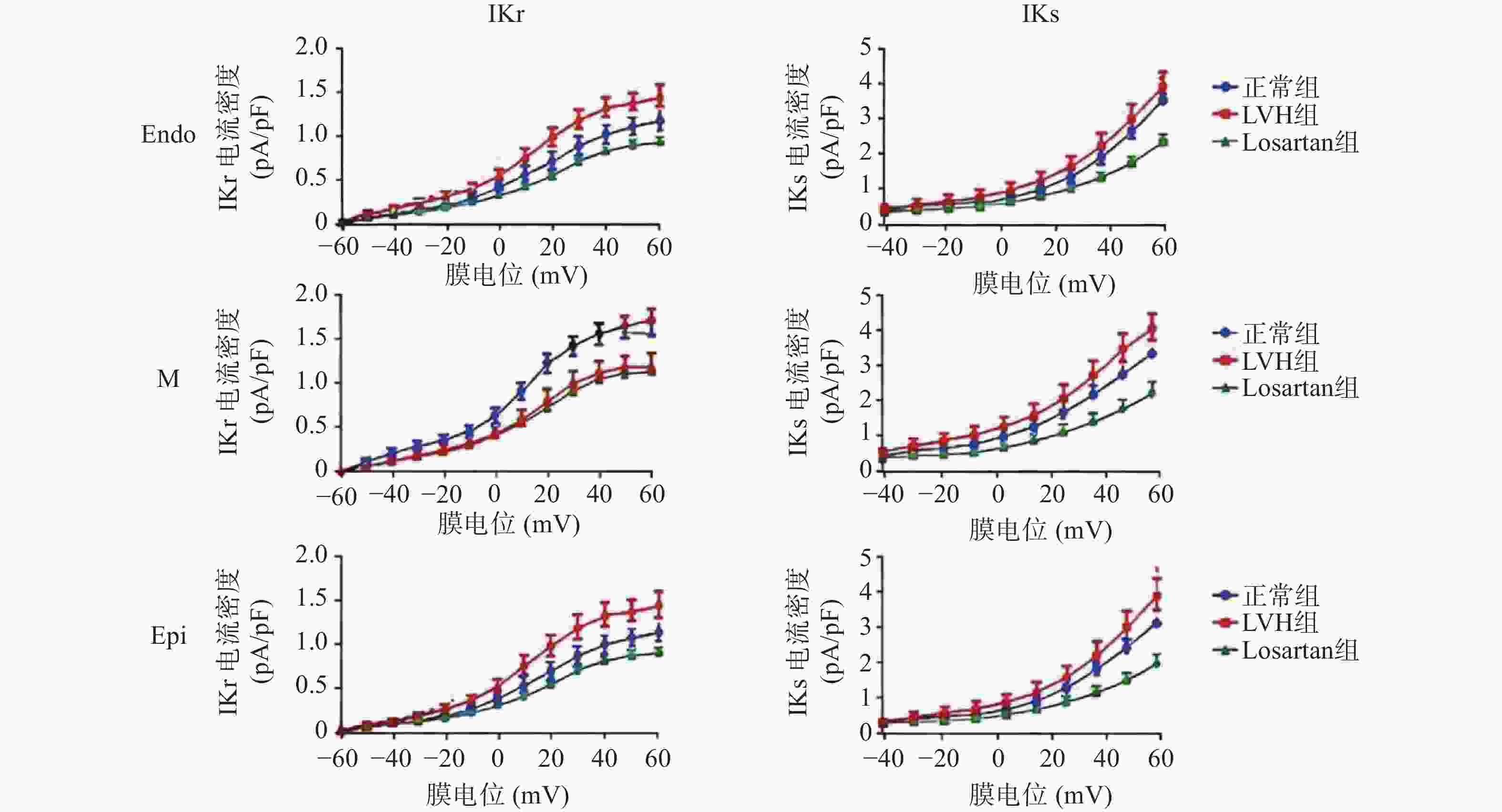
图 3 正常组、LVH组、Losartan组犬同一层心肌细胞IKs、Ikr的I-V曲线
Figure 3. I-V curves of IKs and Ikr in cardiomyocytes from the same layer of the heart in the normal group,LVH group,and losartan group in dogs
表 1 正常组、Losartan组和LVH组术后12周的血压值
Table 1. Blood pressure values at 12 weeks postoperatively in the normal group,the Losartan group,and the LVH group
指标 n 正常组 Losartan组 LVH组 F P SBP(mmHg) 5 119.5 ± 16.92 125.7 ± 22.54 170.0 ±12.84**▲▲ 11.862 <0.001*** DBP(mmHg) 5 76.99 ± 15.54 82.53 ± 19.82 119.5 ±16.92**▲▲ 8.702 0.005** 注:LVH vs 正常组:*P < 0.05,**P < 0.01;LVH vs Losartan组:▲▲P < 0.01。  下载: 导出CSV
表 2 正常组、Losartan组和LVH组术前和术后12周超声心动图检测的指标
Table 2. Echocardiographic parameters in the normal group,the Losartan group,and the LVH group before surgery and at 12 weeks postoperatively
指标 n 时间 正常组 Losartan组 LVH组 F P LVPWd(cm) 5 术前 0.70 ± 0.06 0.70 ± 0.06 0.75 ± 0.12 0.579 0.576 5 术后12周 0.75 ± 0.12 0.78 ± 0.02 1.02 ± 0.06*▲ 17.853 <0.001*** LVPWs(cm) 5 术前 1.45 ± 0.44 1.43 ± 0.42 1.16 ± 0.05 1.056 0.378* 5 术后12周 1.44 ± 0.41 1.45 ± 0.44 1.48 ± 0.19* 0.016 0.984 IVSTd(cm) 5 术前 0.69 ± 0.04 0.69 ± 0.05 0.69 ± 0.02 0.000 1.000 5 术后12周 0.69 ± 0.02 0.76 ± 0.02 0.83 ± 0.02**▲ 61.25 <0.001*** IVSTs(cm) 5 术前 0.98 ± 0.13 0.99 ± 0.14 1.03 ± 0.06 0.262 0.774 5 术后12周 1.03 ± 0.06 1.08 ± 0.06 1.30 ± 0.14*▲ 11.549 0.002** LVEDd(cm) 5 术前 2.85 ± 0.23 2.85 ± 0.23 2.70 ± 0.09 0.988 0.401 5 术后12周 2.70 ± 0.09 3.39 ± 0.59 2.52 ± 0.36 6.512 0.012* LVEDs(cm) 5 术前 1.98 ± 0.23 2.04 ± 0.27 1.76 ± 0.32 1.429 0.278 5 术后12周 1.98 ± 0.20 2.00 ± 0.37 1.97 ± 0.31 0.013 0.987 FS 5 术前 0.30 ± 0.13 0.29 ± 0.08 0.38 ± 0.07 1.294 0.31 5 术后12周 0.27 ± 0.07 0.40 ± 0.01 0.21 ± 0.02▲ 26.204 <0.001*** 注:LVH组:术后vs术前:*P < 0.05,**P < 0.01;LVH术后vs Losartan组:▲P < 0.05。
下载: 导出CSV
表 3 左心室组织病理学定量分析
Table 3. Quantitative histopathological analysis of the left ventricle
组别 n 心肌细胞横截面积(μm2) 胶原容积分数CVF(%) 血管周围纤维化面积(PVF,%) Control组 5 278.6 ± 19.4 2.84 ± 0.52 3.27 ± 0.68 LVH组 5 463.8 ± 34.7** 8.91 ± 1.07** 10.84 ± 1.26** Losartan组 5 338.5 ± 26.3▲▲ 5.13 ± 0.79▲▲ 6.42 ± 0.93▲▲ F — 58.961 69.11 74.414 P — <0.001*** <0.001*** <0.001*** 注:LVH组vs 正常组:**P < 0.01;LVH组vs Losartan组:▲▲P < 0.01。
下载: 导出CSV
表 4 三组犬左心室不同层心肌细胞动作电位复极90%时程(APD90)的比较
Table 4. Comparison of action potential duration at 90% repolarization (APD90) among different myocardial layers and groups
组别 n Endo(ms) M(ms) Epi(ms) F P 正常组 5 257.20 ± 34.24 310.20 ± 37.18* 249.00 ± 44.48▲ 3.733 0.055 LVH组 5 287.50 ± 56.63 518.90 ± 98.47** 414.30 ± 118.60# 7.399 0.008 Losartan组 5 223.20 ± 75.26 276.80 ± 127.50 237.00 ± 93.52 0.355 0.709 F — 1.545 9.446 5.94 — — P — 0.253 0.003** 0.016* — — 注:M层vs Endo层:**P < 0.01;M层 vs Epi层:▲P < 0.05;Endo层 vs Epi层:#P < 0.05。
下载: 导出CSV
表 5 三组犬左心室不同层心肌细胞Ikr、Iks电流密度比较
Table 5. Comparison of Ikr and Iks current densities in myocardial cells from different layers of the left ventricle in three canine groups
指标 组别 n Endo M Epi F P Ikr 正常组 5 0.85 ± 0.64 1.23 ± 0.17 1.05 ± 0.15 1.157 0.347 Losartan组 5 1.48 ± 0.19 1.15 ± 0.27* 1.19 ± 0.33 2.16 0.158 LVH组 5 0.9233 ±0.2282 0.8457 ±0.3671 0.95 ± 0.15 0.214 0.81 F — 3.565 2.599 1.421 — — P — 0.061 0.115 0.279 — — Iks 正常组 5 3.59 ± 0.92 2.38 ± 0.40* 3.76 ± 0.84▲▲ 4.931 0.027 Losartan组 5 3.46 ± 1.23 2.15 ± 0.88* 2.78 ± 0.90 2.057 0.171 LVH组 5 2.90 ± 1.50 1.70 ± 0.70* 3.09 ± 1.55▲ 1.647 0.233 F — 0.43 1.249 0.948 — — P — 0.66 0.322 0.415 — — 注:M vs Endo层:*P < 0.05;M vs Epi层:▲P < 0.05,▲▲P < 0.01。
下载: 导出CSV
表 6 三组犬同一层心肌细胞之间的Ikr、Iks电流变化
Table 6. Changes in Ikr and Iks currents among cardiomyocytes of the same layer across the three canine groups
指标 心肌层 n 正常组 LVH组 Losartan组 F P Ikr Endo 5 0.85 ± 0.64 0.92 ± 0.32 1.48 ± 0.19*# 3.241 0.075 M 5 1.23 ± 0.17 0.85 ± 0.37* 1.15 ± 0.27▲ 2.599 0.115 Epi 5 1.05 ± 0.15 0.97 ± 0.37 1.27 ± 0.28 1.481 0.266 F — 1.157 0.152 2.216 — — P — 0.347 0.86 0.152 — — Iks Endo 5 3.59 ± 0.92 2.90 ± 1.50 3.46 ± 1.23 0.43 0.66 M 5 2.38 ± 0.40 1.70 ± 0.70* 2.15 ± 0.89▲ 1.249 0.322 Epi 5 3.76 ± 0.84 3.09 ± 1.56 2.97 ± 0.87 0.701 0.515 F — 4.931 1.647 2.134 — — P — 0.027* 0.233 0.161 — — 注:LVH vs 正常组:*P < 0.05;LVH vs Losartan组:▲P < 0.05;Losartan vs 正常组:#P < 0.05。
下载: 导出CSV
-
[1] Alexander P M A, Nugent A W, Daubeney P E F, et al. Long-term outcomes of hypertrophic cardiomyopathy diagnosed during childhood: Results from a National Population-Based Study[J]. Circulation, 2018, 138(1): [2] Norrish G, Field E, McLeod K, et al. Clinical presentation and survival of childhood hypertrophic cardiomyopathy: A retrospective study in United Kingdom[J]. Eur Heart J, 2019, 40(12): 986-993. doi: 10.1093/eurheartj/ehy798 [3] Teekakirikul P, Padera R F, Seidman J G, et al. Hypertrophic cardiomyopathy: Translating cellular cross talk into therapeutics[J]. J Cell Biol, 2012, 199(3): 417-421. doi: 10.1083/jcb.201207033 [4] Park H, Kim H K, Jeong M H, et al. Clinical impacts of inhibition of renin–angiotensin system in patients with acute ST-segment elevation myocardial infarction who underwent successful late percutaneous coronary intervention[J]. J Cardiol, 2017, 69(1): 216-221. doi: 10.1016/j.jjcc.2016.03.012 [5] Faria-Costa G, Leite-Moreira A, Henriques-Coelho T. Cardiovascular effects of the angiotensin type 2 receptor[J]. Rev Portuguesa De Cardiol Engl Ed, 2014, 33(7-8): 439-449. doi: 10.1016/j.repc.2014.02.011 [6] Antzelevitch C, Di Diego J M. Tpeak-Tend interval as a marker of arrhythmic risk[J]. Heart Rhythm, 2019, 16(6): 954-955. doi: 10.1016/j.hrthm.2019.01.017 [7] Tashiro N, Muneuchi J, Ezaki H, et al. Ventricular repolarization dispersion is a potential risk for the development of life-threatening arrhythmia in children with hypertrophic cardiomyopathy[J]. Pediatr Cardiol, 2022, 43(7): 1455-1461. doi: 10.1007/s00246-022-02867-3 [8] Lei M, Salvage SC, Jackson AP, et al. Cardiac arrhythmogenesis: roles of ion channels and their functional modification[J]. Frontiers in Physiology, 2024, 15: 1342761. doi: 10.3389/fphys.2024.1342761 [9] Maron B J. Clinical course and management of hypertrophic cardiomyopathy[J]. N Engl J Med, 2018, 379(7): 655-668. doi: 10.1056/NEJMra1710575 [10] Maron B J, Doerer J J, Haas T S, et al. Sudden deaths in young competitive athletes: Analysis of 1866 deaths in the United States, 1980-2006[J]. Circulation, 2009, 119(8): 1085-1092. doi: 10.1161/CIRCULATIONAHA.108.804617 [11] Marian A J, Braunwald E. Hypertrophic cardiomyopathy: Genetics, pathogenesis, clinical manifestations, diagnosis, and therapy[J]. Circ Res, 2017, 121(7): 749-770. doi: 10.1161/CIRCRESAHA.117.311059 [12] Sayer G, Bhat G. The renin-angiotensin-aldosterone system and heart failure[J]. Cardiol Clin, 2014, 32(1): 21-32, vii. doi: 10.1016/j.ccl.2013.09.002 [13] Shimizu M, Ino H, Okeie K, et al. T-peak to T-end interval may be a better predictor of high-risk patients with hypertrophic cardiomyopathy associated with a cardiac troponin I mutation than QT dispersion[J]. Clin Cardiol, 2002, 25(7): 335-339. doi: 10.1002/clc.4950250706 [14] 李峰, 冯耀光. 大鼠心肌肥厚模型概述[J]. 中国现代医生, 2010, 48(32): 6-7, 11. doi: 10.3969/j.issn.1673-9701.2010.32.004 [15] 田作军, 陈长兵, 廖海星, 等. 肾血管性高血压犬中血管紧张素Ⅱ的肾内作用机制探讨[J]. 广东医学, 2013, 34(2): 191-193. doi: 10.3969/j.issn.1001-9448.2013.02.010 [16] 孙琪, 黄荣桂, 郑兴中, 等. 一肾一夹肾血管性高血压动物模型的制作[J]. 临床军医杂志, 2007, 35(6): 812-814. doi: 10.3969/j.issn.1671-3826.2007.06.002 [17] 田作军, 陈长兵, 廖海星, 等. 肾血管性高血压犬中血管紧张素Ⅱ的肾内作用机制探讨[J]. 广东医学, 2013, 34(2): 191-193. [18] Liu D W, Antzelevitch C. Characteristics of the delayed rectifier current (IKr and IKs) in canine ventricular epicardial, midmyocardial, and endocardial myocytes. A weaker IKs contributes to the longer action potential of the M cell[J]. Circ Res, 1995, 76(3): 351-365. doi: 10.1161/01.RES.76.3.351 [19] Flaim S N, Giles W R, McCulloch A D. Contributions of sustained INa and IKv43 to transmural heterogeneity of early repolarization and arrhythmogenesis in canine left ventricular myocytes[J]. Am J Physiol Heart Circ Physiol, 2006, 291(6): H2617-H2629. doi: 10.1152/ajpheart.00350.2006 [20] Viswanathan P C, Shaw R M, Rudy Y. Effects of IKr and IKs heterogeneity on action potential duration and its rate dependence: A simulation study[J]. Circulation, 1999, 99(18): 2466-2474. doi: 10.1161/01.CIR.99.18.2466 [21] Sutherland F J, Shattock M J, Baker K E, et al. Mouse isolated perfused heart: Characteristics and cautions[J]. Clin Exp Pharma Physio, 2003, 30(11): 867-878. doi: 10.1046/j.1440-1681.2003.03925.x [22] 李泱, 马杰, 肖建民, 等. 兔左室壁三层心肌细胞的分离及动作电位、钙和钾电流分布的异质性[J]. 生理学报, 2002, 54(5): 369-374. doi: 10.3321/j.issn:0371-0874.2002.05.003 [23] Yan G X, Rials S J, Wu Y, et al. Ventricular hypertrophy amplifies transmural repolarization dispersion and induces early afterdepolarization[J]. Am J Physiol Heart Circ Physiol, 2001, 281(5): H1968-H1975. doi: 10.1152/ajpheart.2001.281.5.H1968 [24] Khokhlova A, Iribe G, Katsnelson L, et al. The effects of load on transmural differences in contraction of isolated mouse ventricular cardiomyocytes[J]. J Mol Cell Cardiol, 2018, 114: 276-287. doi: 10.1016/j.yjmcc.2017.12.001 [25] White E, Le Guennec J Y, Nigretto J M, et al. The effects of increasing cell length on auxotonic contractions; membrane potential and intracellular calcium transients in single Guinea-pig ventricular myocytes[J]. Exp Physiol, 1993, 78(1): 65-78. [26] Saint D A, Kelly D, MacKenzie L. The contribution of MEF to electrical heterogeneity and arrhythmogenesis[M]//Mechanosensitivity of the Heart. Dordrecht: Springer Netherlands, 2010: 275-300. [27] Zeng T, Bett G C, Sachs F. Stretch-activated whole cell currents in adult rat cardiac myocytes[J]. Am J Physiol Heart Circ Physiol, 2000, 278(2): H548-H557. doi: 10.1152/ajpheart.2000.278.2.H548 [28] Stones R, Calaghan S C, Billeter R, et al. Transmural variations in gene expression of stretch-modulated proteins in the rat left ventricle[J]. Pflügers Arch Eur J Physiol, 2007, 454(4): 545-549. doi: 10.1007/s00424-007-0237-z -

 点击查看大图
计量
- 文章访问数: 5
- HTML全文浏览量: 5
- PDF下载量: 0
- 被引次数: 0
