




The CX3CL1-CX3CR1 Axis Regulates Microglia-Neuron Interactions and Its Role in Neurological Disorders
-
摘要: 小胶质细胞是中枢神经系统的常驻免疫细胞,参与炎症调节、突触修剪、吞噬清除和神经营养支持等。神经元细胞表达的C-X3-C基序趋化因子配体1(C-X3-C motif chemokine ligand 1,CX3CL1)与小胶质细胞表面的C-X3-C基序趋化因子受体1(C-X3-C motif chemokine receptor 1,CX3CR1)共同构成调节两者间相互作用的重要信号系统。CX3CR1表达变化或功能改变可影响小胶质细胞的迁移、炎症反应、吞噬和突触修剪等效应,进而影响神经元存活、突触结构及神经网络功能。既往研究显示,该信号系统异常与孤独症谱系障碍、神经退行性疾病、脑缺血、低氧相关认知损伤和癫痫等疾病的发生和进展相关,且该信号系统受疾病类型、脑区及病程阶段等因素影响。围绕CX3CR1变化,综述神经元和小胶质细胞CX3CL1-CX3CR1信号系统的基本特征、相互作用机制及其在神经系统疾病中的变化,为进一步认识CX3CR1的病理作用和潜在干预价值提供参考。Abstract: Microglia are resident immune cells of the central nervous system, participating in inflammatory regulation, synaptic pruning, phagocytic clearance, and neurotrophic support. C-X3-C motif chemokine ligand 1 (CX3CL1) expressed by neurons and C-X3-C motif chemokine receptor 1 (CX3CR1) on the surface of microglia together constitute an important signaling system that regulates the interactions between the two cell types. Alterations in CX3CR1 expression or function can affect microglial migration, inflammatory responses, phagocytosis, and synaptic pruning, thereby influencing neuronal survival, synaptic structure, and neural network function. Previous studies have shown that abnormalities in this signaling system are associated with the onset and progression of disorders such as autism spectrum disorder, neurodegenerative diseases, cerebral ischemia, hypoxia-related cognitive impairment, and epilepsy, and that this signaling system is influenced by factors including disease type, brain region, and disease stage. Focusing on changes in CX3CR1, this review summarizes the basic characteristics of the neuronal-microglial CX3CL1-CX3CR1 signaling system, the mechanisms underlying their interactions, and its alterations in neurological diseases, with the aim of providing a reference for better understanding the pathological roles of CX3CR1 and its potential value as a therapeutic target.
-
Key words:
- CX3CR1 /
- CX3CL1 /
- Microglia /
- Neurons /
- Neuroinflammation /
- Neurological diseases
-
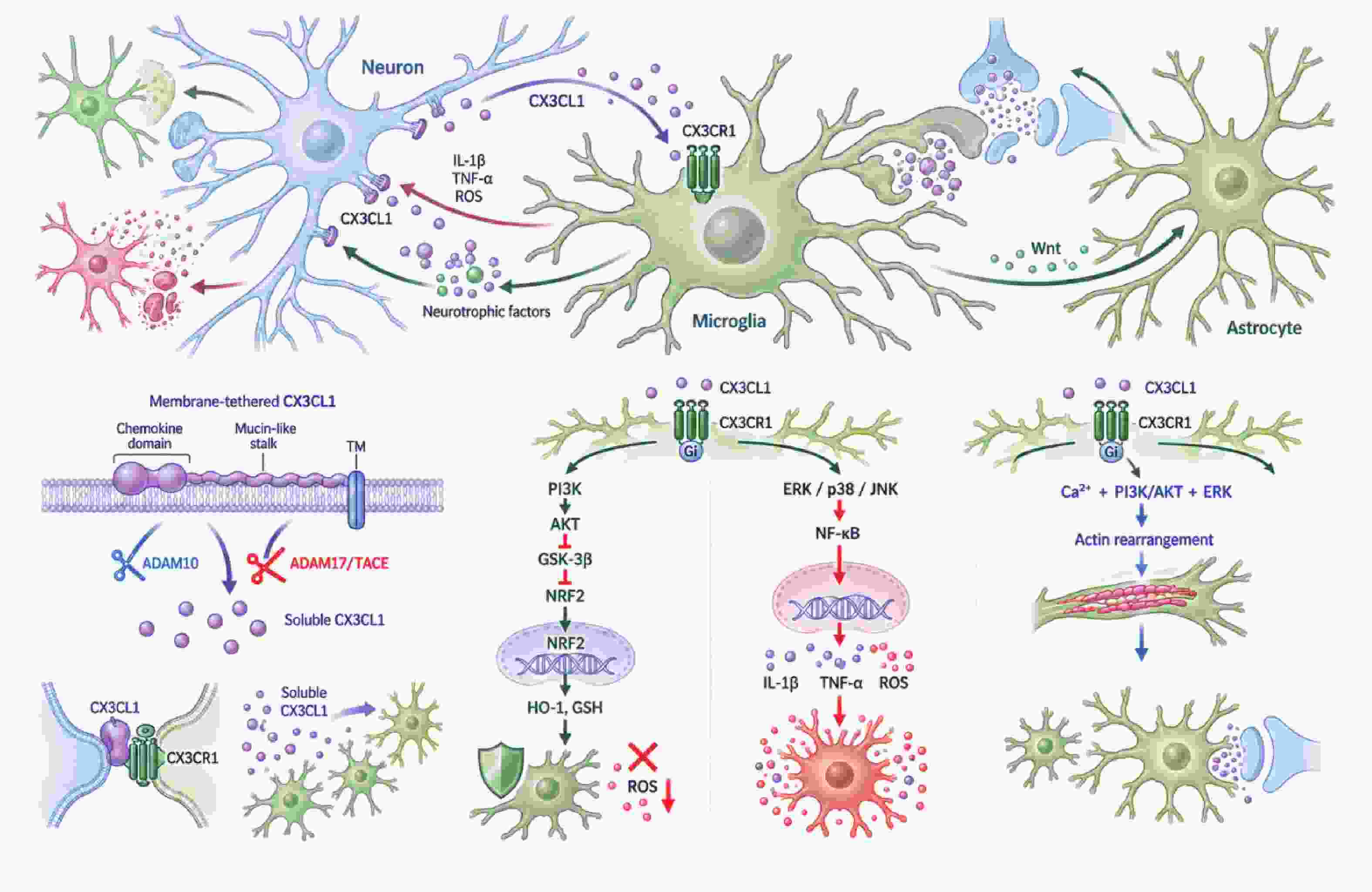
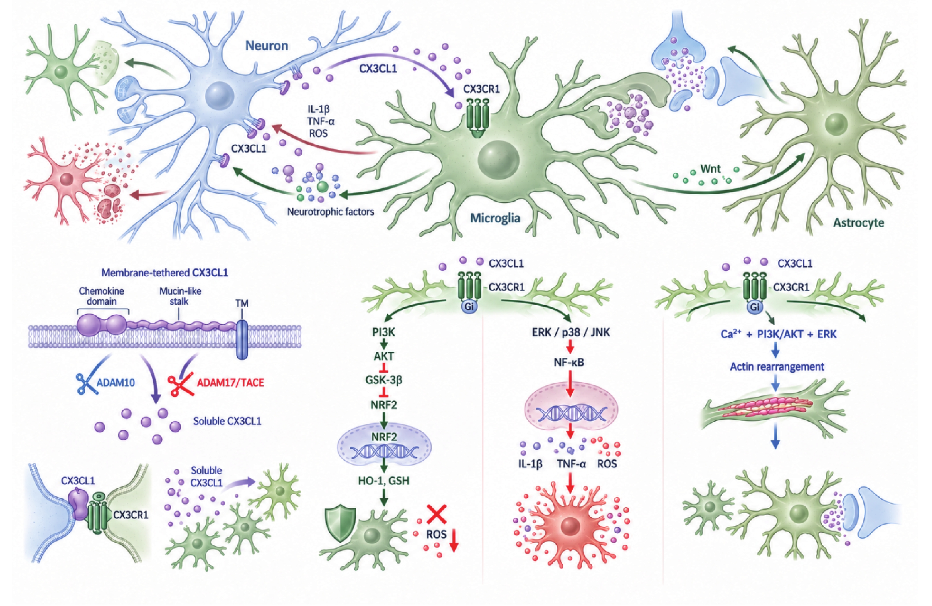
图 1 CX3CL1-CX3CR1信号调控小胶质细胞与神经元相互作用的机制
Figure 1. Mechanisms by which CX3CL1-CX3CR1 signaling regulates microglia-neuron interactions
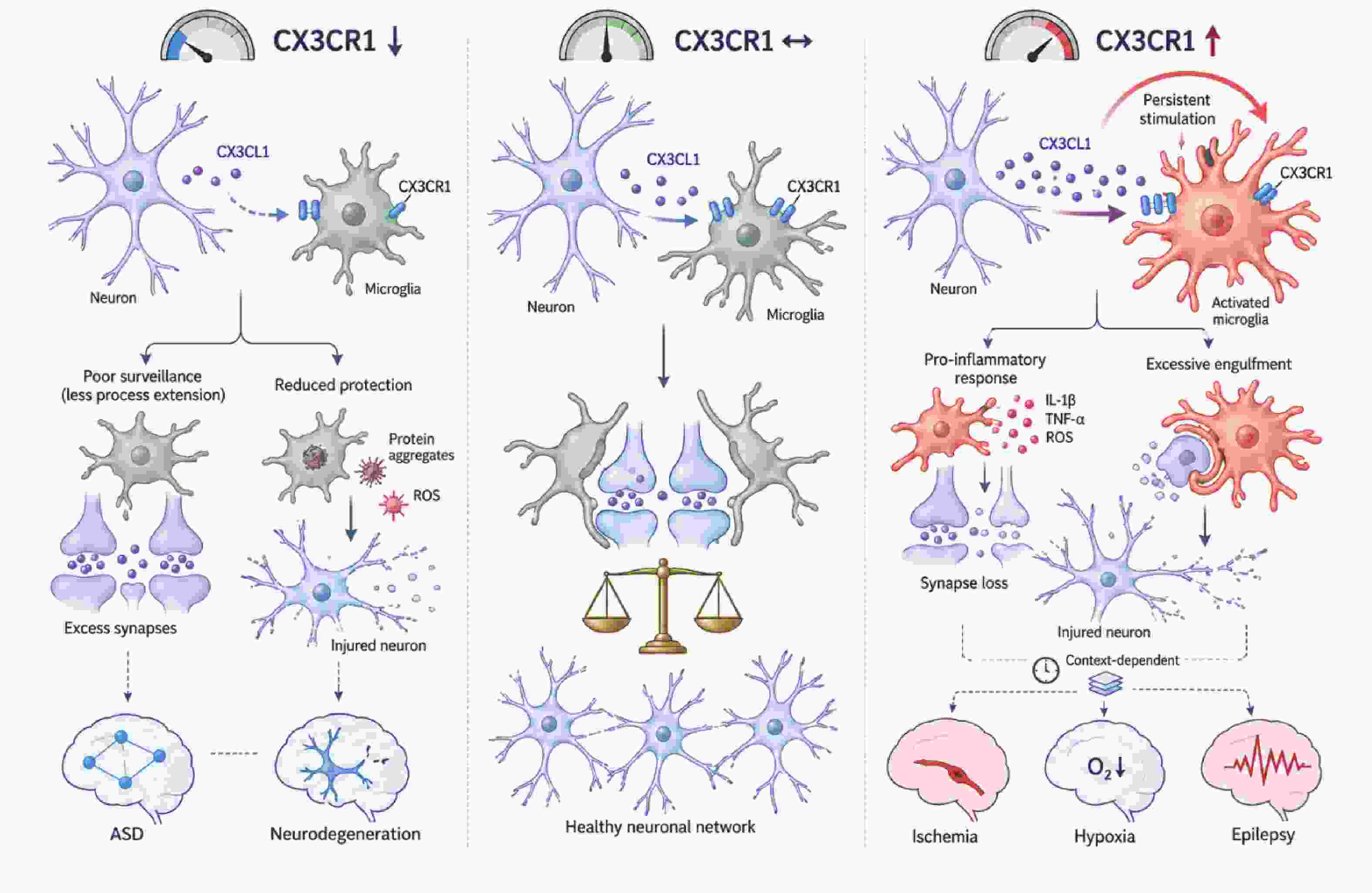
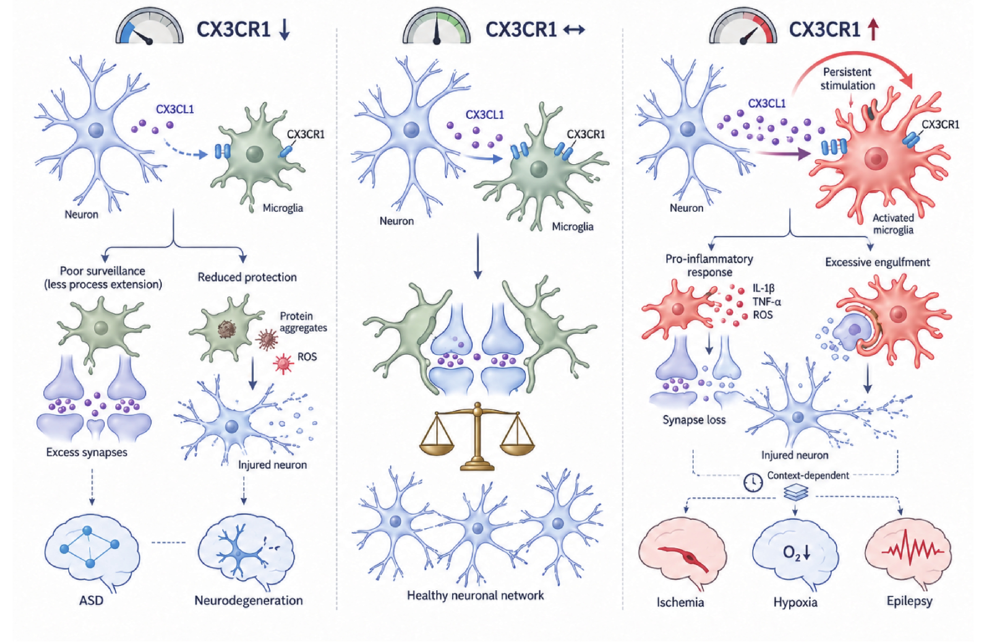
图 2 CX3CR1信号异常在疾病中的双面作用模式图
Figure 2. Dual roles of dysregulated CX3CR1 signaling in disease
表 1 不同神经相关疾病中CX3CR1变化及小胶质细胞–神经元结局汇总
Table 1. Alterations in CX3CR1 and microglia–neuron outcomes across different neurologically related diseases
疾病分类 模型类型 CX3CR1变化方向 小胶质细胞表型 神经元结局 参考文献 孤独症谱系障碍 Cx3cr1敲除小鼠(发育期) CX3CR1缺失 小胶质细胞数量暂时减少,突触募集与吞噬/修剪延迟 树突棘密度升高,突触传递与脑网络成熟异常 [19] 孤独症谱系障碍 Cx3cr1敲除小鼠 CX3CR1缺失 神经元—小胶质细胞信号交流受损 功能连接减弱,社会交往减少并出现重复行为 [29] 孤独症谱系障碍 人群遗传学及细胞功能实验 Ala55Thr罕见变异;功能降低 CX3CL1诱导的Akt磷酸化减弱,细胞应答受损 与孤独症谱系障碍表型风险相关,但变异罕见 [30] 孤独症谱系障碍 孕期丙戊酸暴露大鼠 海马CX3CR1及CX3CL1无明显变化 未发现可归因于该信号轴的明确改变 未支持CX3CR1改变是该模型的共同特征 [31] 阿尔茨海默病 淀粉样蛋白驱动的小鼠模型/Cx3cr1缺失 CX3CR1缺失 与Aβ斑块接触减少,Aβ吞噬及溶酶体酸化受损,炎症和氧化反应异常 Aβ负荷及Tau磷酸化增加,突触后结构和神经元减少,工作记忆恶化 [35] 阿尔茨海默病 Tau摄取实验及Cx3cr1缺失模型 CX3CR1缺失或减少 小胶质细胞对细胞外Tau的内化能力下降 Tau清除受损,可能促进Tau病理积累 [36] 阿尔茨海默病 晚发性阿尔茨海默病人群 CX3CR1-V249I功能变异 未直接评价小胶质细胞表型 与神经原纤维病理进展相关 [37] 帕金森病 α-突触核蛋白A53T小鼠/Cx3cr1缺失 CX3CR1缺失 小胶质细胞活化及神经炎症增强 黑质多巴胺能神经元丢失加重 [38] 肌萎缩侧索硬化症 SOD1G93A小鼠/Cx3cr1缺失 CX3CR1缺失 小胶质细胞活化增强,异常SOD1聚集增加 运动神经元丢失及终末期疾病表现加重 [39] 肌萎缩侧索硬化症 SOD1G93A小鼠(症状期) CX3CL1降低、CX3CR1升高 促炎反应增强;受体升高可能为代偿性变化 运动神经元减少 [40] 脑缺血/脑卒中 实验性缺血性脑卒中小鼠/Cx3cr1信号缺失 CX3CR1信号缺失 小胶质细胞焦亡及炎症反应受到抑制 卒中后认知功能恢复改善 [42] 脑缺血/脑卒中 脑缺血模型及神经元培养 缺血诱导神经元CX3CR1升高 该研究重点为神经元CX3CR1,未直接界定小胶质细胞表型 CX3CR1介导Ca2+内流和兴奋性毒性;敲除或中和后神经元死亡减少 [43] 脑缺血/脑卒中 全脑缺血及低氧后处理小鼠 胶质细胞CX3CR1降低;CX3CL1相关保护信号并存 海马CA1区小胶质细胞活化及TNF-α、IL-1β表达减少 CA1神经元存活增加;作用具有细胞来源和时相差异 [44] 低氧相关认知损伤 高海拔/低压低氧暴露小鼠及体外低氧模型 CX3CL1-CX3CR1信号增强 CD16、IL-1β表达及海马CA1突触吞噬增加;Cx3cr1缺失可减轻上述改变 PSD95等突触成分减少、突触丢失并出现记忆损害 [45] 癫痫 海人酸诱导癫痫持续状态;CX3CR1拮抗剂AZD8797干预 发作后CX3CR1相关信号增强;药理性抑制 拮抗CX3CR1后小胶质细胞活化及炎症反应减轻 海马神经元损伤减轻;作用可能随发作后阶段变化 [46] 注:CX3CR1表达变化不等同于受体功能变化;同一疾病中的作用可随模型、受累细胞、脑区及病程阶段而改变。Aβ,淀粉样β蛋白;ROS,活性氧。  下载: 导出CSV
-
[1] Salter M W, Stevens B. Microglia emerge as central players in brain disease[J]. Nat Med, 2017, 23(9): 1018-1027. doi: 10.1038/nm.4397 [2] Zhu H, Guan A, Liu J, et al. Noteworthy perspectives on microglia in neuropsychiatric disorders[J]. J Neuroinflammation, 2023, 20: 223. doi: 10.1186/s12974-023-02901-y [3] Colonna M, Butovsky O. Microglia function in the central nervous system during health and neurodegeneration[J]. Annu Rev Immunol, 2017, 35: 441-468. doi: 10.1146/annurev-immunol-051116-052358 [4] Zhou M, Cornell J, Salinas S, et al. Microglia regulation of synaptic plasticity and learning and memory[J]. Neural Regen Res, 2022, 17(4): 705. doi: 10.4103/1673-5374.322423 [5] Nimmerjahn A, Kirchhoff F, Helmchen F. Resting microglial cells are highly dynamic surveillants of brain parenchyma in vivo[J]. Science, 2005, 308(5726): 1314-1318. doi: 10.1126/science.1110647 [6] McNamara N B, Munro D A D, Bestard-Cuche N, et al. Microglia regulate central nervous system myelin growth and integrity[J]. Nature, 2023, 613(7942): 120-129. doi: 10.1038/s41586-022-05534-y [7] Lin S, Yin Q, Zhong Q, et al. Heme activates TLR4-mediated inflammatory injury via MyD88/TRIF signaling pathway in intracerebral hemorrhage[J]. Journal of Neuroinflammation, 2012, 9: 46. doi: 10.1186/1742-2094-9-46 [8] Ulland T K, Song WM, Huang SC, et al. TREM2 Maintains Microglial Metabolic Fitness in Alzheimer's Disease [J]. Cell, 2017, 170(4): 649-663. e13. [9] Wang S, Sudan R, Peng V, et al. TREM2 drives microglia response to amyloid-β via SYK-dependent and -independent pathways [J]. Cell, 2022, 185(22): 4153-4169. e19. [10] Keren-Shaul H, Spinrad A, Weiner A, et al. A Unique Microglia Type Associated with Restricting Development of Alzheimer's Disease [J]. Cell, 2017, 169(7): 1276-1290. e17. [11] Masuda T, Sankowski R, Staszewski O, et al. Spatial and temporal heterogeneity of mouse and human microglia at single-cell resolution[J]. Nature, 2019, 566(7744): 388-392. doi: 10.1038/s41586-019-0924-x [12] Camacho-Hernández N P, Peña-Ortega F. Fractalkine/CX3CR1-dependent modulation of synaptic and network plasticity in health and disease[J]. Neural Plast, 2023, 2023: 4637073. doi: 10.1155/2023/4637073 [13] Harrison J K, Jiang Y, Chen S, et al. Role for neuronally derived fractalkine in mediating interactions between neurons and CX3CR1-expressing microglia[J]. Proc Natl Acad Sci U S A, 1998, 95(18): 10896-10901. doi: 10.1073/pnas.95.18.10896 [14] Maciejewski-Lenoir D, Chen S, Feng L, et al. Characterization of fractalkine in rat brain cells: Migratory and activation signals for CX3CR-1-expressing microglia[J]. J Immunol, 1999, 163(3): 1628-1635. [15] Gunner G, Cheadle L, Johnson K M, et al. Sensory lesioning induces microglial synapse elimination via ADAM10 and fractalkine signaling[J]. Nat Neurosci, 2019, 22(7): 1075-1088. doi: 10.1038/s41593-019-0419-y [16] Rodriguez D, Church K A, Pietramale A N, et al. Fractalkine isoforms differentially regulate microglia-mediated inflammation and enhance visual function in the diabetic retina[J]. J Neuroinflammation, 2024, 21(1): 42. doi: 10.1186/s12974-023-02983-8 [17] Eugenín J, Eugenín-von Bernhardi L, von Bernhardi R. Age-dependent changes on fractalkine forms and their contribution to neurodegenerative diseases[J]. Front Mol Neurosci, 2023, 16: 1249320. doi: 10.3389/fnmol.2023.1249320 [18] Mizuno T, Kawanokuchi J, Numata K, et al. Production and neuroprotective functions of fractalkine in the central nervous system[J]. Brain Res, 2003, 979(1-2): 65-70. doi: 10.1016/S0006-8993(03)02867-1 [19] Paolicelli R C, Bolasco G, Pagani F, et al. Synaptic pruning by microglia is necessary for normal brain development[J]. Science, 2011, 333(6048): 1456-1458. doi: 10.1126/science.1202529 [20] Rogers J T, Morganti J M, Bachstetter A D, et al. CX3CR1 deficiency leads to impairment of hippocampal cognitive function and synaptic plasticity[J]. J Neurosci, 2011, 31(45): 16241-16250. doi: 10.1523/JNEUROSCI.3667-11.2011 [21] Wang L, Ling H, He H, et al. Dysfunctional synaptic pruning by microglia correlates with cognitive impairment in sleep-deprived mice: Involvement of CX3CR1 signaling[J]. Neurobiol Stress, 2023, 25: 100553. doi: 10.1016/j.ynstr.2023.100553 [22] Faust T E, Lee Y H, O’Connor C D, et al. Microglia-astrocyte crosstalk regulates synapse remodeling via Wnt signaling [J]. Cell, 2025, 188(19): 5212-5230. e21. [23] Castro-Sánchez S, García-Yagüe Á J, Kügler S, et al. CX3CR1-deficient microglia shows impaired signalling of the transcription factor NRF2: Implications in tauopathies[J]. Redox Biol, 2019, 22: 101118. doi: 10.1016/j.redox.2019.101118 [24] Liu Y, Wu X M, Luo Q Q, et al. CX3CL1/CX3CR1-mediated microglia activation plays a detrimental role in ischemic mice brain via p38MAPK/PKC pathway[J]. J Cereb Blood Flow Metab, 2015, 35(10): 1623-1631. doi: 10.1038/jcbfm.2015.97 [25] Huang J M, Zhao N, Hao X N, et al. CX3CL1/CX3CR1 signaling mediated neuroglia activation is implicated in the retinal degeneration: A potential therapeutic target to prevent photoreceptor death[J]. Invest Ophthalmol Vis Sci, 2024, 65(1): 29. doi: 10.1167/iovs.65.1.29 [26] Bhaskar K, Konerth M, Kokiko-Cochran O N, et al. Regulation of tau pathology by the microglial fractalkine receptor[J]. Neuron, 2010, 68(1): 19-31. doi: 10.1016/j.neuron.2010.08.023 [27] Lyons A, Lynch A M, Downer E J, et al. Fractalkine-induced activation of the phosphatidylinositol-3 kinase pathway attentuates microglial activation in vivo and in vitro[J]. J Neurochem, 2009, 110(5): 1547-1556. doi: 10.1111/j.1471-4159.2009.06253.x [28] Hoshiko M, Arnoux I, Avignone E, et al. Deficiency of the microglial receptor CX3CR1 impairs postnatal functional development of thalamocortical synapses in the barrel cortex[J]. J Neurosci, 2012, 32(43): 15106-15111. doi: 10.1523/JNEUROSCI.1167-12.2012 [29] Zhan Y, Paolicelli R C, Sforazzini F, et al. Deficient neuron-microglia signaling results in impaired functional brain connectivity and social behavior[J]. Nat Neurosci, 2014, 17(3): 400-406. doi: 10.1038/nn.3641 [30] Ishizuka K, Fujita Y, Kawabata T, et al. Rare genetic variants in CX3CR1 and their contribution to the increased risk of schizophrenia and autism spectrum disorders[J]. Transl Psychiatry, 2017, 7(8): e1184. doi: 10.1038/tp.2017.173 [31] Xu X, Tan L, Zhang X. Prenatal exposure to valproic acid may alter CD200/CD200R signaling pathways in a rat model of autism spectrum disorder[J]. Alpha Psychiatry, 2025, 26(2): 39444. doi: 10.31083/AP39444 [32] 马自蓉, 张少川, 曹雪, 等. 阿尔茨海默病中神经炎症生物标志物及相关药物治疗研究进展[J]. 昆明医科大学学报, 2026, 47(2): 1-13. doi: 10.12259/j.issn.2095-610X.S20260201 [33] Subbarayan M S, Joly-Amado A, Bickford P C, et al. CX3CL1/CX3CR1 signaling targets for the treatment of neurodegenerative diseases[J]. Pharmacol Ther, 2022, 231: 107989. doi: 10.1016/j.pharmthera.2021.107989 [34] Bivona G, Iemmolo M, Ghersi G. CX3CL1 pathway as a molecular target for treatment strategies in Alzheimer’s disease[J]. Int J Mol Sci, 2023, 24(9): 8230. doi: 10.3390/ijms24098230 [35] Puntambekar S S, Moutinho M, Lin P B, et al. CX3CR1 deficiency aggravates amyloid driven neuronal pathology and cognitive decline in Alzheimer’s disease[J]. Mol Neurodegener, 2022, 17(1): 47. doi: 10.1186/s13024-022-00545-9 [36] Bolós M, Llorens-Martín M, Perea J R, et al. Absence of CX3CR1 impairs the internalization of Tau by microglia[J]. Mol Neurodegener, 2017, 12(1): 59. doi: 10.1186/s13024-017-0200-1 [37] López-López A, Gelpi E, Lopategui D M, et al. Association of the CX3CR1-V249I variant with neurofibrillary pathology progression in late-onset Alzheimer’s disease[J]. Mol Neurobiol, 2018, 55(3): 2340-2349. doi: 10.1007/s12035-017-0489-3 [38] Castro-Sánchez S, García-Yagüe Á J, López-Royo T, et al. Cx3cr1-deficiency exacerbates alpha-synuclein-A53T induced neuroinflammation and neurodegeneration in a mouse model of Parkinson’s disease[J]. Glia, 2018, 66(8): 1752-1762. doi: 10.1002/glia.23338 [39] Liu C, Hong K, Chen H, et al. Evidence for a protective role of the CX3CL1/CX3CR1 axis in a model of amyotrophic lateral sclerosis[J]. Biol Chem, 2019, 400(5): 651-661. doi: 10.1515/hsz-2018-0204 [40] Zhang J, Liu Y, Liu X, et al. Dynamic changes of CX3CL1/CX3CR1 axis during microglial activation and motor neuron loss in the spinal cord of ALS mouse model[J]. Transl Neurodegener, 2018, 7: 35. doi: 10.1186/s40035-018-0138-4 [41] 李智勇, 陈政刚, 彭俊, 等. miR-16-5p通过介导GPR30表达促进OGD模型小胶质细胞的炎症反应和凋亡[J]. 昆明医科大学学报, 2025, 46(10): 23-31. doi: 10.12259/j.issn.2095-610X.S20251003 [42] Ge Y, Yang J, Chen J, et al. Absence in CX3CR1 receptor signaling promotes post-ischemic stroke cognitive function recovery through suppressed microglial pyroptosis in mice[J]. CNS Neurosci Ther, 2024, 30(2): e14551. doi: 10.1111/cns.14551 [43] Wang J, Gan Y, Han P, et al. Ischemia-induced neuronal cell death is mediated by chemokine receptor CX3CR1[J]. Sci Rep, 2018, 8(1): 556. doi: 10.1038/s41598-017-18774-0 [44] Zhan L, Qiu M, Zheng J, et al. Fractalkine/CX3CR1 axis is critical for neuroprotection induced by hypoxic postconditioning against cerebral ischemic injury[J]. Cell Commun Signal, 2024, 22(1): 457. doi: 10.1186/s12964-024-01830-4 [45] Wang X, Xie Y, Niu Y, et al. CX3CL1/CX3CR1 signal mediates M1-type microglia and accelerates high-altitude-induced forgetting[J]. Front Cell Neurosci, 2023, 17: 1189348. doi: 10.3389/fncel.2023.1189348 [46] Shih H L, Cheng K H, Chen C H, et al. Upregulation of cathepsin S expression contributes to neuronal damage following kainic acid-induced status epilepticus[J]. Journal of Neurochemistry, 2025, 169(3): e70037. doi: 10.1111/jnc.70037 -

 点击查看大图
图(2) / 表(1)
计量
- 文章访问数: 4
- HTML全文浏览量: 2
- PDF下载量: 0
- 被引次数: 0
