The Analysis of 19 Cases of Odontoma Diagnosed by CBCT Imagining
-
摘要:
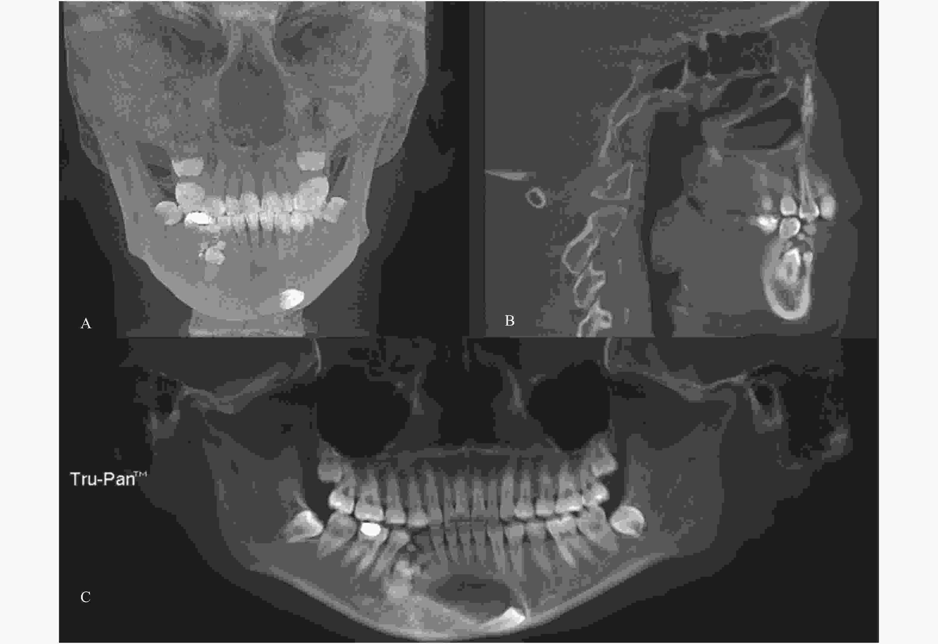
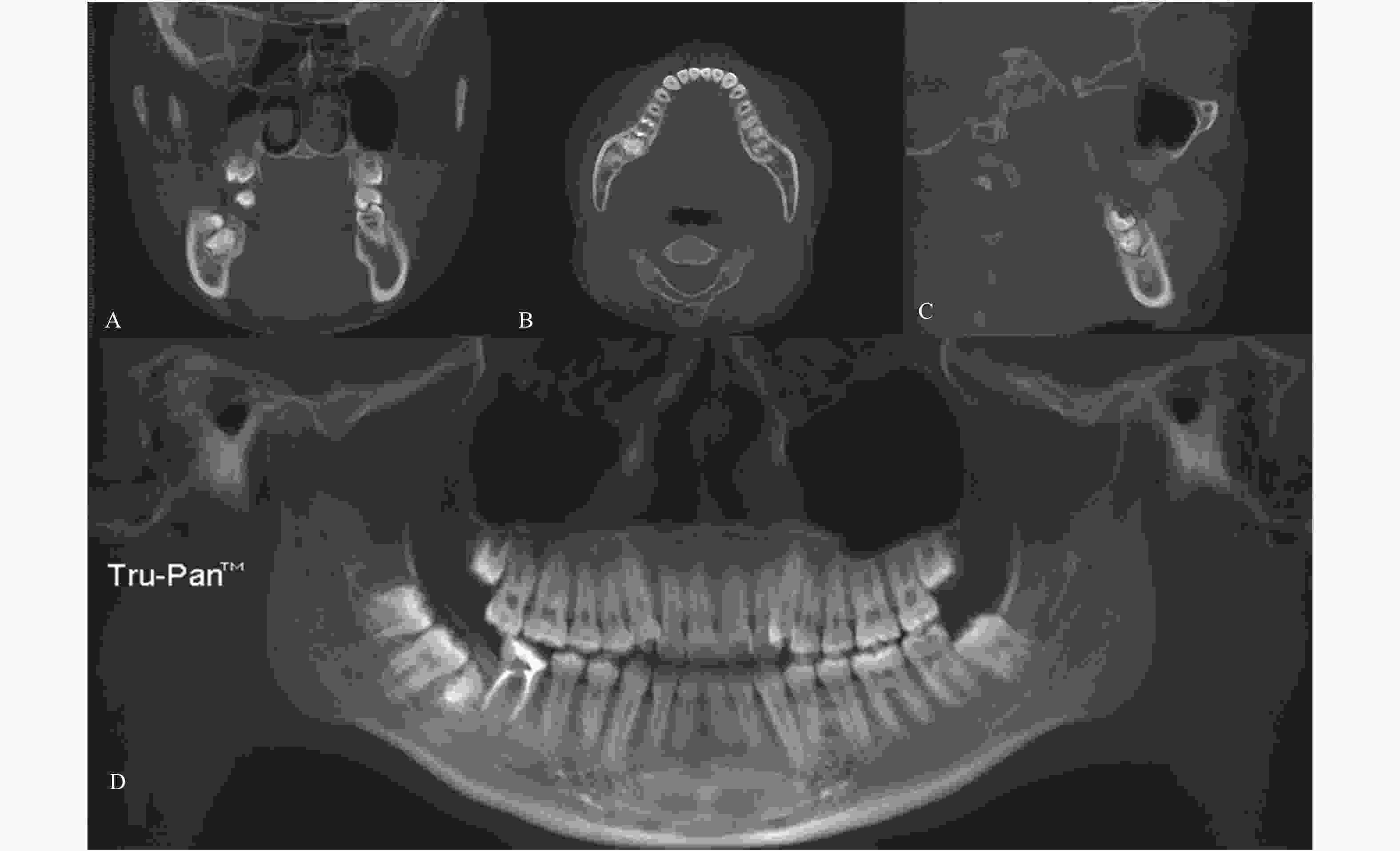
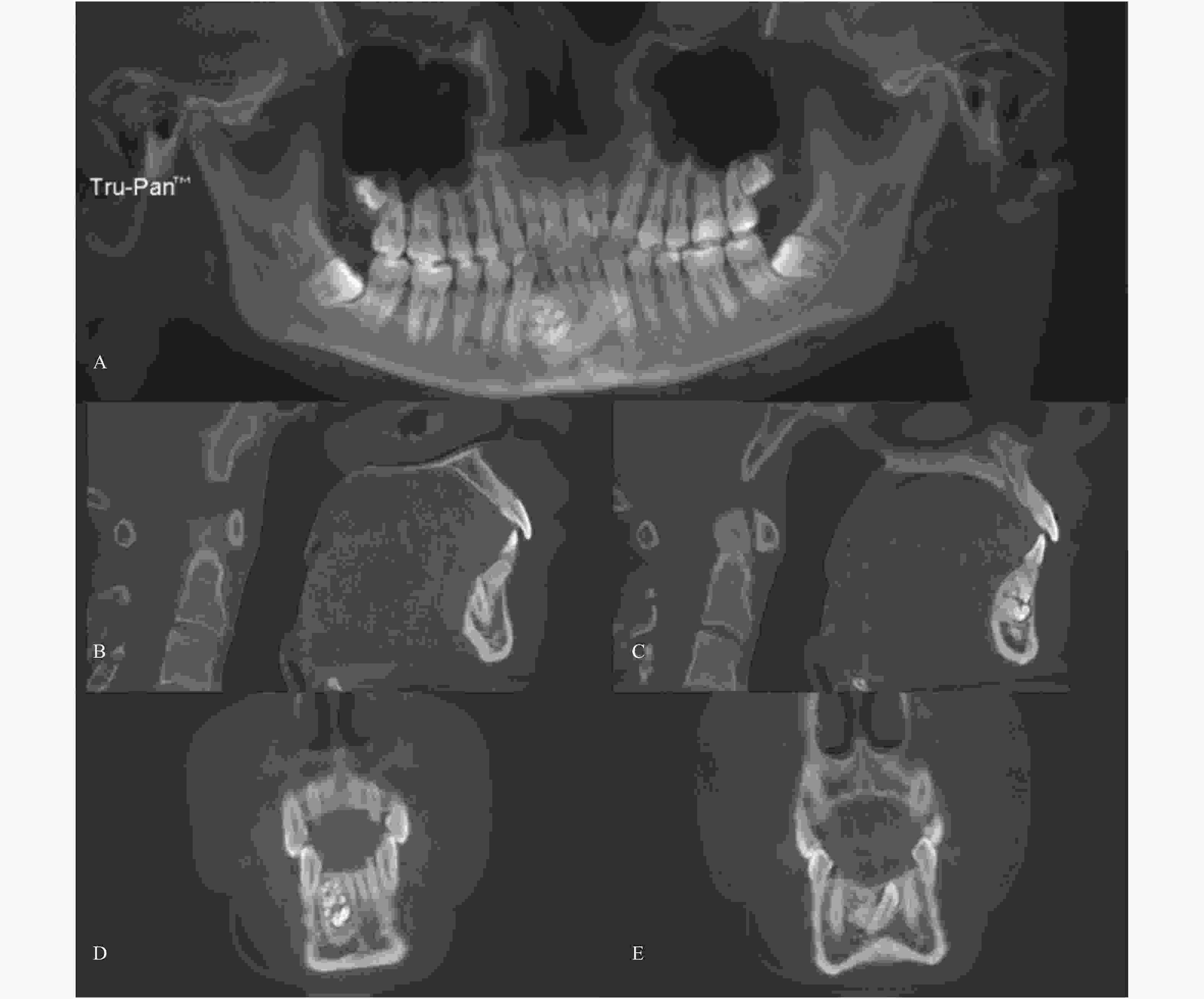
目的 通过探讨CBCT对牙瘤的诊疗价值来总结牙瘤的影像学特点,从而提高对牙瘤的临床认识。 方法 采用回顾性研究,收集2015年5月至2022年2月昆明市延安医院口腔科经CBCT( KavoiCAT 17-19,德国)诊断为牙瘤的19例门诊患者的影像资料,从牙瘤发生年龄、空间位置、牙位、类型等方面进行分析。 结果 19例牙瘤患者发生年龄为8~23岁,其中男性6例,女性13例。牙瘤的发生区域为上颌10例,下颌9例,前牙区16例,前磨牙区2例,磨牙区1例;发生牙位为上颌中切牙位4例,上颌侧切牙位3例,上颌尖牙位3例;下颌侧切牙位1例,尖牙位2例,第一前磨牙位1例,其余5例均发生在相邻两牙位之间。牙瘤的发生类型为组合型14例,混合型5例(包括囊性牙瘤2例),其中牙瘤发生牙位牙埋伏阻生12例。 结论 牙瘤多发现于青少年,女性患者多见;患者多为出现牙列不齐、牙齿迟萌、发病区域疼痛等临床症状时摄片检查发现,少部分患者常规影像检查时意外发现。牙瘤的发生位置多位于前牙区,其中尖牙位、切牙位居多,并常常伴有尖牙、切牙埋伏阻生;其中组合型牙瘤较多见,囊性牙瘤相对少见。CBCT的广泛应用对于全面了解牙瘤的发病情况、临床表现和鉴别诊断具有重要的临床价值。 Abstract:Objective To summarize the imaging characteristics of odontoma based on detailed discussion of the value of CBCT in the diagnosis and treatment of odontoma, thereby improving the clinical understanding of odontoma. Methods A retrospective study was undertaken on the imaging data of 19 outpatients who were diagnosed as odontoma by CBCT (KavoiCAT 17-19, Germany) in the Department of Stomatology, Yan’an Hospitalof Kunming City between May 2015 and February 2022, from which the age of the patient, thespatial location, the tooth positionand the type of the odontoma were analyzed. Results The age of the 19 patients with odontoma ranged from 8 to 23, including 6 males and 13 females. With respect to the location of odontoma, there were 10 cases in the upper jawand 9 cases in the lower jaw. Alternatively, these consisted of 16 cases in the anterior area, 2 case in the premolar area, and 1 case in the molar area. As to the tooth position of odontoma, there were 4 cases in the central incisor position of maxillary, 3 cases in the lateral incisor position of maxillary, 3 cases in the canine tooth position of maxillary, 1 case in the lateral incisor position of mandibule, 2 cases in the canine tooth positionof mandibule, 1 case in the first premolar tooth position of mandibule, and 5 cases between two adjacent tooth positions. Divided by the occurrence type, there were 14 cases of compound type and 5 cases of complex type (2 case of cystic odontoma), of which 12 cases were accompanied byimpacted teeth. Conclusions Odontoma are mostly found in adolescents, with commoner occurrence in females. Most of the patientswere found by radiographic examination when clinical symptoms such as irregular dentition, delayed teeth eruption, and pain of the affected area appeared. The rest patients were accidentally discovered during routine radiographic examination. Odontomas were mostly located in the anterior tooth area, usually the canines and incisors and often accompanied by impacted canines and incisors. Compound odontoma are commoner, while cystic odontoma are relatively rare.The wide application of CBCT would be important for comprehensive understanding of the incidence, clinical manifestations and differential diagnosis of odontoma. -
Key words:
- CBCT /
- Odontoma /
- Impacted tooth /
- Cystic odontoma
-
表 1 19例牙瘤牙位及类型列表[n(%)]
Table 1. List of location and the types of odontoma within 19 patients [n(%)]
区域/牙位 组合性 混合性 合计 上颌 8(42.1) 2(10.5) 10(52.6) 中切牙 3(15.8) 1(5.3) 4(21.0) 侧切牙 3(15.8) 0(0) 3(15.8) 尖牙 2(10.5) 1(5.3) 3(15.8) 下颌 6(31.6) 3(15.8) 9(47.4) 双侧中切牙间 1(5.3) 0(0) 1(5.3) 中切牙、侧切牙间 1(5.3) 0(0) 1(5.3) 侧切牙 1(5.3) 0(0) 1(5.3) 侧切牙、尖牙间 1(5.3) 0(0) 1(5.3) 尖牙 0(0) 2(10.5) 2(10.5) 尖牙、第一前磨牙间 1(5.3) 0(0) 1(5.3) 第一前磨牙 1(5.3) 0(0) 1(5.3) 第一磨牙、第二磨牙间 0(0) 1(5.3) 1(5.3) 合计 14(73.7) 5(26.3) 19(100) -
[1] 张祖燕. 口腔颌面影像诊断学[M]. 第7版, 北京: 人民卫生出版社, 2020: 100. [2] 李金儒,甄国朋,郭滨,等. 老年人下颌后牙区外周型组合性牙瘤1例[J]. 中华老年口腔医学杂志,2020,18(6):335-336. [3] 朱远平,王华,洪丽. 罕见下颌双侧第二磨牙远中对称萌出混合型牙瘤[J]. 口腔医学研究,2016,32(10):1100-1102. [4] 张志愿. 口腔颌面外科学[M]. 第7版, 北京: 人民卫生出版社, 2017: 312. [5] 吴燕玲,谭勇华,贺佳倩. 学龄期儿童混合牙列牙齿数目异常分析[J]. 口腔医学,2019,39(8):724-726. [6] 凌豫琦,张琼,邹静. 混合牙列期儿童的牙齿数目及形态异常的分析[J]. 华西口腔医学杂志,2015,33(6):597-601. doi: 10.7518/hxkq.2015.06.010 [7] 赵松波,李睿弢,叶胜强,等. 牙瘤的临床病理特征及其影像表现[J]. 医学影像学杂志,2018,28(9):1442-1445. [8] 林聪,邹亚楠. 牙瘤的X线曲面断层全景片和CT表现的临床应用[J]. 中华实用诊断与治疗杂志,2009,23(4):363-365. [9] 陈秋秋,吕儒雅,刘海霞. 年轻女性多发性髓石伴多生牙及牙瘤1例[J]. 临床口腔医学杂志,2016,32(6):350-351. doi: 10.3969/j.issn.1003-1634.2016.06.012 [10] 郝作琦. 乳牙滞留合并牙瘤1例[J]. 牙体牙髓牙周病学杂志,2017,27(4):231. [11] 郭文巧,尹峥嵘,张琳,等. 组合性牙瘤:附1例报道并文献复习[J]. 口腔疾病防治,2018,26(2):117-119. [12] Niharika P,Reddy B V,Kiran M J,et al. Super odontoma-a destructive swarm entity[J]. J Clin Diagn Res,2015,9(3):ZJ01. [13] 王虎. 口腔临床CBCT影像诊断学[M]. 第1版, 北京: 人民卫生出版社, 2014: 162. [14] 陈建国. 右下颌骨囊性牙瘤1例报告[J]. 口腔医学,1997,17(1):3. [15] 徐其章,张红亮,王晓宇,等. 混合性牙瘤合并含牙囊肿1例[J]. 华西口腔医学杂志,2014,32(6):616-617. doi: 10.7518/hxkq.2014.06.020 [16] 于鸿滨,黄月苏,杜志琴,等. 迁徙还是漂移:5例下颌尖牙病案分析[J]. 口腔医学,2019,39(3):250-253. [17] 张国来,潘在兴,陈光辉,等. 64层螺旋CT成像在牙瘤诊断中的应用(附9例分析)[J]. 福建医药杂志,2014,36(2):126-128. doi: 10.3969/j.issn.1002-2600.2014.02.057 [18] 司振忠,田昭俭,杨新国,等. 良性成牙骨质细胞瘤的影像诊断(附1例报告及文献复习)[J]. 实用医学影像杂志,2007,8(3):161-163. doi: 10.3969/j.issn.1009-6817.2007.03.010 [19] 于世凤. 口腔组织病理学[M]. 第7版, 北京: 人民卫生出版社, 2018: 352-354. -


 下载:
下载:


