

Predictive Value of Nomogram Model Based on Clinicopathological Factors and Radiomics in Predicting Mortality Risk of Death in Elderly Patients with Advanced Non-small Cell Lung Cancer
-
摘要:
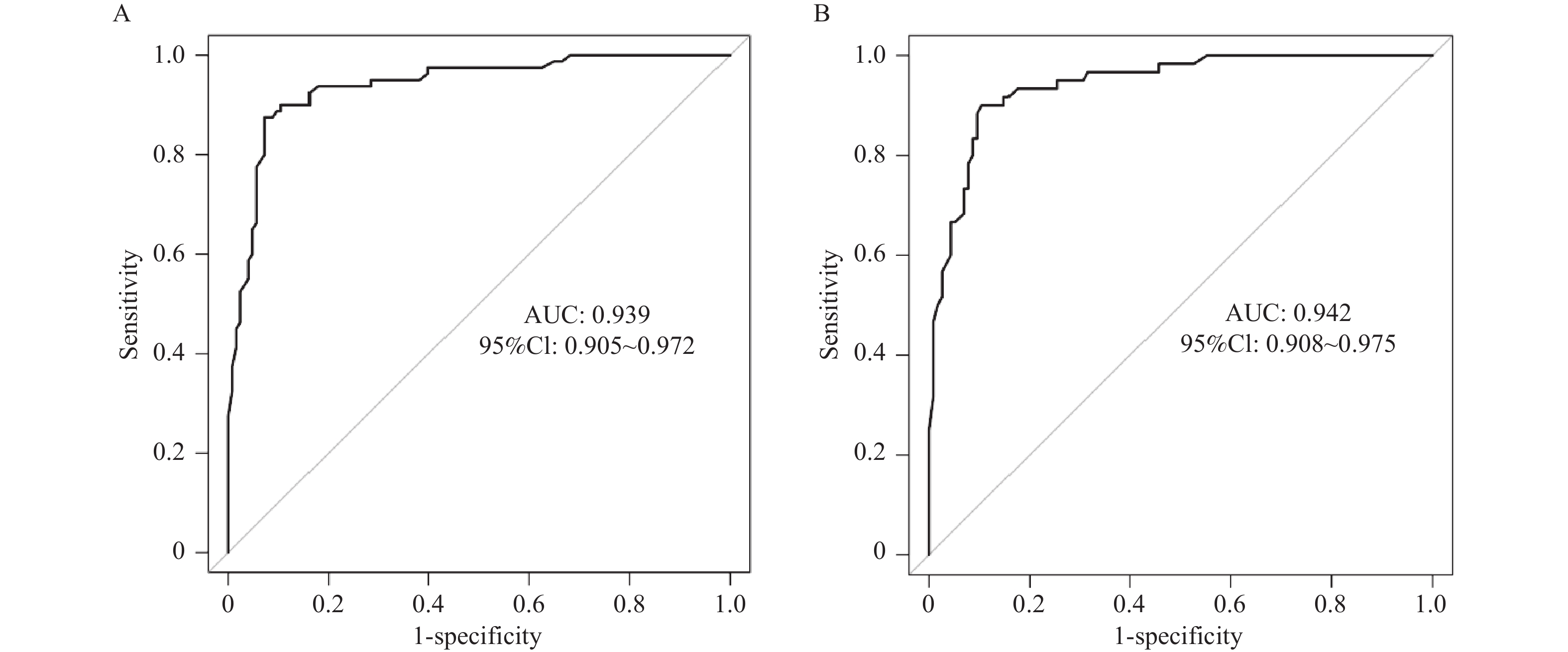
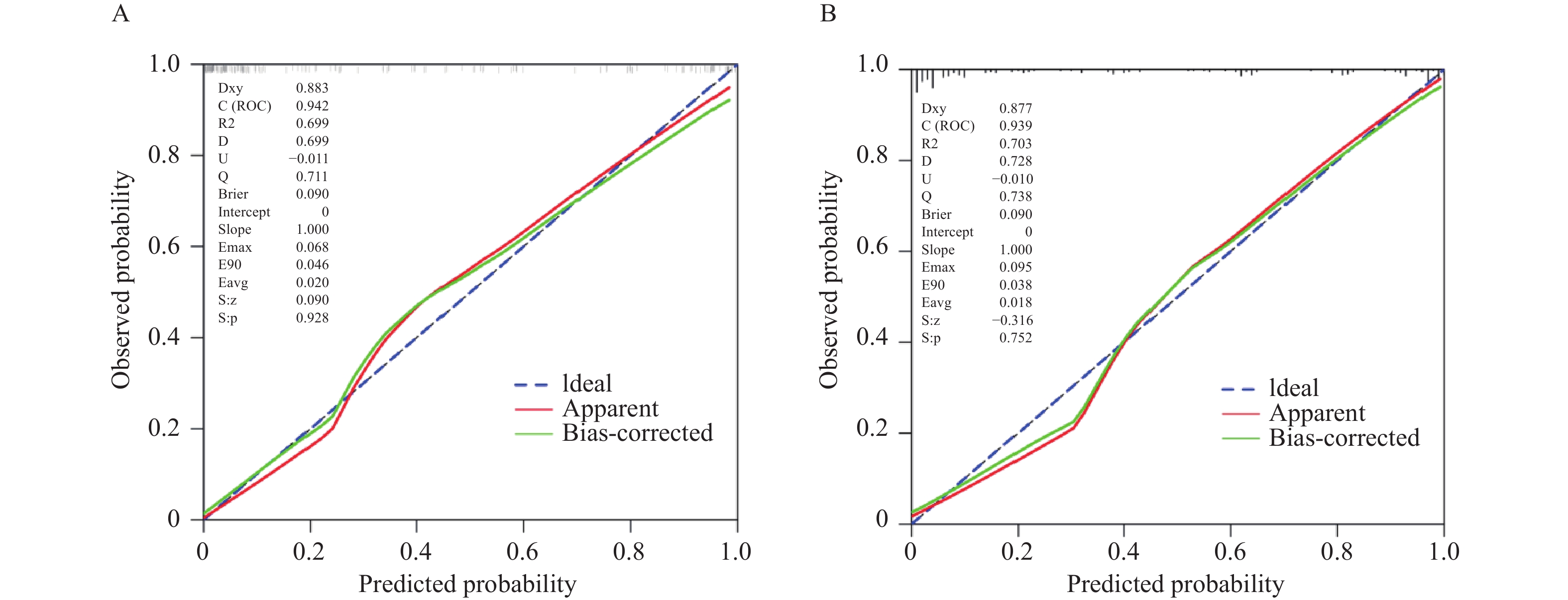
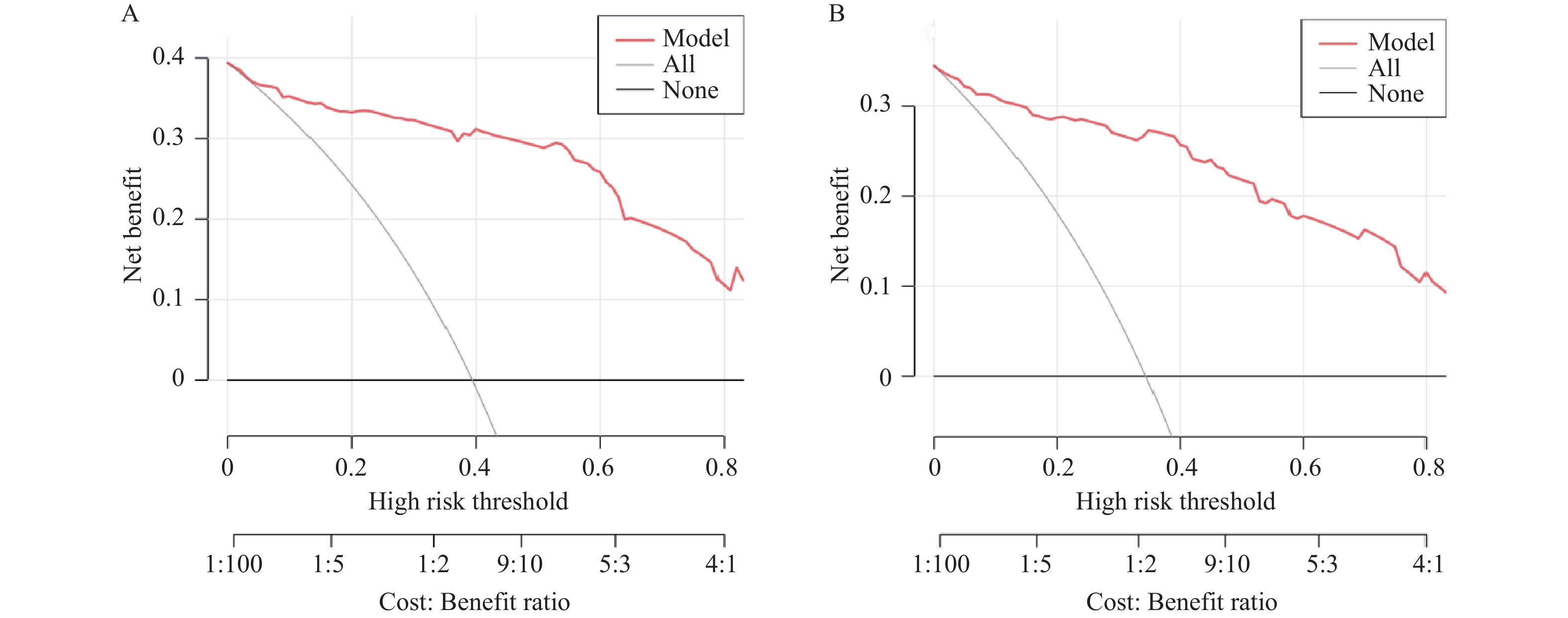
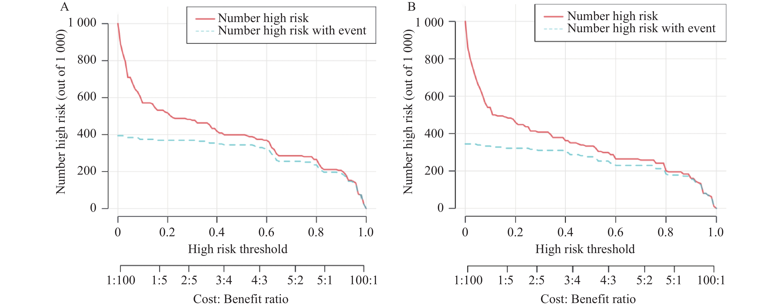
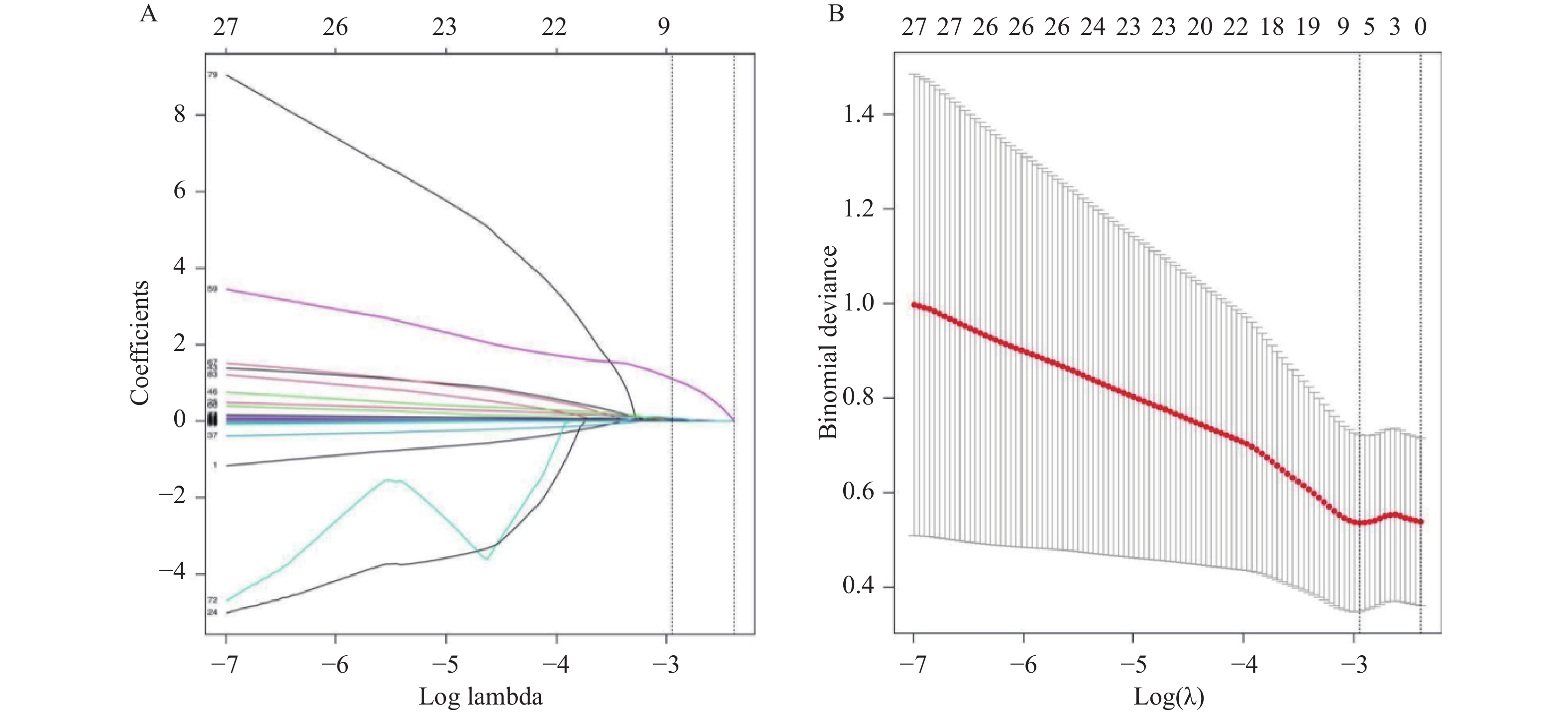
目的 探讨基于临床病理因素、影像组学的列线图模型预测老年晚期非小细胞肺癌(non-small cell lung cancer,NSCLC)患者死亡风险的价值。 方法 回顾性选取2021年3月至2024年3月石家庄市中医院290例老年晚期NSCLC患者,按照7∶3分为训练集(n = 203)与验证集(n = 87)。统计训练集患者1年内预后生存情况,比较不同预后患者个人基本情况、临床病理资料、X线计算机断层成像(computed tomography,CT)影像组学特征及评分,COX回归分析老年晚期NSCLC患者预后生存的影响因素,构建基于影像组学的列线图模型预测老年晚期NSCLC患者预后生存情况,并采用受试者工作特征(receiver operating characteristic,ROC)曲线、校准曲线、决策曲线分析(decision curve analysis,DCA)、临床影响曲线(clinical impact curve,CIC)检验模型预测效能。 结果 训练集中203例老年晚期NSCLC患者1年内死亡率为39.41%(80/203);训练集中死亡组患者美国东部肿瘤协作组(eastern cooperative oncology group,ECOG)评分、营养不良占比、病灶直径、分化程度、临床分期、微波消融(microwave ablation,MWA)反应与生存组比较,差异有统计学意义(P < 0.05);Lasso回归筛选8个最佳特征值,死亡组CT影像组学评分为(0.58±0.15)分,高于生存组(-0.42±0.13)分,差异有统计学意义(P < 0.05);COX回归分析显示,ECOG评分、病灶直径、分化程度、临床分期、MWA反应、CT影像组学评分均为预后生存的独立影响因素(P < 0.05);构建基于影像组学的列线图预后预测模型,ROC曲线显示,该模型在训练集、验证集中预测预后生存的曲线下面积(area under curve,AUC)分别为0.939(95%CI:0.905~0.972)、0.942(95%CI:0.908~0.975),校准曲线显示,该模型预测结果与实际结果的一致性良好,DCA曲线显示,0.2~0.8阈值范围内,在训练集、验证集中使用该模型预测预后生存的临床净获益较高,CIC曲线显示,横轴从0.8~1.0,在训练集、验证集中使用该模型预测预后生存与实际情况高度重合。 结论 基于临床病理因素、影像组学构建的列线图模型对老年晚期NSCLC患者死亡风险具有一定预测价值,且具有良好的区分度、校准度及临床实用性,可作为临床预测死亡风险的有效模型。 Abstract:Objective To explore the value of a nomogram model based on clinicopathological factors and radiomics in predicting mortality risk in elderly patients with advanced non-small cell lung cancer (NSCLC). Methods A retrospective cohort of 290 elderly patients with advanced NSCLC admitted to Shijiazhuang Hospital of Traditional Chinese Medicine from March 2021 to March 2024 was enrolled and divided into a training set (n = 203) and validation set (n = 87) in a 7:3 ratio. One-year prognostic survival outcomes were assessed in the training set. Personal baseline characteristics, clinicopathological data, computed tomography (CT) radiomics features and scores were compared between patients with different prognoses. Cox regression analysis was performed to identify prognostic factors affecting survival in elderly patients with advanced NSCLC. A nomogram model based on radiomics was constructed to predict the prognosis survival in these patients. The predictive performance was evaluated using receiver operating characteristic (ROC) curves, calibration curves, decision curve analysis (DCA), and clinical impact curves (CIC). Results The Eastern Cooperative Oncology Group (ECOG) score, proportion of malnutrition, lesion diameter, differentiation grade, clinical stage, and microwave ablation (MWA) response in the deceased group showed statistically significant differences compared with the survival group (P < 0.05). Lasso regression identified 8 optimal features. The CT radiomics score in the deceased group (0.58±0.15) was significantly higher than in the survival group (-0.42±0.13) (P < 0.05). COX regression analysis showed that the ECOG score, lesion diameter, differentiation grade, clinical stage, MWA response, and CT radiomics score were independent prognostic factors (P < 0.05). The nomogram model demonstrated area under the curve (AUC) values of 0.939 (95%CI: 0.905~0.972) in the training set and 0.942 (95%CI: 0.908~0.975) in the validation set. Calibration curves showed good agreement between predicted and actual outcomes. DCA curves demonstrated high clinical net benefit within the 0.2~0.8 threshold range in both sets. CIC curves showed substantial overlap between predicted and actual prognoses across the 0.8~1.0 range. Conclusion The nomogram model based on clinicopathological factors and radiomics demonstrates predictive value for mortality risk in elderly patients with advanced NSCLC, with good discrimination, calibration and clinical practicability. It can be used as an effective model for clinical prediction of mortality risk. -
Key words:
- Old age /
- Advanced non-small cell lung cancer /
- Clinicopathological factors /
- Radiomics /
- Risk of death /
- Nomogram model /
- Prediction
-
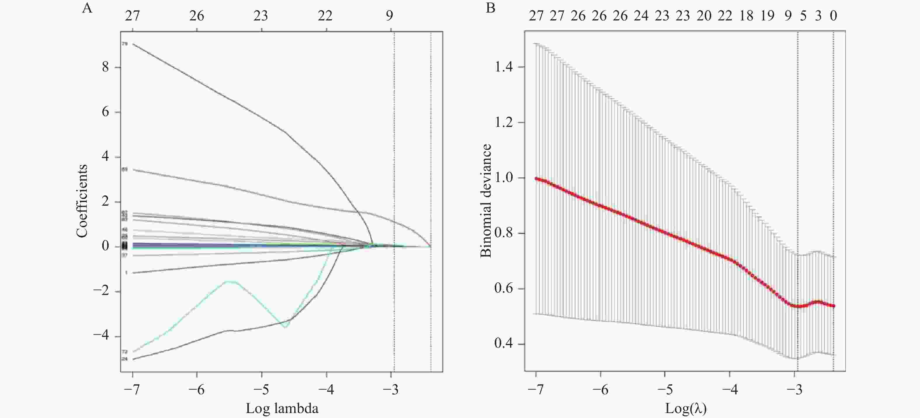
图 1 Lasso回归筛选影像组学特征的交叉验证过程
A:Lasso回归系数路径图;B:Lasso回归交叉验证图。
Figure 1. Cross-validation process of lasso regression for screening radiomics features
表 1 不同预后患者个人基本情况比较[($\bar x \pm s $)/n(%)]
Table 1. Comparison of basic personal information of patients with different prognosis [($\bar x \pm s $)/n(%)]
资料 死亡组
(n = 80)生存组
(n = 123)t/χ2 P 性别 0.134 0.715 男 52(65.00) 83(67.48) 女 28(35.00) 40(32.52) 年龄(岁) 68.43 ± 4.12 67.79 ± 3.35 1.213 0.226 BMI(kg/m2) 21.85 ± 2.52 22.39 ± 2.31 −1.570 0.118 吸烟史 1.840 0.175 有 50(62.50) 65(52.85) 无 30(37.50) 58(47.15) 饮酒史 0.240 0.624 有 32(40.00) 45(36.59) 无 48(60.00) 78(63.41) PS评分(分) 2.499 0.114 0~1 30(37.50) 60(48.78) ≥2 50(62.50) 63(51.22) ECOG评分(分) 36.212 < 0.001* 0~1 28(35.00) 95(77.24) ≥2 52(65.00) 28(22.76) 营养状态 22.165 < 0.001* 良好 25(31.25) 80(65.04) 不良 55(68.75) 43(34.96) *P < 0.05。  下载: 导出CSV
表 2 不同预后患者临床病理资料比较[($ \bar x \pm s $)/n(%)]
Table 2. Comparison of clinicopathological data of patients with different prognosis [($ \bar x \pm s $)/n(%)]
资料 死亡组
(n = 80)生存组
(n = 123)t/χ2 P 病理类型 1.718 0.190* 腺癌 42(52.50) 76(61.79) 鳞癌 38(47.50) 47(38.21) 病灶直径(cm) 9.160 0.002* ≤5 25(31.25) 65(52.85) > 5 55(68.75) 58(47.15) 分化程度 15.328 < 0.001* 低分化 55(68.75) 50(40.65) 中高分化 25(31.25) 73(59.35) 临床分期 14.465 < 0.001* ⅢC期 15(18.75) 55(44.72) Ⅳ期 65(81.25) 68(55.28) MWA反应 53.858 < 0.001* 完全 20(25.00) 95(77.24) 不完全 60(75.00) 28(22.76) *P < 0.05。
下载: 导出CSV
表 3 变量赋值情况
Table 3. Variable assignment
变量 变量说明 因变量 预后生存 死亡 = 1,生存 = 0 自变量 ECOG评分(分) 0~1 = 1,≥2 = 2 营养状态 良好 = 0,不良 = 1 病灶直径(cm) ≤5 = 1, > 5 = 2 分化程度 中高分化 = 1,低分化 = 2 临床分期 ⅢC期 = 1,Ⅳ期 = 2 MWA反应 完全 = 0,不完全 = 1 CT影像组学评分 原值带入
下载: 导出CSV
表 4 老年晚期NSCLC患者预后生存的COX回归分析
Table 4. COX regression analysis of prognostic survival in elderly patients with advanced NSCLC
变量 β S.E. Waldχ2 P HR 95%CI 下限 上限 ECOG评分 0.728 0.221 10.856 < 0.001* 2.071 1.343 3.194 病灶直径 0.621 0.195 10.134 < 0.001* 1.860 1.269 2.726 分化程度 0.884 0.251 12.404 < 0.001* 2.421 1.480 3.959 临床分期 0.964 0.295 10.671 < 0.001* 2.621 1.470 4.673 MWA反应 0.772 0.243 10.102 < 0.001* 2.165 1.345 3.486 CT影像组学评分 0.310 0.084 13.580 < 0.001* 1.363 1.156 1.607 *P < 0.05。
下载: 导出CSV
-
[1] 谢芳芳, 樊静, 李建梅. miR-217、USP10、LZTS1在晚期非小细胞肺癌癌组织与癌旁组织中的表达及与临床疗效的关系[J]. 临床误诊误治, 2025, 38(8): 73-79. doi: 10.3969/j.issn.1002-3429.2025.08.014 [2] Rami-Porta R, Nishimura K K, Giroux D J, et al. The international association for the study of lung cancer lung cancer staging project: Proposals for revision of the TNM stage groups in the forthcoming (ninth) edition of the TNM classification for lung cancer[J]. J Thorac Oncol, 2024, 19(7): 1007-1027. doi: 10.1016/j.jtho.2024.02.011 [3] Bourdillon A T. Computer vision—Radiomics & pathognomics[J]. Otolaryngol Clin N Am, 2024, 57(5): 719-751. doi: 10.1016/j.otc.2024.05.003 [4] Warkentin M T, Al-Sawaihey H, Lam S, et al. Radiomics analysis to predict pulmonary nodule malignancy using machine learning approaches[J]. Thorax, 2024, 79(4): 307-315. doi: 10.1136/thorax-2023-220226 [5] 李曼曼, 符益纲, 肖勇, 等. CT影像组学列线图预测结直肠癌肿瘤沉积和预后[J]. CT理论与应用研究, 2025, 34(4): 694-702. [6] Ettinger D S, Wood D E, Aisner D L, et al. Non–small cell lung cancer, version 5.2017, NCCN clinical practice guidelines in oncology[J]. J Natl Compr Canc Netw, 2017, 15(4): 504-535. [7] 孙军, 郑皆红, 曾悦, 等. 老年晚期非小细胞肺癌患者PD-1抑制剂临床疗效及预后与淋巴细胞亚群水平的关系研究[J]. 中国肿瘤外科杂志, 2024, 16(4): 372-376. [8] 秦茵茵, 张德华, 林心情, 等. 36例体力状况评分≥2分的晚期非小细胞肺癌患者的临床分析[J]. 中华肿瘤杂志, 2017, 39(11): 855-861. doi: 10.3760/cma.j.issn.0253-3766.2017.11.009 [9] Young J, Badgery-Parker T, Dobbins T, et al. Comparison of ECOG/WHO performance status and ASA score as a measure of functional status[J]. J Pain Symptom Manag, 2015, 49(2): 258-264. doi: 10.1016/j.jpainsymman.2014.06.006 [10] Meyer M L, Fitzgerald B G, Paz-Ares L, et al. New promises and challenges in the treatment of advanced non-small-cell lung cancer[J]. Lancet, 2024, 404(10454): 803-822. doi: 10.1016/S0140-6736(24)01029-8 [11] Kim M N, Kim B K, Han K H, et al. Evolution from WHO to EASL and mRECIST for hepatocellular carcinoma: Considerations for tumor response assessment[J]. Expert Rev Gastroenterol Hepatol, 2015, 9(3): 335-348. doi: 10.1586/17474124.2015.959929 [12] 李海斌, 吴振虎, 丁建峰. 肿瘤相关巨噬细胞内Notch-1通过抑制组织蛋白酶S过表达影响非小细胞肺癌腺癌侵袭转移的机制研究[J]. 临床误诊误治, 2024, 37(16): 88-95. doi: 10.3969/j.issn.1002-3429.2024.16.016 [13] Russo L, Charles-Davies D, Bottazzi S, et al. Radiomics for clinical decision support in radiation oncology[J]. Clin Oncol, 2024, 36(8): e269-e281. doi: 10.1016/j.clon.2024.03.003 [14] Kong Y, Xu M, Wei X, et al. CT imaging-based radiomics signatures improve prognosis prediction in postoperative colorectal cancer[J]. J X Ray Sci Technol Clin Appl Diagn Ther, 2023, 31(6): 1281-1294. doi: 10.3233/XST-230090 [15] Yoshiyasu N, Kojima F, Hayashi K, et al. Low-dose CT screening of persistent subsolid lung nodules: First-order features in radiomics[J]. Thorac Cardiovasc Surg, 2024, 72(7): 542-549. doi: 10.1055/a-2158-1364 [16] Salazar P, Cheung P, Ganeshan B, et al. Predefined and data-driven CT radiomics predict recurrence-free and overall survival in patients with pulmonary metastases treated with stereotactic body radiotherapy[J]. PLoS One, 2024, 19(12): e0311910. doi: 10.1371/journal.pone.0311910 [17] Qian L D, Zhou Z A, Li S Q, et al. 18F-fluorodeoxyglucose (18F-FDG) positron emission tomography/computed tomography (PET/CT) imaging of pediatric neuroblastoma: A multi-omics parameters method to predict MYCN copy number category[J]. Quant Imaging Med Surg, 2024, 14(4): 3131-3145. doi: 10.21037/qims-23-494 [18] Li J, Qiu Z, Zhang C, et al. ITHscore: Comprehensive quantification of intra-tumor heterogeneity in NSCLC by multi-scale radiomic features[J]. Eur Radiol, 2023, 33(2): 893-903. doi: 10.1007/s00330-022-09055-0 [19] Nayak P, Sinha S, Goda J S, et al. Computerized tomography-based first order tumor texture features in non-small cell lung carcinoma treated with concurrent chemoradiation: A simplistic and potential surrogate imaging marker for survival[J]. J Cancer Res Ther, 2023, 19(2): 366-375. doi: 10.4103/jcrt.jcrt_2317_21 [20] Zhang Y R, Lu Y H, Lin C M, et al. Pretreatment CT texture analysis for predicting survival outcomes in advanced nonsmall cell lung cancer patients receiving immunotherapy: A systematic review and meta-analysis[J]. Thorac Cancer, 2025, 16(15): e70144. doi: 10.1111/1759-7714.70144 [21] 徐刚, 陈鹏, 纪伟, 等. 基于CT影像组学列线图预测实性非小细胞肺癌组织PD-L1蛋白表达状态[J]. 现代肿瘤医学, 2024, 32(5): 913-920. [22] Zhang K, Zhou X, Xi Q, et al. Outcome prediction of spontaneous supratentorial intracerebral hemorrhage after surgical treatment based on non-contrast computed tomography: A multicenter study[J]. J Clin Med, 2023, 12(4): 1580. doi: 10.3390/jcm12041580 [23] Miranda J, Horvat N, Assuncao A N, et al. MRI-based radiomic score increased mrTRG accuracy in predicting rectal cancer response to neoadjuvant therapy[J]. Abdom Radiol, 2023, 48(6): 1911-1920. doi: 10.1007/s00261-023-03898-x [24] Shimozono T, Shiiba T, Takano K. Radiomics score derived from T1-w/T2-w ratio image can predict motor symptom progression in Parkinson’s disease[J]. Eur Radiol, 2024, 34(12): 7921-7933. doi: 10.1007/s00330-024-10886-2 [25] Bhargavan R, Philip F A, Km J K, et al. Comparison of modified frailty index, clinical frailty scale, ECOG score, and ASA PS score in predicting postoperative outcomes in cancer surgery: A prospective study[J]. Indian J Surg Oncol, 2024, 15(4): 938-945. doi: 10.1007/s13193-024-01995-x [26] Caii W, Wu X, Guo K, et al. Integration of deep learning and habitat radiomics for predicting the response to immunotherapy in NSCLC patients[J]. Cancer Immunol Immunother, 2024, 73(8): 153. doi: 10.1007/s00262-024-03724-3 -

 点击查看大图
计量
- 文章访问数: 158
- HTML全文浏览量: 136
- PDF下载量: 70
- 被引次数: 0
