

Regulatory Mechanisms of Vascular Smooth Muscle Cell Phenotype Switching in Segmental Heterogeneity of Aortic Aneurysm and Dissection and Its Diagnostic and Therapeutic Applications
-
摘要: 主动脉瘤与夹层是致命性心血管疾病,其发生发展与血管平滑肌细胞(vascular smooth muscle cell,VSMCs)的表型转换密切相关。近年研究揭示,主动脉疾病存在显著的节段异质性,即从主动脉根部至腹主动脉的不同节段,在胚胎起源、组织结构、分子机制及病理表现上存在本质差异。这种差异直接影响了疾病易感性,而VSMCs表型转换在不同的节段中也表现出不同的调控网络,其分子层面的差异构成了各节段独特的“转录表型指纹”。本综述的核心观点认为,明确在不同节段中调控VSMCs表型转换的分子机制,绘制其节段特异性调控网络,是推动主动脉疾病治疗从“均质化”走向“节段精准化”的关键,系统总结了各主动脉节段在流行病学、VSMCs表型转换调控机制、分子诊断及靶向治疗策略等方面的进展。深入解析VSMCs表型转换的节段特异性调控网络,将为主动脉疾病的个体化精准治疗提供新的见解。
-
关键词:
- 主动脉瘤 /
- 主动脉夹层 /
- 血管平滑肌细胞表型转换 /
- 异质性 /
- 精准治疗
Abstract: Aortic aneurysm and aortic dissection are life-threatening cardiovascular diseases whose occurrence and development are closely associated with phenotypic switching of vascular smooth muscle cells (VSMCs). Recent studies have revealed significant segmental heterogeneity in aortic diseases; that is, different segments of the aorta from the aortic root to the abdominal aorta exhibit essential differences in embryologic origin, tissue architecture, molecular mechanisms, and pathological manifestations. This heterogeneity directly influence disease susceptibility, and VSMC phenotypic switching exhibits distinct regulatory networks across different segments, with molecular-level differences constituting unique "transcriptional phenotype fingerprints" for each segment. The central thesis of this review posits that elucidating the molecular mechanisms regulating VSMC phenotypic switching in different aortic segments and mapping their segment-specific regulatory networks are key to advancing the treatment of aortic disease from a "homogeneous" approach toward "segment-specific precision medicine." This review systematically summarizes recent advances in epidemiology, regulatory mechanisms of VSMC phenotypic switching, molecular diagnostics, and targeted therapeutic strategies across various aortic segments. In-depth analysis of the segment-specific regulatory networks governing VSMC phenotypic switching will provide new insights for individualized precision treatment of aortic disease. -
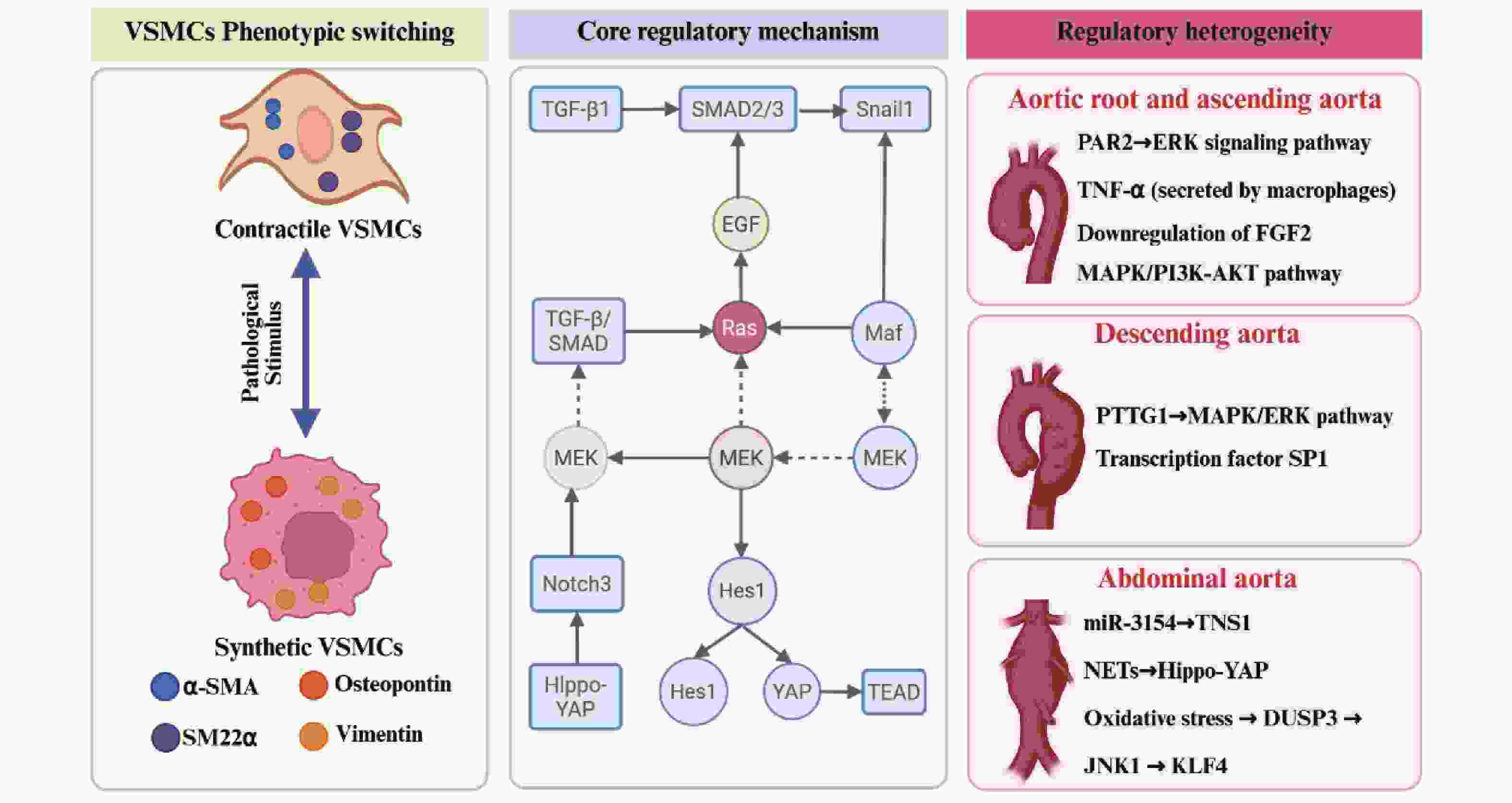
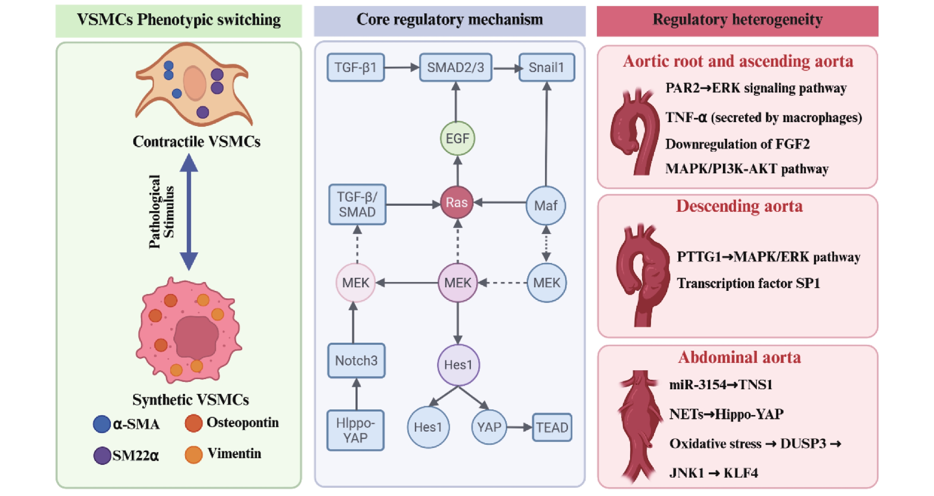
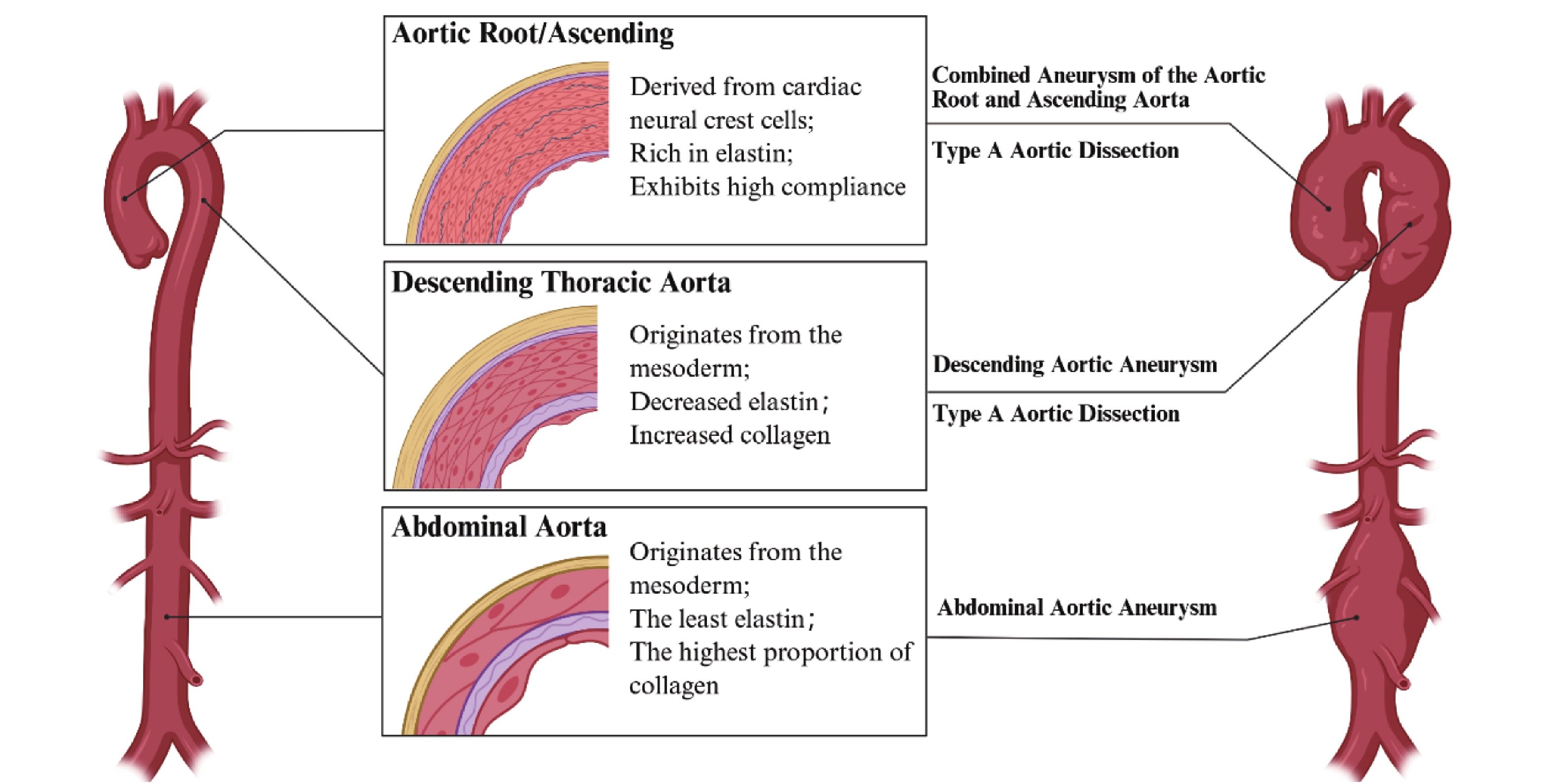
图 2 主动脉不同节段血管平滑肌细胞表型转换异质性
Figure 2. Segmental heterogeneity in phenotypic switching of vascular smooth muscle cells in the aorta
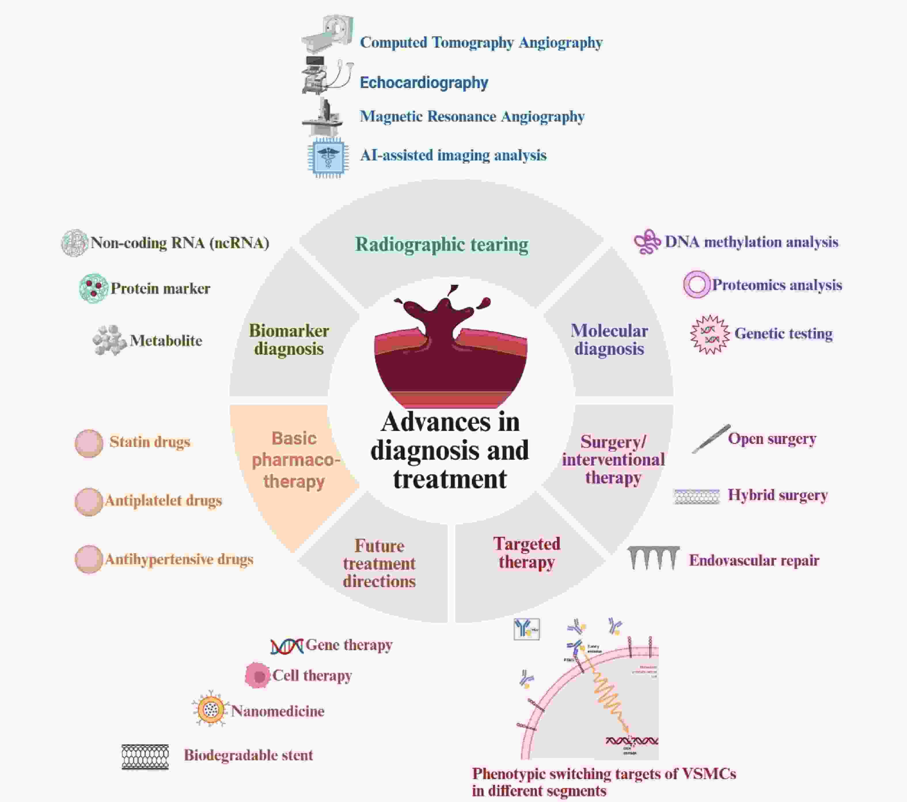
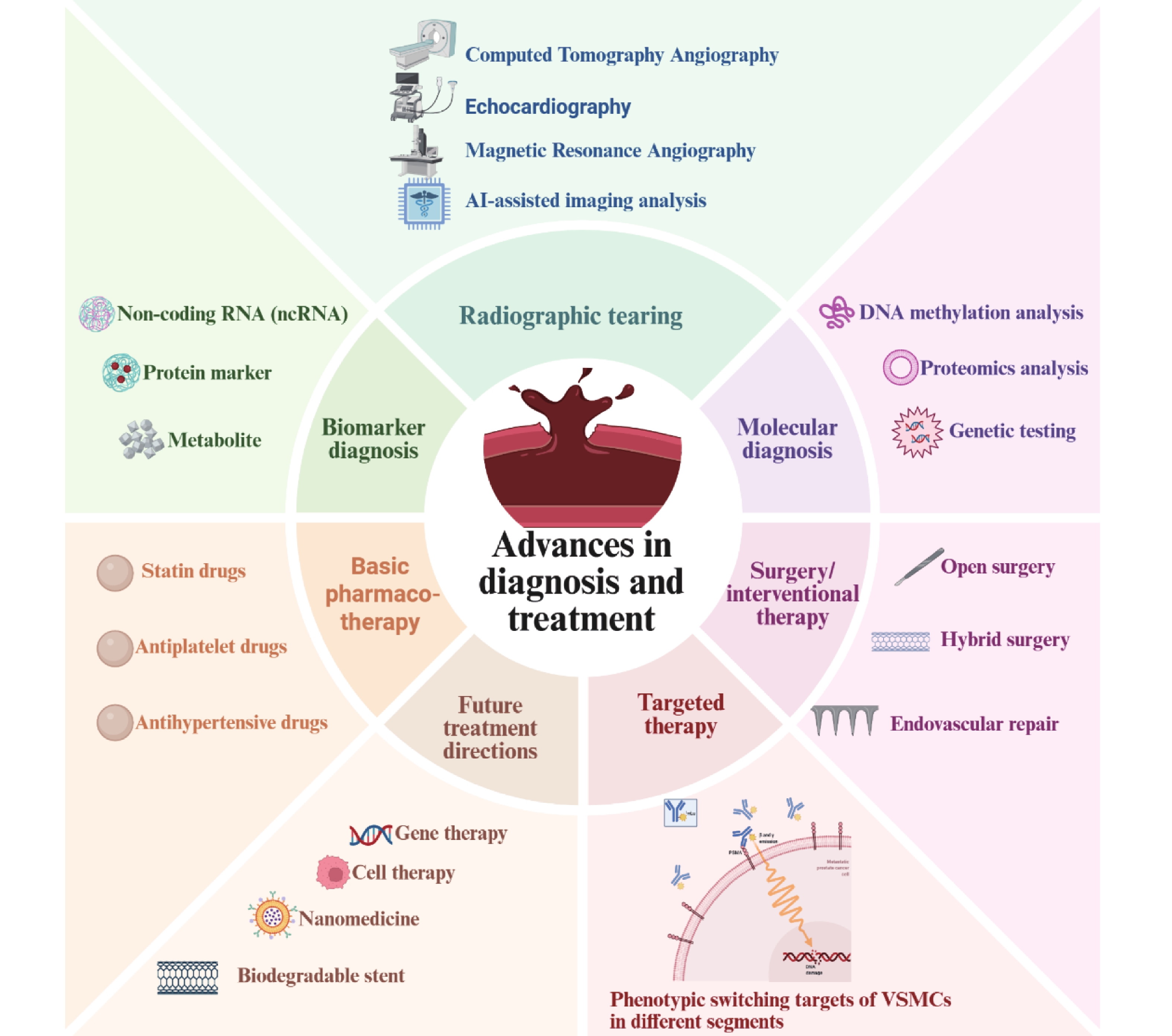
图 3 主动脉瘤及夹层诊断及治疗进展
Figure 3. Advances in the diagnosis and treatment of aortic aneurysm and dissection
表 1 主动脉不同节段动脉瘤与夹层的流行病学、分子机制、诊断标志物及治疗策略对比
Table 1. Comparison of epidemiology,molecular mechanisms,diagnostic biomarkers,and treatment strategies for aortic aneurysms and dissections across different aortic segments
特征分类 主动脉根部瘤 (ARA) 升主动脉瘤/夹层 (AsAA/TAAD) 降主动脉瘤/夹层 (DTAA/TBAD) 腹主动脉瘤 (AAA) 流行病学 发病率:1~2/10万人/年;男:女≈1.2:1;40~60岁发病;与LDS、Marfan综合征相关 发病率:6~10/10万人/年;男:女≈2~3:1;50~70岁发病;与二叶式主动脉瓣、高血压相关 发病率:3~6/10万人/年;男:女≈1.5~2:1;60~75岁发病;与高血压、动脉粥样硬化相关 发病率:10~20/10万人/年;男:女≈4~6:1;糖尿病是保护因素;种族差异明显 VSMCs表型转换核心机制 PAR2-ERK信号轴;PAR2激活ERK通路,促进合成型转换;炎症细胞浸润加剧 FGF2-MAPK/PI3K通路;FGF2缺乏导致收缩型标志物下调;周细胞表型转换参与 SP1-PTTG1-MAPK轴;PTTG1激活ERK通路;SP1转录激活PTTG1 多通路:miR-3154/TNS1、DUSP3-JNK1-KLF4、NETs-Hippo-YAP;IL-10/M2巨噬细胞极化调节 特异性分子标志物 PAR2↑;α-SMA↓;OPN↑ FGF2↓;SM22α↓ PTTG1↑;血浆PTTG1可能反映病变活动 miR-3154↑;NETs相关蛋白↑;IL-10↓ 药物治疗策略 β受体阻滞剂/ARB延缓根部扩张 β受体阻滞剂/ACEI控制血压心率;他汀类药物降低增长率 急性期严格降压;β受体阻滞剂+ACEI 无明确延缓进展药物;控制心血管风险因素、戒烟 靶向VSMCs表型转换治疗 PAR2拮抗剂;ERK抑制剂(PD98059) 外源性FGF2重组蛋白/基因治疗 PTTG1 siRNA;SP1抑制剂;ERK抑制剂(PD98059) miR-3154拮抗剂;IL-10过表达;NETs形成阻断;YAP核转位抑制;DUSP3抑制剂 手术/介入治疗 干预阈值:直径>55 mm;David手术 干预阈值:直径>55 mm;急性TAAD手术死亡率15%~20%(未手术20%~30%) 干预阈值:直径≥55 mm;TEVAR首选 干预阈值:男≥55 mm,女≥50 mm,囊状≥45 mm;EVAR 诊断技术特点 经胸超声心动图敏感;CTA精确评估 CTA首选;超声可用于根部初筛 CTA为核心手段;超声显像困难 超声筛查;CTA术前精确评估  下载: 导出CSV
-
[1] Shi Y, Xie X, Sun Y, et al. Vascular smooth muscle cell metabolic disorders in the occurrence and development of aortic aneurysms and dissections: Implications for therapy[J]. Biomedicines, 2025, 13(12): 3072. doi: 10.3390/biomedicines13123072 [2] Elmarasi M, Elmakaty I, Elsayed B, et al. Phenotypic switching of vascular smooth muscle cells in atherosclerosis, hypertension, and aortic dissection[J]. J Cell Physiol, 2024, 239(4): e31200. doi: 10.1002/jcp.31200 [3] Tang H Y, Chen A Q, Zhang H, et al. Vascular smooth muscle cells phenotypic switching in cardiovascular diseases[J]. Cells, 2022, 11(24): 4060. doi: 10.3390/cells11244060 [4] Hu Y, Cai Z, He B. Smooth muscle heterogeneity and plasticity in health and aortic aneurysmal disease[J]. Int J Mol Sci, 2023, 24(14): 11701. doi: 10.3390/ijms241411701 [5] Rombouts K B, van Merrienboer T A R, Ket J C F, et al. The role of vascular smooth muscle cells in the development of aortic aneurysms and dissections[J]. Eur J Clin Investig, 2022, 52(4): e13697. doi: 10.1111/eci.13697 [6] Liu G, Li J, Ming Y, et al. A hiPSC-derived lineage-specific vascular smooth muscle cell-on-a-chip identifies aortic heterogeneity across segments[J]. Lab Chip, 2023, 23(7): 1835-1851. doi: 10.1039/D2LC01158A [7] Ciavarella C, Motta I, Capri M, et al. Heterogeneity and differentiation of the human arterial tree: Focus on microRNA expression in vascular disease[J]. Biomolecules, 2024, 14(3): 343. doi: 10.3390/biom14030343 [8] Cao G, Xuan X, Li Y, et al. Single-cell RNA sequencing reveals the vascular smooth muscle cell phenotypic landscape in aortic aneurysm[J]. Cell Commun Signal, 2023, 21(1): 113. doi: 10.1186/s12964-023-01120-5 [9] Vellarikkal S, Atri D, Lee-Kim V, et al. Abstract 13348: Single nuclear RNA-seq of human thoracic aortic dissection identifies expansion of vascular smooth muscle cells with a distinct transcriptional phenotype[J]. Circulation, 2021, 144(Suppl_1): A13348-A13348. doi: 10.1161/circ.144.suppl_1.13348 [10] Shi D, Zhang M, Zhang Y, et al. The pathophysiological role of vascular smooth muscle cells in abdominal aortic aneurysm[J]. Cells, 2025, 14(13): 1009. doi: 10.3390/cells14131009 [11] Qian G, Adeyanju O, Olajuyin A, et al. Abdominal aortic aneurysm formation with a focus on vascular smooth muscle cells[J]. Life, 2022, 12(2): 191. doi: 10.3390/life12020191 [12] Jauhiainen S, Kiema M, Hedman M, et al. Large vessel cell heterogeneity and plasticity: Focus in aortic aneurysms[J]. Arterioscler Thromb Vasc Biol, 2022, 42(7): 811-818. doi: 10.1161/ATVBAHA.121.316237 [13] 马思伟, 董伟, 陈思. LncRNA MIAT在颅内动脉瘤患者血清中的表达及靶向调节miR-331-3p对血管平滑肌细胞增殖和凋亡的影响[J]. 贵州医科大学学报, 2024, 49(7): 997-1004. doi: 10.19367/j.cnki.2096-8388.2024.07.008 [14] Milewicz D M, Braverman A C, De Backer J, et al. Marfan syndrome[J]. Nat Rev Dis Primers, 2021, 7: 64. doi: 10.1038/s41572-021-00298-7 [15] Yamabe T, Zhao Y, Kurlansky P A, et al. Assessment of long-term outcomes: Aortic valve reimplantation versus aortic valve and root replacement with biological valved conduit in aortic root aneurysm with tricuspid valve[J]. Eur J Cardiothorac Surg, 2021, 59(3): 658-665. doi: 10.1093/ejcts/ezaa389 [16] Guo M H, Appoo J J, Saczkowski R, et al. Association of mortality and acute aortic events with ascending aortic aneurysm: A systematic review and meta-analysis[J]. JAMA Netw Open, 2018, 1(4): e181281. doi: 10.1001/jamanetworkopen.2018.1281 [17] Hawatmeh A, Abu Arqoub A, Isbitan A, et al. A case of ascending aortic dissection mimicking acute myocardial infarction and complicated with pericardial tamponade[J]. Cardiovasc Diagn Ther, 2016, 6(2): 166-171. doi: 10.21037/cdt.2015.11.06 [18] Schuster V, Eggersmann T K, Eifert S, et al. Ascending aortic disease is associated with earlier menopause and shorter reproductive life span[J]. J Women’s Health, 2016, 25(9): 912-919. doi: 10.1055/s-0036-1593095 [19] 杨铭铭, 何垚, 于绍梅. Sievers分型0型与1型二叶式主动脉瓣患者超声心动图特征及升主动脉扩张的影响因素[J]. 贵州医科大学学报, 2023, 48(4): 422-429. [20] Gegouskov V, Manchev G, Danov V, et al. Direct cannulation of ascending aorta versus standard femoral artery cannulation in acute aortic dissection type A[J]. Heart Surg Forum, 2018, 21(3): 139. doi: 10.1532/hsf.1956 [21] Biancari F, Mariscalco G, Mariani S, et al. Endovascular treatment of degenerative aneurysms involving only the descending thoracic aorta: Systematic review and meta-analysis[J]. J Endovasc Ther, 2016, 23(2): 387-392. doi: 10.1177/1526602815626560 [22] Nauta F J, Trimarchi S, Kamman A V, et al. Update in the management of type B aortic dissection[J]. Vasc Med, 2016, 21(3): 251-263. doi: 10.1177/1358863X16642318 [23] Ziganshin B A, Theodoropoulos P, Salloum M N, et al. Simple renal cysts as markers of thoracic aortic disease[J]. J Am Heart Assoc, 2016, 5: e002248. doi: 10.1161/JAHA.115.002248 [24] Gifford S M, Duncan A A, Greiten L E, et al. The natural history and outcomes for thoracic and abdominal penetrating aortic ulcers[J]. J Vasc Surg, 2016, 63(5): 1182-1188. doi: 10.1016/j.jvs.2015.11.050 [25] Bartek M A, Kessler L G, Talbott J M, et al. Washington State abdominal aortic aneurysm-related mortality shows a steady decline between 1996 and 2016[J]. J Vasc Surg, 2019, 70(4): 1115-1122. doi: 10.1016/j.jvs.2018.12.040 [26] Zommorodi S, Bottai M, Hultgren R. Sex differences in repair rates and outcomes of patients with ruptured abdominal aortic aneurysm[J]. BJS Br J Surg, 2019, 106(11): 1480-1487. doi: 10.1002/bjs.11258 [27] Takagi H, Umemoto T. Negative association of diabetes with rupture of abdominal aortic aneurysm[J]. Diabetes Vasc Dis Res, 2016, 13(5): 341-347. doi: 10.1177/1479164116651389 [28] Salata K, Syed M, Hussain M A, et al. Renin-angiotensin system blockade does not attenuate abdominal aortic aneurysm growth, rupture rate, or perioperative mortality after elective repair[J]. J Vasc Surg, 2018, 67(2): 629-636. e2. [29] Ballantyne M D, Pinel K, Dakin R, et al. Smooth muscle enriched long noncoding RNA (SMILR) regulates cell proliferation[J]. Circulation, 2016, 133(21): 2050-2065. doi: 10.1161/CIRCULATIONAHA.115.021019 [30] Lee S J, Won S Y, Park S L, et al. Rosa hybrida extract suppresses vascular smooth muscle cell responses by the targeting of signaling pathways, cell cycle regulation and matrix metalloproteinase-9 expression[J]. Int J Mol Med, 2016, 37(4): 1119-1126. doi: 10.3892/ijmm.2016.2504 [31] Jin M, Wu Y, Wang Y, et al. microRNA-29a promotes smooth muscle cell differentiation from stem cells by targeting YY1[J]. Stem Cell Res, 2016, 17(2): 277-284. doi: 10.1016/j.scr.2016.07.011 [32] van der Pluijm I, van Vliet N, von der Thusen J H, et al. Defective connective tissue remodeling in Smad3 mice leads to accelerated aneurysmal growth through disturbed downstream TGF-β signaling[J]. EBioMedicine, 2016, 12: 280-294. doi: 10.1016/j.ebiom.2016.09.006 [33] Li B, Wang Z, Hu Z, et al. P38 MAPK signaling pathway mediates angiotensin II-induced miR143/145 gene cluster downregulation during aortic dissection formation[J]. Ann Vasc Surg, 2017, 40: 262-273. doi: 10.1016/j.avsg.2016.09.016 [34] Hsu M Y, Yang M H, Schnegg C I, et al. Notch3 signaling-mediated melanoma–endothelial crosstalk regulates melanoma stem-like cell homeostasis and niche morphogenesis[J]. Lab Investig, 2017, 97(6): 725-736. doi: 10.1038/labinvest.2017.1 [35] Hu C, Huang W, Xiong N, et al. SP1-mediated transcriptional activation of PTTG1 regulates the migration and phenotypic switching of aortic vascular smooth muscle cells in aortic dissection through MAPK signaling[J]. Arch Biochem Biophys, 2021, 711: 109007. doi: 10.1016/j.abb.2021.109007 [36] Hou Q, Liu Y, Hou J, et al. miR-3154: Novel pathogenic and therapeutic target in abdominal aortic aneurysm[J]. Circ Res, 2025, 137(5): 587-604. doi: 10.1161/CIRCRESAHA.124.325256 [37] Das A A, Waldeck-Weiermair M, Yadav S, et al. Differential aortic aneurysm formation provoked by chemogenetic oxidative stress[J]. J Clin Investig, 2025, 135(9): e188743. doi: 10.1172/JCI188743 [38] Wang M, Tang Z, Zeng H, et al. Protease activated receptor 2 deficiency retards progression of abdominal aortic aneurysms by modulating phenotypic transformation of vascular smooth muscle cells via ERK signaling[J]. Exp Cell Res, 2024, 443(1): 114286. doi: 10.1016/j.yexcr.2024.114286 [39] Huang W, Hill J C, Patel S, et al. Deficiency of fibroblast growth factor 2 promotes contractile phenotype of pericytes in ascending thoracic aortic aneurysm[J]. Am J Physiol Heart Circ Physiol, 2025, 328(5): H1130-H1143. doi: 10.1152/ajpheart.00834.2024 [40] Zhu H, Qu X, Zhang C, et al. Interleukin-10 promotes proliferation of vascular smooth muscle cells by inhibiting inflammation in rabbit abdominal aortic aneurysm[J]. Int J Clin Exp Pathol, 2019, 12(4): 1260-1271. [41] Yang S, Chen L, Wang Z, et al. Neutrophil extracellular traps induce abdominal aortic aneurysm formation by promoting the synthetic and proinflammatory smooth muscle cell phenotype via Hippo-YAP pathway[J]. Transl Res, 2023, 255: 85-96. doi: 10.1016/j.trsl.2022.11.010 [42] He L, Wang S, Liu R, et al. A model fusion method based DAT-DenseNet for classification and diagnosis of aortic dissection[J]. Phys Eng Sci Med, 2024, 47(4): 1537-1546. doi: 10.1007/s13246-024-01466-1 [43] Qiu Z H, He J, Chai T C, et al. miR-145 attenuates phenotypic transformation of aortic vascular smooth muscle cells to prevent aortic dissection[J]. J Clin Lab Anal, 2021, 35(12): e23773. doi: 10.1002/jcla.23773 [44] Wu X, Ye J, Cai W, et al. LDHA mediated degradation of extracellular matrix is a potential target for the treatment of aortic dissection[J]. Pharmacol Res, 2022, 176: 106051. doi: 10.1016/j.phrs.2021.106051 [45] Pan S, Lai H, Shen Y, et al. DNA methylome analysis reveals distinct epigenetic patterns of ascending aortic dissection and bicuspid aortic valve[J]. Cardiovasc Res, 2017, 113(6): 692-704. doi: 10.1093/cvr/cvx050 [46] Régent A, Ly K H, Lofek S, et al. Proteomic analysis of vascular smooth muscle cells in physiological condition and in pulmonary arterial hypertension: Toward contractile versus synthetic phenotypes[J]. Proteomics, 2016, 16(20): 2637-2649. doi: 10.1002/pmic.201500006 [47] Salata K, Syed M, Hussain M A, et al. Statins reduce abdominal aortic aneurysm growth, rupture, and perioperative mortality: A systematic review and meta-analysis[J]. J Am Heart Assoc, 2018, 7(19): e008657. doi: 10.1161/JAHA.118.008657 [48] Schuster V, Eggersmann T K, Eifert S, et al. Ascending aortic disease is associated with earlier menopause and shorter reproductive life span[J]. J Womens Health, 2016, 25(9): 912-919. doi: 10.1089/jwh.2015.5559 [49] Biancari F, Mariscalco G, Mariani S, et al. Endovascular treatment of degenerative aneurysms involving only the descending thoracic aorta: Systematic review and meta-analysis[J]. J Endovasc Ther, 2016, 23(2): 387-392. doi: 10.1177/1526602815626560 [50] Yamawaki-Ogata A, Mutsuga M, Narita Y. A review of current status of cell-based therapies for aortic aneurysms[J]. Inflamm Regen, 2023, 43(1): 40. doi: 10.1186/s41232-023-00280-8 [51] Bi C X, Jin K Q, Yan J, et al. Nanofiber-based stretchable electrodes for oriented culture and mechanotransduction monitoring of smooth muscle cells[J]. ACS Sens, 2023, 8(8): 3248-3256. doi: 10.1021/acssensors.3c01135 [52] Wang Z, Chen Y E, Chang L. Unleashing PD-1: A duel of immunity in aortic aneurysm formation[J]. J Clin Investig, 2024, 134(15): e182554. doi: 10.1172/JCI182554 -

 点击查看大图
计量
- 文章访问数: 62
- HTML全文浏览量: 51
- PDF下载量: 38
- 被引次数: 0
