Analysis of Clinical and Radiological Outcomes between UBE Procedure and Conventional Open Surgery in the Treatment of Lumbar Disc Herniation
-
摘要:
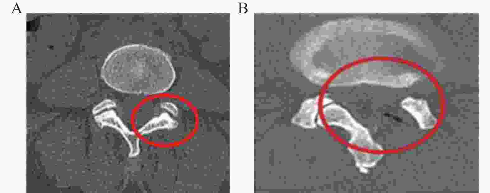
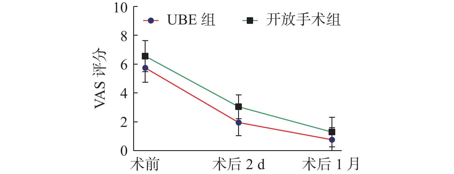
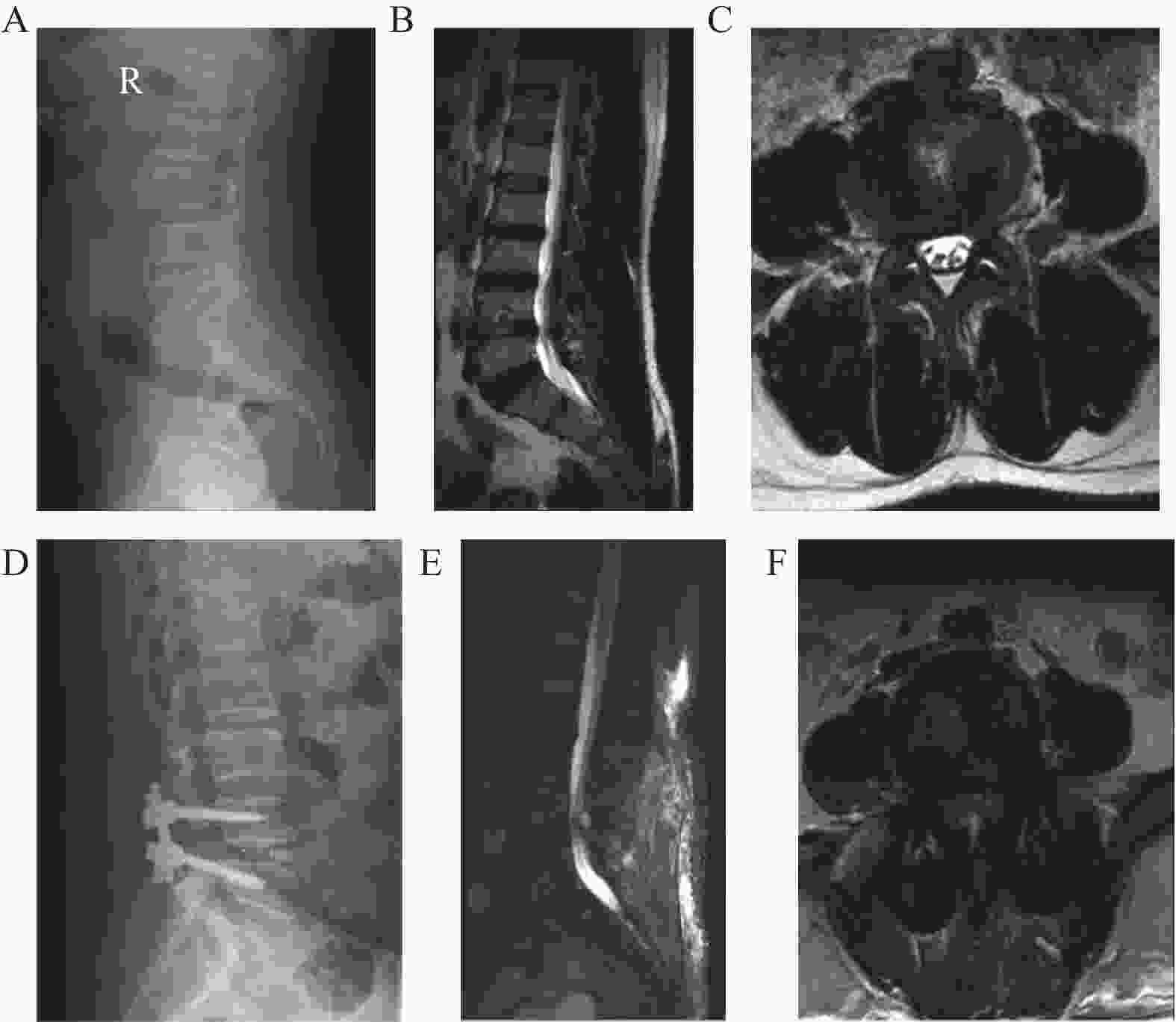
目的 对比单边双通道脊柱内镜(unilateral biportal endoscopy,UBE)与传统开放手术治疗腰椎间盘突出症的临床疗效及影像学结果。 方法 回顾分析2022年1月至2023年3月在昆明医科大学第一附属医院收治的84例单节段腰椎间盘突出患者,其中44例接受单边双通道脊柱内镜手术(UBE组),40例接受传统开放手术。记录患者的年龄、性别、椎间盘突出部位、手术节段、手术时间、术中出血量、住院天数等信息。术前、术后2d和术后1月进行视觉模拟量表(VAS)评分及术后1月采用改良的Macnab评价指标评估疗效。比较2组术前和术后关节突保留率以及椎间盘高度变化。 结果 2组患者在年龄、性别、手术节段及椎间盘突出类型上的差异无统计学意义(P > 0.05)。所有患者均顺利完成手术,相对于开放组,UBE组手术耗时较短,出血量更少,术后住院时间缩短(P < 0.05),同时UBE组围术期并发症发生率显著低于开放组(P < 0.05)。2组患者术前、术后2d时VAS评分明显下降(P < 0.05),但术后1月时2组差异无统计学意义(P > 0.05);且组内术前、术后2d及术后1月时VAS评分差异有统计学意义(P < 0.05)。末次随访患者时改良 Macnab 疗效评定标准结果中UBE组优、良、可、差依次为 40、2、2 与 0 例,总体优良率高达 95.4%。在开放手术组中,优良可差分别为29、7、4与 0例,整体优良率达到90%。术前和术后2组患者的椎间盘高度进行比较,差异有统计学意义(P < 0.05)。在UBE组中,术前和术后椎间盘高度之间比较,差异无统计学意义(P > 0.05),而开放组术后椎间盘高度明显增加(P < 0.05)。UBE组的关节突保留率为63.6%,而开放手术组的关节突保留率仅为10%。 结论 UBE可以直达靶点解除神经压迫,是一种微创、灵活、创伤小、学习曲线平缓、对脊柱活动度影响小、有利于术后康复的新技术,可彻底摘除突出髓核,临床治疗效果理想。 Abstract:Objective To compare the clinical efficacy and imaging results of unilateral biportal endoscopic discectomy (UBE) with traditional open surgery for the treatment of lumbar disc herniation. Methods We retrospectively analyzed 84 patients with single-segment lumbar disc herniation admitted to the First Affiliated Hospital of Kunming Medical University from January 2022 to March 2023, 44 cases in the UBE group and 40 cases in the open surgery group, and recorded the patients' age, gender, disc herniation site, operation segment, operation time, intraoperative bleeding, and hospitalization days, respectively. Visual analog scale (VAS) scores were performed preoperatively, 2 days postoperatively, and at follow-up at 1 month postoperatively. Efficacy was evaluated using the modified Macnab Treatment Effectiveness Evaluation Index at 1 month of surgery. The preoperative and postoperative articular process preservation rate and disc height changes were compared between the two groups. Results There were no statistically significant differences between the two groups of patients in terms of age, gender, operative segment and type of disc herniation (P > 0.05). All patients completed the surgery. Compared with the open group, the UBE group had a shorter operation time, less bleeding, and a shorter postoperative hospitalization (P < 0.05), and the perioperative complication rate was lower in the UBE group than in the open group (P < 0.05). The VAS scores of patients in the two groups decreased significantly at preoperation and 2 days postoperation (P < 0.05), but the difference between the two groups was not significant at 1 month postoperation (P > 0.05); and the difference in VAS scores at preoperation, 2 days postoperation and 1 month postoperation within the groups was statistically significant (P < 0.05). The results of the modified Macnab efficacy evaluation criteria in the UBE group were 40, 2, 2 and 0 cases in order of excellent, good, acceptable and poor at the last follow-up, and the overall excellent rate was as high as 95.4%. In the open surgery group, there were 29, 7, 4 and 0 cases of excellent, good, feasible and poor, with an overall excellent rate of 90%. The difference in disc height between the two groups was statistically significant when comparing preoperative and postoperative disc heights (P < 0.05). For the UBE group, there was no statistically significant difference between the preoperative and postoperative disc heights within the group (P > 0.05), while the postoperative disc height in the open group was significantly increased compared with that of the preoperative period, with a statistically significant difference (P < 0.05).The preservation rate of the articular eminence in the UBE group was 63.6%, while the preservation rate of the articular eminence in the open surgery group was 10%. Conclusion UBE can directly reach the target point to release nerve compression, and is a new technique that is minimally invasive, flexible, less traumatic, has a gentle learning curve, has little effect on spinal mobility, and is conducive to postoperative rehabilitation, which can completely remove the protruding nucleus pulposus, and has an ideal clinical therapeutic effect. -
表 1 2组患者基线数据比较[($\bar x \pm s $)/n(%)]
Table 1. Comparison of baseline data between the two groups of patients [($\bar x \pm s $)/n(%)]
指标 UBE组 开放手术组 t/χ2 P 平均年龄(岁) 48.75±7.95 51.80±6.37 −1.931 0.061 性别 男 21(47.73) 21(52.50) 0.191 0.658 女 23(52.27) 19(47.50) 手术节段 L4/5 19(43.18) 29(72.50) 1.912 0.830 L5/S1 25(58.62) 11(27.50) 椎间盘突出位置 中央型 15(34.10) 16(40.00) 0.473 0.642 旁中央型 17(38.60) 14(35.00) 外侧型 12(27.30) 10(25.00) 表 2 2组患者围手术期相关指标比较[($\bar x \pm s $)/n(%)]
Table 2. Comparison of perioperative related indicators between the two groups of patients [($\bar x \pm s $)/n(%)]
指标 UBE组 开放手术组 t/χ2 P 手术时间(min) 172.32±50.03 202.07±45.18 −2.851 0.006* 术中出血量(mL) 72.84±52.94 274.77±70.43 −14.732 <0.001* 术后住院时间(d) 3.95±1.92 6.75±2.11 −5.937 <0.001* 围术期并发症 4(9.1%) 12(30%) −2.362 0.022* *P < 0.05。 表 3 组患者围术期并发症比较[n(%)]
Table 3. Perioperative complications comparison between two groups of patients[n(%)]
组别 硬脊膜撕裂 神经根刺激症状 并发症率 t P UBE组(n=44) 1(2.3) 3(6.8) 4(9.100) −2.430 0.017* 开放手术组(n=40) 6(15) 5(12.5) 11(27.500) *P < 0.05。 表 4 2组患者腰腿痛VAS评分比较($\bar x \pm s $)
Table 4. Comparison of VAS scores for lower back and leg pain between two groups of patients ($\bar x \pm s $)
指标 UBE组 开放手术组 t P VAS 术前 5.79±1.02 6.60±1.08 −3.501 <0.001* 术后2 d 1.97±0.92 3.07±0.83 −5.686 <0.001* 术后1月 0.77±0.83 1.30±1.04 −2.541 0.131 F 692.730 352.311 P <0.001* <0.001* 组内效应 F=950.791 ,P < 0.001* 组间效应 F=27.120 ,P < 0.001* 组内×组间 F=2.740,P=0.069* *P < 0.05。 表 5 2组患者改进后的MacNab量表评分比较[n(%)]
Table 5. Comparison of Improved MacNab Scale Scores between Two Groups of Patients [n(%)]
组别 优 良 可 差 优良率 χ2 P UBE组(n = 44) 40(90.9) 2(4.5) 2(4.5) 0(0) 95.4 5.019 0.081 开放手术组(n = 40) 29(72.5) 7(17.5) 4(10.0) 0(0) 90 表 6 2组患者影像学结果比较($\bar x \pm s $,mm)
Table 6. Comparison of imaging results between the two groups of patients ($\bar x \pm s $,mm)
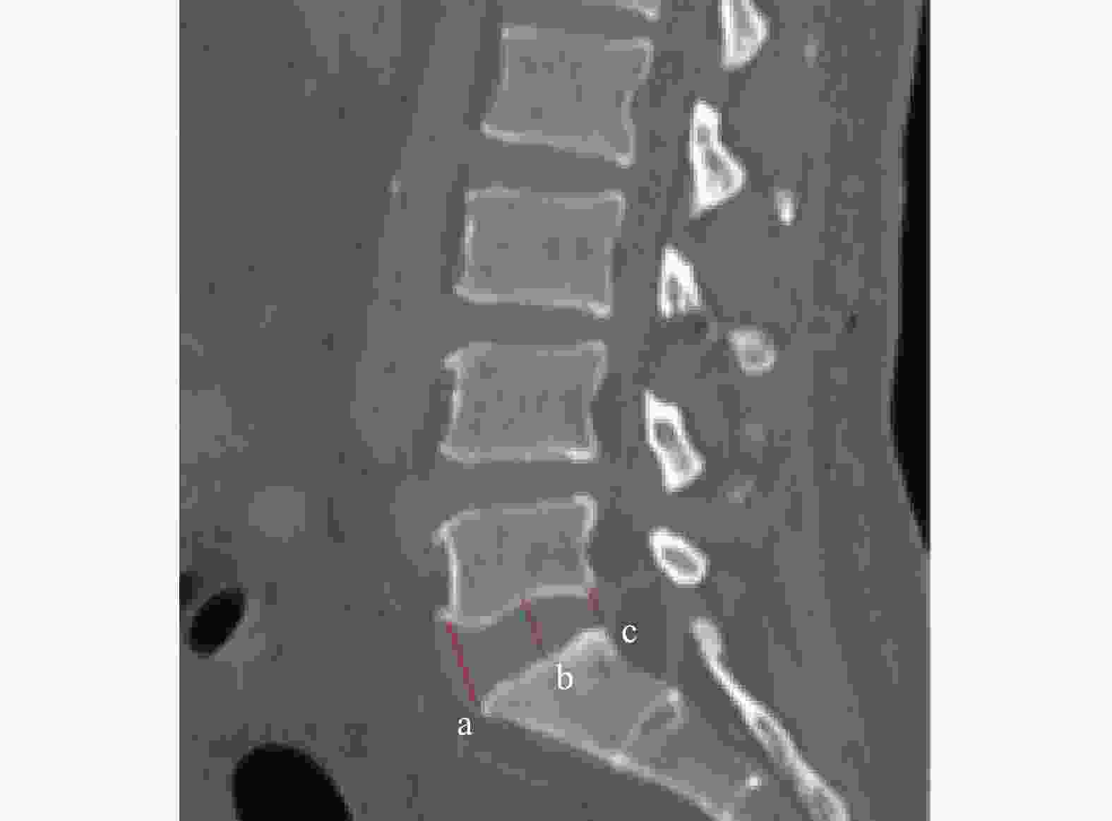
指标 UBE组 开放手术组 t P 椎间盘高度 术前 8.58±2.06 9.79±3.02 −2.121 0.038* 术后 8.46±2.18 10.24±2.68 −3.340 0.001* t 0.472 7.171 P 0.487 0.010* *P < 0.05。 -
[1] Lindbäck Y,Tropp H,Enthoven P,et al. PREPARE: Presurgery physiotherapy for patients with degenerative lumbar spine disorder: A randomized controlled trial[J]. Spine J,2018,18(8):1347-1355. doi: 10.1016/j.spinee.2017.12.009 [2] 陈洋,赵红卫,王谦. 单侧双通道内窥镜技术治疗腰椎退行性疾病的研究进展[J]. 脊柱外科杂志,2023,21(4):284-288. doi: 10.3969/j.issn.1672-2957.2023.04.013 [3] Yue J J,Long W. Full endoscopic spinal surgery techniques: advancements,indications,and outcomes[J]. Int J Spine Surg,2015,9:17. doi: 10.14444/2017 [4] Heo D H,Son S K,Eum J H,et al. Fully endoscopic lumbar interbody fusion using a percutaneous unilateral biportal endoscopic technique: Technical note and preliminary clinical results[J]. Neurosurg Focus,2017,43(2):E8. doi: 10.3171/2017.5.FOCUS17146 [5] Macnab I. Negative disc exploration. An analysis of the causes of nerve-root involvement in sixty-eight patients[J]. J Bone Joint Surg Am,1971,53(5):891-903. doi: 10.2106/00004623-197153050-00004 [6] Yoon W W,Koch J. Herniated discs: When is surgery necessary?[J]. EFORT Open Rev,2021,6(6):526-530. doi: 10.1302/2058-5241.6.210020 [7] Zhang Y,Feng B,Hu P,et al. One-hole split endoscopy technique versus unilateral biportal endoscopy technique for L5-S1 lumbar disk herniation: analysis of clinical and radiologic outcomes[J]. J Orthop Surg Res,2023,18(1):668. doi: 10.1186/s13018-023-04159-9 [8] 杨书情,张世民,吴冠男,等. 两种不同入路经皮椎间孔镜技术治疗高位腰椎间盘突出症[J]. 中国骨伤,2020,33(7):7. [9] Stanuszek A,Jędrzejek A,Gancarczyk-Urlik E,et al. Preoperative paraspinal and psoas major muscle atrophy and paraspinal muscle fatty degeneration as factors influencing the results of surgical treatment of lumbar disc disease[J]. Arch Orthop Trauma Surg,2022,142(7):1375-1384. doi: 10.1007/s00402-021-03754-x [10] Shim H K,Choi K C,Cha K H,et al. Interlaminar endoscopic lumbar discectomy using a new 8.4-mm endoscope and nerve root retractor[J]. Clin Spine Surg,2020,33(7):265-270. doi: 10.1097/BSD.0000000000000878 [11] Goudman L, Pilitsis JG, Billet B, et al. The level of agreement between the numerical rating scale and visual analogue scale for assessing pain intensity in adults with chronic pain[J]. Anaesthesia,2024,79(2):128-138. [12] Kambin P,Gellman H. Percutaneous lateral discectomy of the lumbar spine: A preliminary report[J]. Clin Orthop Relat Res,1983,174(174):127-132. [13] De Antoni D J,Claro M L,Poehling G G,et al. Translaminar lumbar epidural endoscopy: Anatomy,technique,and indications[J]. Arthroscopy,1996,12(3):330-334. doi: 10.1016/S0749-8063(96)90069-9 [14] Choi K C,Shim H K,Hwang J S,et al. Comparison of surgical invasiveness between microdiscectomy and 3 different endoscopic discectomy techniques for lumbar disc herniation[J]. World Neurosurg,2018,116:e750-e758. doi: 10.1016/j.wneu.2018.05.085 [15] Kinaci A,Moayeri N,van der Zwan A,et al. Effectiveness of sealants in prevention of cerebrospinal fluid leakage after spine surgery: A systematic review[J]. World Neurosurg,2019,127: 567-575. e1. [16] Park H J,Kim S K,Lee S C,et al. Dural tears in percutaneous biportal endoscopic spine surgery: Anatomical location and management[J]. World Neurosurg,2020,136:e578-e585. doi: 10.1016/j.wneu.2020.01.080 [17] 温冰涛,张西峰,王岩,等. 经皮内窥镜治疗腰椎间盘突出症的并发症及其处理[J]. 中华外科杂志,2011,49(12):5. [18] Wu B,Zhan G,Tian X,et al. Comparison of transforaminal percutaneous endoscopic lumbar discectomy with and without foraminoplasty for lumbar disc herniation: A 2-year follow-up[J]. Pain Research & Management,2019,2019:1-12. [19] Ahn Y,Lee H Y,Lee S H,et al. Dural tears in percutaneous endoscopic lumbar discectomy[J]. Eur Spine J,2011,20(1):58-64. doi: 10.1007/s00586-010-1493-8 [20] Pfirrmana C W,Metzdorf A,Zanetti M,et al. Magnetic resonance classification of lumbar intervertebral discdegeneration[J]. Spine(Phila Pa 1976),2001,26(17): 1873-1878. [21] 冯德伟,逄树婷,孙盼,等. 两种手术方式治疗不同Pfirrmann分级腰椎间盘突出症的疗效分析[J]. 中国骨与关节损伤杂志,2022,37(5):458-463. doi: 10.7531/j.issn.1672-9935.2022.05.003 -


 下载:
下载:


