




Immunopathogenesis of Lichen Planus
-
摘要: 扁平苔藓是一种慢性、免疫介导的炎症性疾病,以苔藓样界面皮炎为特征。扁平苔藓发病是遗传易感性与环境因素交互作用的结果。免疫机制以T细胞介导为主:IFN-γ、TNF-α及IL-23/Th17轴驱动炎症,CD8+细胞毒性T细胞通过穿孔素/颗粒酶B及Fas/FasL途径诱导角质形成细胞凋亡;JAK-STAT信号通路及代谢异常进一步放大炎症。对LP病因学、免疫发病机制及临床关联进行系统综述,为精准分型与靶向治疗提供理论依据。Abstract: Lichen planus is a chronic, immune-mediated inflammatory disease characterized by lichenoid interface dermatitis. Its development results from interactions between genetic susceptibility and environmental factors. The immune mechanisms are predominantly T-cell mediated: IFN-γ, TNF-α, and the IL-23/Th17 axis drive inflammation, while CD8+cytotoxic T cells induce keratinocyte apoptosis via the perforin/granzyme B and Fas/FasL pathways. JAK-STAT signaling and metabolic abnormalities further amplify the inflammatory response. A systematic review of the etiology, immunopathogenesis, and clinical associations of LP provides a theoretical basis for precision subtyping and targeted therapy.
-
Key words:
- Lichen planus /
- Immunity /
- Pathogenesis
-
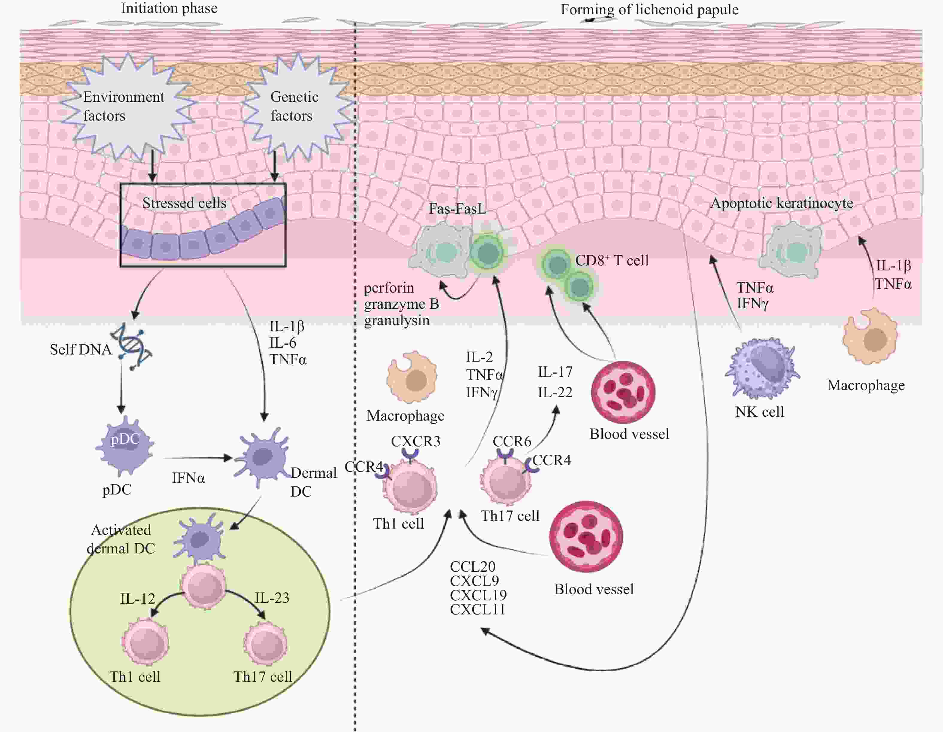
图 2 LP复杂炎症网络中包括的主要效应细胞和信号通路
注:遗传与环境因素驱动表皮基底细胞应激,激活pDC与真皮DC引发IL-12/23-Th1/Th17信号轴极化,并在CCL20/CXCL趋化因子受体轴的招募下靶向真皮,继而通过Th1/Th17协同活化CD8+T细胞,经由Fas-FasL通路及穿孔素/颗粒酶释放触发细胞毒性效应,最终诱导基底角质形成细胞凋亡,形成扁平苔藓样丘疹。
Figure 2. Major effector cells and signaling pathways involved in the complex inflammatory network of lichen planus
表 1 与LP相关的主要免疫细胞亚群及其功能
Table 1. Main immune cell subsets associated with LP and their functions
细胞类型 主要亚型/标记 病变中变化
(vs健康对照)核心功能与效应 关键分子 参考文献 树突状细胞 朗格汉斯细胞(CD1a+/CD207+)、髓系DC、浆细胞样DC 各亚型数量增加 抗原呈递,分泌IFN-α、IL-12、IL-23,启动Th1/Th17应答 CD1a,CD207,IFN-α,IL-12,IL-23 [50−53] 巨噬细胞 M1型、CD68+ 数量增加,聚集于基底膜附近(距表皮≤125μm) 分泌促炎因子及趋化因子,维持慢性炎症,介导CD8+T细胞趋化 TNF-α,IL-1β,CCL5,MMPs,MIP-1α/β(外泌体) [54−56] CD4+T细胞 Th1、Th17、Tfh 真皮浸润增加,CD4+/CD8+比值约1.75:1 分泌IFN-γ、IL-17等,辅助CD8+T细胞活化,促进慢性炎症及体液免疫 IFN-γ,IL-17,IL-22,IL-21 [60−62] CD8+T细胞 Tc1、Tc17、MAIT、γδT 表皮基底层浸润为主,随疾病进展增多 识别MHC-I呈递的抗原,通过穿孔素/颗粒酶B、FasL、TNF-α诱导角质形成细胞凋亡 穿孔素,颗粒酶B,颗粒溶素,FasL,IFN-γ [45,58−59] 调节性T细胞 FoxP3+Treg 病变组织及血液中比例升高(可占浸润细胞17.7%) 免疫抑制功能受损,可重编程为促炎表型(异常分泌IFN-γ、IL-17) FoxP3,IL-17(异常) [63−66] NK细胞 CD56dimCD16- 占皮肤LP浸润细胞约10% 产生TNF-α、IFN-γ、IL-17、IL-22,可能促进T细胞动员1 TNF-α,IFN-γ,IL-17,IL-22 [67−68] 角质形成细胞 基底角质形成细胞 凋亡增加(靶细胞) 通过MHC-I/II呈递抗原,分泌促炎因子,维持基底膜;被CTL诱导凋亡 MHC-I/II,IL-1β,IL-6,TNF-α,聚丝蛋白 [69−71] 肥大细胞 类胰蛋白酶/乳糜酶阳性 基底膜破坏区数量增加,60%呈脱颗粒状态 释放RANTES、蛋白酶等,激活MMPs,形成正反馈放大炎症 RANTES,类胰蛋白酶,乳糜酶,TNF-α [57] 中性粒细胞 多形核中性粒细胞 血清钙卫蛋白水平升高 形成NETs,N2表型可能参与OLP向鳞癌的恶性转化 钙卫蛋白,BAFF [72−73]  下载: 导出CSV
-
[1] Tekin B, Xie F, Lehman J S. Lichen planus: What is new in diagnosis and treatment[J]. Am J Clin Dermatol, 2024, 25(5): 735-764. doi: 10.1007/s40257-024-00878-9 [2] Pelet Del Toro N, Strunk A, Garg A, et al. Prevalence and treatment patterns of lichen planus[J]. J Am Acad Dermatol, 2025, 92(4): 717-723. doi: 10.1016/j.jaad.2024.09.081 [3] González-Moles M Á, Warnakulasuriya S, González-Ruiz I, et al. Worldwide prevalence of oral lichen planus: A systematic review and meta-analysis[J]. Oral Dis, 2021, 27(4): 813-828. doi: 10.1111/odi.13323 [4] Lu S L, Qi X M, Dong G, et al. Clinical characteristics and analysis of familial oral lichen planus in eight Chinese families[J]. Exp Ther Med, 2016, 12(4): 2281-2284. [5] Gorouhi F, Davari P, Fazel N. Cutaneous and mucosal lichen planus: A comprehensive review of clinical subtypes, risk factors, diagnosis, and prognosis[J]. Scientific World Journal, 2014, 2014: 742826. [6] Reeve M P, Vehviläinen M, Luo S, et al. Oral and non-oral lichen planus show genetic heterogeneity and differential risk for autoimmune disease and oral cancer[J]. Am J Hum Genet, 2024, 111(6): 1047-1060. doi: 10.1016/j.ajhg.2024.04.020 [7] Nagao Y, Nishida N, Toyo-oka L, et al. Genome-wide association study identifies risk variants for lichen planus in patients with hepatitis C virus infection[J]. Clin Gastroenterol Hepatol, 2017, 15(6): 937-944. e5. [8] Ritari J, Reeve M P, FinnGen M S, et al. Fine-mapping of HLA effects in oral and non-oral lichen planus[J]. HLA, 2026, 108(1): e70809. [9] Long X, Zhang G, Wang Q, et al. Correlations between lichen planus and autoimmune diseases: A Mendelian randomization study[J]. Arch Dermatol Res, 2024, 317(1): 36. [10] Cao J, Du T, Li J, et al. Picolinate-mediated immunomodulation: Insights from Mendelian randomization on the role of NK cell percentage in the pathogenesis of lichen planus[J]. Front Immunol, 2024, 15: 1464479. [11] Chen X, Zhang S, Wu X, et al. Inflammatory cytokines and oral lichen planus: A Mendelian randomization study[J]. Front Immunol, 2024, 15: 1332317. doi: 10.3389/fimmu.2024.1332317 [12] Guida S, Tamburelli M, Podo-Brunetti A, et al. Lichen planus is associated with hepatitis C but not with hepatitis B virus: a systematic review and meta-analysis with a focus on Italian data[J]. Ital J Dermatol Venerol, 2026, 161(1): 57-64. [13] Carrozzo M, Francia Di Celle P, Gandolfo S, et al. Increased frequency of HLA-DR6 allele in Italian patients with hepatitis C virus-associated oral lichen planus[J]. Br J Dermatol, 2001, 144(4): 803-808. doi: 10.1046/j.1365-2133.2001.04136.x [14] Raybaud H, Olivieri C V, Lupi-Pegurier L, et al. Epstein-Barr virus-infected plasma cells infiltrate erosive oral lichen planus[J]. J Dent Res, 2018, 97(13): 1494-1500. doi: 10.1177/0022034518788282 [15] de Vries H J C, Teunissen M B M, Zorgdrager F, et al. Lichen planus remission is associated with a decrease of human herpes virus type 7 protein expression in plasmacytoid dendritic cells[J]. Arch Dermatol Res, 2007, 299(4): 213-219. doi: 10.1007/s00403-007-0750-0 [16] Mizukawa Y, Horie C, Yamazaki Y, et al. Detection of Varicella-zoster virus antigens in lesional skin of zosteriform lichen planus but not in that of linear lichen planus[J]. Dermatology, 2012, 225(1): 22-26. doi: 10.1159/000339771 [17] Choi Y S, Kim Y, Yoon H J, et al. The presence of bacteria within tissue provides insights into the pathogenesis of oral lichen planus[J]. Sci Rep, 2016, 6: 29186. doi: 10.1038/srep29186 [18] Israyani, Rovani C A, Marlina E, et al. Complexities of Candida colonization and oral microbiome in oral lichen planus: A systematic review and meta-analysis[J]. Dent J, 2025, 13(7): 310. doi: 10.3390/dj13070310 [19] Pignatelli P, Curia M C, Tenore G, et al. Oral bacteriome and oral potentially malignant disorders: A systematic review of the associations[J]. Arch Oral Biol, 2024, 160: 105891. doi: 10.1016/j.archoralbio.2024.105891 [20] Zhou Q, Liu P, Chen M, et al. Microbiological mechanisms of oral mucosal disease: Oral-intestinal crosstalk and probiotic therapy[J]. Front Oral Health, 2025, 6: 1485028. doi: 10.3389/froh.2025.1485028 [21] Maul J T, Guillet C, Oschmann A, et al. Cutaneous lichenoid drug eruptions: A narrative review evaluating demographics, clinical features and culprit medications[J]. J Eur Acad Dermatol Venereol, 2023, 37(5): 965-975. doi: 10.1111/jdv.18879 [22] Somasundaram A, Avanija G S, Munisamy M, et al. An isoradiotopic response of lichen planus: A case report and review of literature[J]. Radiat Oncol J, 2025, 43(2): 99-103. doi: 10.3857/roj.2024.00668 [23] Li K, He W, Hua H. Characteristics of the psychopathological status of oral lichen planus: A systematic review and meta-analysis[J]. Aust Dent J, 2022, 67(2): 113-124. doi: 10.1111/adj.12896 [24] Aghamajidi A, Raoufi E, Parsamanesh G, et al. The attentive focus on T cell-mediated autoimmune pathogenesis of psoriasis, lichen planus and vitiligo[J]. Scand J Immunol, 2021, 93(4): e13000. doi: 10.1111/sji.13000 [25] El-Howati A, Thornhill M H, Colley H E, et al. Immune mechanisms in oral lichen planus[J]. Oral Dis, 2023, 29(4): 1400-1415. doi: 10.1111/odi.14142 [26] Kawamura E, Nakamura S, Sasaki M, et al. Accumulation of oligoclonal T cells in the infiltrating lymphocytes in oral lichen planus[J]. J Oral Pathol Med, 2003, 32(5): 282-289. doi: 10.1034/j.1600-0714.2003.00143.x [27] de Carvalho G C, Domingues R, Almeida de Sousa Nogueira M, et al. Up-regulation of proinflammatory genes and cytokines induced by S100A8 in CD8+ T cells in lichen planus[J]. Acta Derm Venereol, 2016, 96(4): 485-489. [28] Kersh A E, Sati S, Huang J, et al. CXCL9, CXCL10, and CCL19 synergistically recruit T lymphocytes to skin in lichen planus[J]. JCI Insight, 2024, 9(20): e179899. doi: 10.1172/jci.insight.179899 [29] Vičić M, Hlača N, Kaštelan M, et al. Comprehensive insight into lichen planus immunopathogenesis[J]. Int J Mol Sci, 2023, 24(3): 3038. doi: 10.3390/ijms24033038 [30] Grolleau C, Poisot T, Roux C, et al. Single-cell profiling of lichen planus reveals type I IFN response triggering the interface dermatitis reaction[J]. J Investig Dermatol, 2025, 145(12): 3025-3036. e12. [31] Didona D, Caposiena Caro R D, Calabrese L, et al. Use of JAK inhibitors in lichen planus: An update[J]. Medicina, 2025, 61(6): 1056. doi: 10.3390/medicina61061056 [32] Walton L J, Thornhill M H, Macey M G, et al. Cutaneous lymphocyte associated antigen (CLA) and alpha e beta 7 integrins are expressed by mononuclear cells in skin and oral lichen planus[J]. J Oral Pathol Med, 1997, 26(9): 402-407. doi: 10.1111/j.1600-0714.1997.tb00239.x [33] Shiohara T, Mizukawa Y, Takahashi R, et al. Pathomechanisms of lichen planus autoimmunity elicited by cross-reactive T cells[J]. Curr Dir Autoimmun, 2008, 10: 206-226. [34] Zargaran M, Jamshidi S, Eshghyar N, et al. Suitability/unsuitability of cell proliferation as an indicator of malignant potential in oral lichen planus: An immunohistochemical study[J]. Asian Pac J Cancer Prev, 2013, 14(11): 6979-6983. doi: 10.7314/APJCP.2013.14.11.6979 [35] de Brito Monteiro B V, Dos Santos Pereira J, Nonaka C F W, et al. Immunoexpression of Th17-related cytokines in oral lichen planus[J]. Appl Immunohistochem Mol Morphol, 2015, 23(6): 409-415. doi: 10.1097/PAI.0000000000000096 [36] Georgescu S R, Mitran C I, Mitran M I, et al. Oxidative stress in cutaneous lichen planus—a narrative review[J]. J Clin Med, 2021, 10(12): 2692. doi: 10.3390/jcm10122692 [37] Lauffer F, Jargosch M, Krause L, et al. Type I immune response induces keratinocyte necroptosis and is associated with interface dermatitis[J]. J Invest Dermatol, 2018, 138(8): 1785-1794. doi: 10.1016/j.jid.2018.02.034 [38] Vičić M, Sotošek V, Brajac I, et al. The possible involvement of granulysin mediated cytotoxicity in keratinocytes disruption in lichen planus[J]. Med Hypotheses, 2021, 149: 110546. doi: 10.1016/j.mehy.2021.110546 [39] de Saint Basile G, Ménasché G, Fischer A. Molecular mechanisms of biogenesis and exocytosis of cytotoxic granules[J]. Nat Rev Immunol, 2010, 10(8): 568-579. doi: 10.1038/nri2803 [40] Ammar M, Mokni M, Boubaker S, et al. Involvement of granzyme B and granulysin in the cytotoxic response in lichen planus[J]. J Cutan Pathol, 2008, 35(7): 630-634. doi: 10.1111/j.1600-0560.2007.00892.x [41] Sklavounou A, Chrysomali E, Scorilas A, et al. TNF-alpha expression and apoptosis-regulating proteins in oral lichen planus: A comparative immunohistochemical evaluation[J]. J Oral Pathol Med, 2000, 29(8): 370-375. [42] Tudi M, Aierken Z, Tuerxun M, et al. Deletion of NFIA leads to activation of S100A7 and inflammatory response-induced apoptosis of keratinocytes in oral lichen planus progression[J]. Kaohsiung J Med Sci, 2025, 41(9): e70052. doi: 10.1002/kjm2.70052 [43] Kurzen N, Mubarak M, Eigemann J, et al. Death-associated protein kinase 1 dampens keratinocyte necroptosis and expression of inflammatory genes in lichen planus[J]. J Investig Dermatol, 2025, 145(8): 1921-1929. e13. [44] Dorrego M V, Correnti M, Delgado R, et al. Oral lichen planus: Immunohistology of mucosal lesions[J]. J Oral Pathol Med, 2002, 31(7): 410-414. doi: 10.1034/j.1600-0714.2002.00097.x [45] Sugerman P B, Satterwhite K, Bigby M. Autocytotoxic T-cell clones in lichen planus[J]. Br J Dermatol, 2000, 142(3): 449-456. doi: 10.1046/j.1365-2133.2000.03355.x [46] Hu J Y, Zhang J, Cui J L, et al. Increasing CCL5/CCR5 on CD4+ T cells in peripheral blood of oral lichen planus[J]. Cytokine, 2013, 62(1): 141-145. doi: 10.1016/j.cyto.2013.01.020 [47] Yamamoto T. Tissue-resident memory T cells in lichen planus on a surgical scar[J]. Clin Exp Dermatol, 2024, 49(9): 1076-1078. doi: 10.1093/ced/llae125 [48] Trucci V M, Salum F G, Figueiredo M A, et al. Interrelationship of dendritic cells, type 1 interferon system, regulatory T cells and toll-like receptors and their role in lichen planus and lupus erythematosus —a literature review[J]. Arch Oral Biol, 2013, 58(10): 1532-1540. doi: 10.1016/j.archoralbio.2013.06.016 [49] Podolsky A S, Lipner S R. Mechanisms underlying lichen planus in association with biologic therapy[J]. J Inflamm Res, 2025, 18: 14961-14970. doi: 10.2147/JIR.S554375 [50] Santoro A, Majorana A, Roversi L, et al. Recruitment of dendritic cells in oral lichen planus[J]. J Pathol, 2005, 205(4): 426-434. doi: 10.1002/path.1699 [51] Ferrisse T M, de Oliveira A B, Palaçon M P, et al. Immunohistochemical evaluation of Langerhans cells in oral lichen planus and oral lichenoid lesions[J]. Arch Oral Biol, 2021, 124: 105027. doi: 10.1016/j.archoralbio.2020.105027 [52] Cardozo A L, Moura-Castro C, Figueiredo M, et al. Oral lichen planus and dermal dendrocytes[J]. Actas Dermo Sifiliográficas Engl Ed, 2009, 100(1): 46-52. doi: 10.1016/S0001-7310(09)70056-0 [53] Wenzel J, Scheler M, Proelss J, et al. Type I interferon-associated cytotoxic inflammation in lichen planus[J]. J Cutan Pathol, 2006, 33(10): 672-678. doi: 10.1111/j.1600-0560.2006.00527.x [54] Ferrisse T M, de Oliveira A B, Palaçon M P, et al. The role of CD68+ and CD163+ macrophages in immunopathogenesis of oral lichen planus and oral lichenoid lesions[J]. Immunobiology, 2021, 226(3): 152072. doi: 10.1016/j.imbio.2021.152072 [55] Sutera S, Furchì O A, Pentenero M. Macrophages and the immune microenvironment in OPMDs: A systematic review of the literature[J]. Front Oral Health, 2025, 6: 1605978. doi: 10.3389/froh.2025.1605978 [56] Yang J Y, Zhang J, Lu R, et al. T cell-derived exosomes induced macrophage inflammatory protein-1α/β drive the trafficking of CD8+ T cells in oral lichen planus[J]. J Cell Mol Med, 2020, 24(23): 14086-14098. doi: 10.1111/jcmm.16020 [57] Żychowska M, Woźniak Z, Baran W. Immunohistochemical analysis of the expression of selected cell lineage markers (CD4, CD8, CD68, c-Kit, Foxp3, CD56, CD20) in cutaneous variant of lichen planus[J]. Int J Dermatol, 2021, 60(9): 1097-1101. doi: 10.1111/ijd.15437 [58] Zhou X J, Sugerman P B, Savage N W, et al. Intra-epithelial CD8+ T cells and basement membrane disruption in oral lichen planus[J]. J Oral Pathol Med, 2002, 31(1): 23-27. doi: 10.1046/j.0904-2512.2001.10063.x [59] Le Bourhis L, Martin E, Péguillet I, et al. Antimicrobial activity of mucosal-associated invariant T cells[J]. Nat Immunol, 2010, 11(8): 701-708. doi: 10.1038/ni.1890 [60] Khan A, Farah C S, Savage N W, et al. Th1 cytokines in oral lichen planus[J]. J Oral Pathol Med, 2003, 32(2): 77-83. doi: 10.1034/j.1600-0714.2003.00077.x [61] Gupta R, Singh S, Mohanty S, et al. Localization of T-cell subsets in cutaneous lichen planus: An insight into pathogenetic mechanism[J]. Indian J Dermatol Venereol Leprol, 2010, 76(6): 707. doi: 10.4103/0378-6323.72452 [62] Qian H, Jiao L, Fan Z, et al. Analysis of immunologic function changes in lichen planus after clinical treatment[J]. Med Sci Monit, 2018, 24: 8716-8721. doi: 10.12659/msm.910931 [63] Tao X A, Xia J, Chen X B, et al. FOXP3+ T regulatory cells in lesions of oral lichen planus correlated with disease activity[J]. Oral Dis, 2010, 16(1): 76-82. doi: 10.1111/j.1601-0825.2009.01608.x [64] Zhou L, Cao T, Wang Y, et al. Frequently increased but functionally impaired CD4+CD25+ regulatory T cells in patients with oral lichen planus[J]. Inflammation, 2016, 39(3): 1205-1215. doi: 10.1007/s10753-016-0356-9 [65] Xue N, Wang Y, Cheng H, et al. Regulatory T cell therapy suppresses inflammation of oral mucosa[J]. Front Immunol, 2022, 13: 1009742. doi: 10.3389/fimmu.2022.1009742 [66] . Gatzka M, Scharffetter-Kochanek K. T-cell plasticity in inflammatory skin diseases--the good, the bad, and the chameleons[J]. J Dtsch Dermatol Ges, 2015, 13(7): 647-652. [67] Carbone T, Nasorri F, Pennino D, et al. CD56 highCD16 - NK cell involvement in cutaneous lichen planus[J]. Eur J Dermatol, 2010, 20(6): 724-730. [68] Parolini S, Santoro A, Marcenaro E, et al. The role of chemerin in the colocalization of NK and dendritic cell subsets into inflamed tissues[J]. Blood, 2007, 109(9): 3625-3632. doi: 10.1182/blood-2006-08-038844 [69] Shao S, Tsoi L, Sarkar M K, et al. 044 IFN-γ enhances cell-mediated cytotoxicity against keratinocytes via JAK2/STAT1 in lichen planus[J]. J Investig Dermatol, 2019, 139(5): S8. [70] Ge X, Xie H, Wang L, et al. microRNA-122 promotes apoptosis of keratinocytes in oral lichen planus through suppressing VDR expression[J]. J Cell Mol Med, 2021, 25(7): 3400-3407. doi: 10.1111/jcmm.16418 [71] Yamamoto T, Osaki T, Yoneda K, et al. Cytokine production by keratinocytes and mononuclear infiltrates in oral lichen planus[J]. J Oral Pathology Medicine, 1994, 23(7): 309-315. doi: 10.1111/j.1600-0714.1994.tb00067.x [72] Khattab F M, Ghonaim R, Samir M A. Estimation of neutrophil activation marker in lichen planus patients[J]. J Cosmet Dermatol, 2022, 21(4): 1625-1628. doi: 10.1111/jocd.14252 [73] Jablonska E, Garley M, Surazynski A, et al. Neutrophil extracellular traps (NETs) formation induced by TGF-β in oral lichen planus–Possible implications for the development of oral cancer[J]. Immunobiology, 2020, 225(2): 151901. doi: 10.1016/j.imbio.2019.151901 [74] Mozaffari H R, Sharifi R, Hayati M, et al. Evaluation of serum and salivary interferon-γ levels in patients with oral lichen planus: A systematic review and meta-analysis of case-control studies[J]. Oral Surg Oral Med Oral Pathol Oral Radiol, 2019, 127(3): 210-217. doi: 10.1016/j.oooo.2018.11.006 [75] Hama P N, Ahmed K M. Evaluation of salivary interleukin 6 interleukin 1 beta and interferon gamma in oral and mucocutaneous lichen planus patients[J]. Sci Rep, 2026, 16: 5678. doi: 10.1038/s41598-026-36506-1 [76] Abduelmula A, Bagit A, Mufti A, et al. The use of Janus kinase inhibitors for lichen planus: An evidence-based review[J]. J Cutan Med Surg, 2023, 27(3): 271-276. doi: 10.1177/12034754231156100 [77] Nakahira M, Ahn H J, Park W R, et al. Synergy of IL-12 and IL-18 for IFN-γ gene expression: IL-12-induced STAT4 contributes to IFN-γ promoter activation by up-regulating the binding activity of IL-18-induced activator protein 1[J]. J Immunol, 2002, 168(3): 1146-1153. [78] Pietschke K, Holstein J, Meier K, et al. The inflammation in cutaneous lichen planus is dominated by IFN-ϒ and IL-21—a basis for therapeutic JAK1 inhibition[J]. Exp Dermatol, 2021, 30(2): 262-270. doi: 10.1111/exd.14226 -

 点击查看大图
图(2) / 表(1)
计量
- 文章访问数: 8
- HTML全文浏览量: 3
- PDF下载量: 0
- 被引次数: 0
