




Research on Risk Assessment of High-risk Plaque Risk Based on Pericoronary Fat Quantification via CCTA
-
摘要:
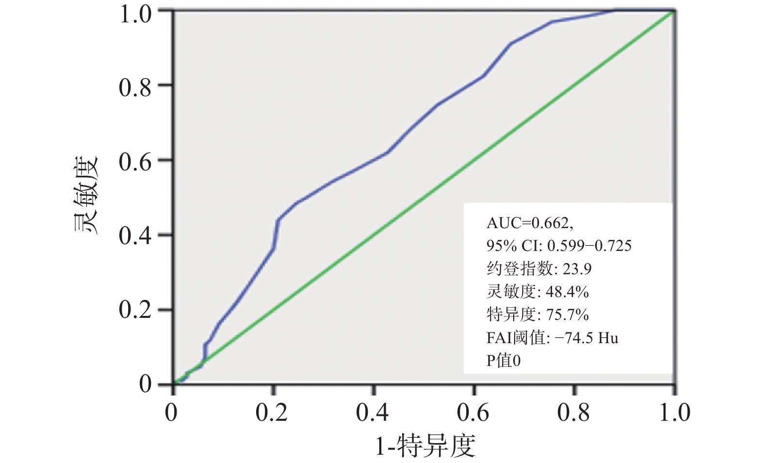
目的 以斑块冠周脂肪衰减指数(fat attenuation index,FAI)值为参考标准,探讨不同高危特征数量及组合的高危斑块风险程度。 方法 回顾性收集2022年9月至2024年6月行冠状动脉CT血管成像(coronary CT angiography,CCTA)且单支冠脉仅存在高危斑块或非高危斑块的患者331例,共纳入399处斑块,其中高危斑块289处、非高危斑块110处。根据斑块是否具有高危特征、高危特征数量、特征组合及是否包含点状钙化或餐巾环征进行分组,测量斑块周围FAI。以FAI值为评估斑块风险性的参考标准,确定识别高危斑块的FAI阈值,并比较不同特征数量及组成的高危斑块风险差异,最终对患者进行随访及对所得结果进行外部验证。 结果 高危斑块的FAI识别阈值为-74.5 Hu。含2种高危特征的斑块FAI值低于含3种或4种高危特征的斑块,差异具有统计学意义(P < 0.05);3种与4种高危特征组间FAI值差异无统计学意义(P > 0.05)。低密度合并正性重构斑块的FAI值高于点状钙化合并正性重构斑块,差异显著(P < 0.01);有餐巾环征组FAI值高于无餐巾环征组(P < 0.05);有点状钙化组FAI值低于无点状钙化组,但差异无统计学意义(P > 0.05);外部验证高危组FAI值显著高于非高危组(P < 0.001),MACE 事件全部发生于高危组,低密度斑块+正性重构特征及含餐巾环征特征的分布亦呈现相似趋势,且均有统计学意义(P < 0.05)。 结论 结合冠脉斑块高危特征的数量与组合,可对斑块发生急性冠脉综合征的风险进行分层评估。 -
关键词:
- 冠状动脉CT血管成像 /
- 脂肪衰减指数 /
- 高危斑块 /
- 风险分层 /
- 急性冠脉综合征
Abstract:Objective To investigate the risk levels of high-risk plaques with different numbers and combinations of high-risk features, using the pericoronary fat attenuation index (FAI) as a reference standard. Methods A total of 331 patients who underwent coronary CT angiography (CCTA) between September 2022 and June 2024 and had either high-risk or non-high-risk plaques in a single coronary artery were retrospectively enrolled. A total of 399 plaques were analyzed, including 289 high-risk plaques and 110 non-high-risk plaques. Patients were grouped based on the presence of high-risk features, the number and composition of these features, and the presence of spotty calcification or napkin-ring sign. The FAI surrounding the plaques was measured. Using the FAI value as a reference standard for assessing plaque risk, the optimal FAI threshold for identifying high-risk plaques was determined, and risk differences among plaques with varying high-risk features were compared. Finally, patients were followed up and the obtained results were externally validated. Results The optimal FAI threshold for identifying high-risk plaques was -74.5 HU. The FAI values of plaques containing two high-risk features were significantly lower than those of plaques containing three or four features (P < 0.05). No significant difference was found between the three and four features groups(P > 0.05). Plaques with low attenuation and positive remodeling showed significantly higher FAI values than those with spotty calcification and positive remodeling (P < 0.01). The group with napkin-ring sign had significantly higher FAI values than those of the group without the napkin-ring sign(P < 0.05). No significant difference in FAI was found between plaques with and without spotty calcification (P > 0.05).The high-risk group exhibited a significantly higher FAI values (P < 0.001). Notably, all MACE events occurred in the high-risk group, and the distribution of the low-density plaque plus positive remodeling feature and the napkin-ring sign feature also showed similar trends, all with statistical significance (P < 0.05). Conclusion Combining the number and combination of high-risk features of coronary plaques allows for stratified assessment of the risk of acute coronary syndrome associated with plaques. -
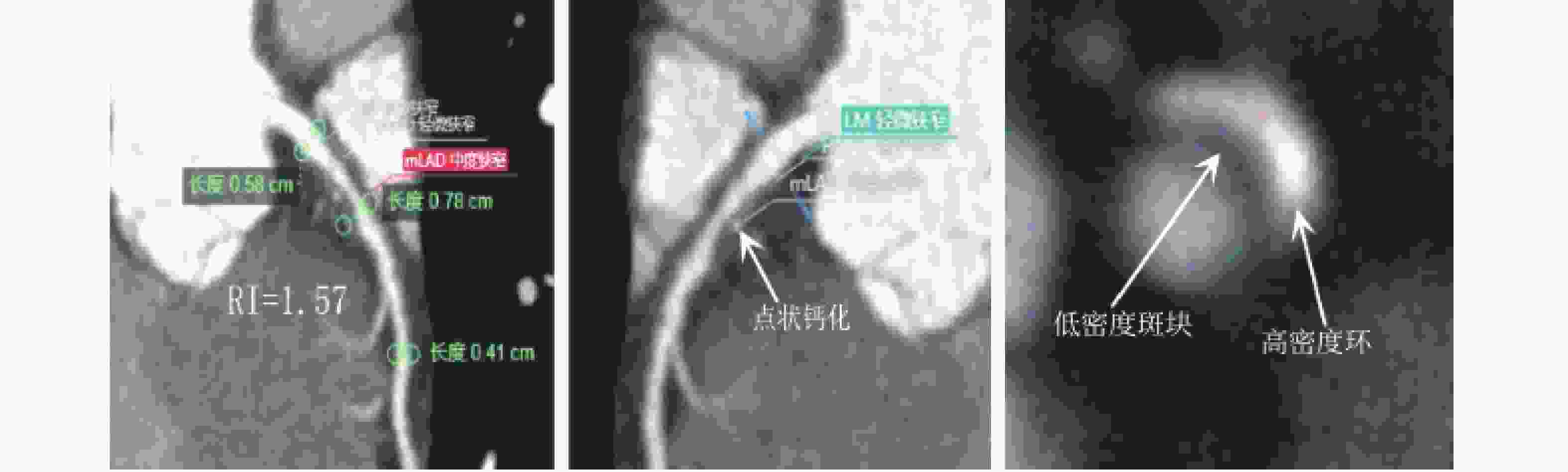
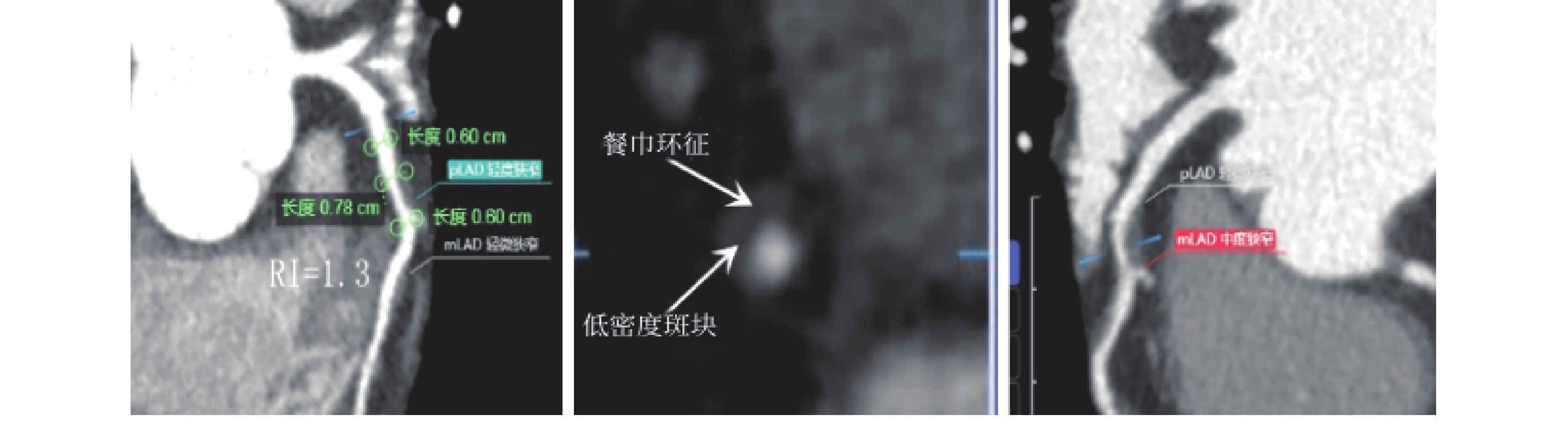
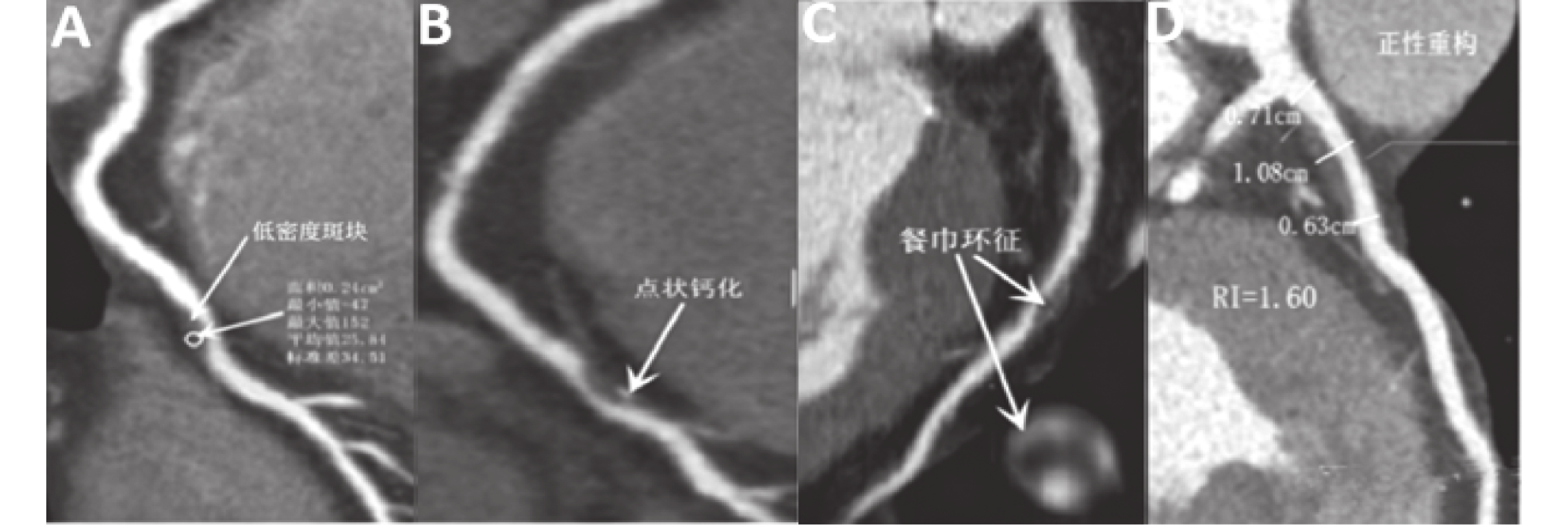
图 4 4项高危特征
注: 低密度斑块合并正性重构、餐巾环征及点状钙化。FAI测量方法:选取目标斑块后,以斑块为中心设置矩形ROI:径向直径为血管外壁外扩5 mm,长度覆盖斑块两端管腔无狭窄处。由软件自动计算FAI值(Hu)。由1名心血管影像研究生在基线(第1周)进行测量2次并取平均值,间隔1周后(第2周)再次进行测量2次并取平均值,以2次平均值的均值作为测量结果。最终以第1周的平均值和第2周的平均值计算得到ICC为0.92,且P= 0.019。
Figure 4. Four high-risk features
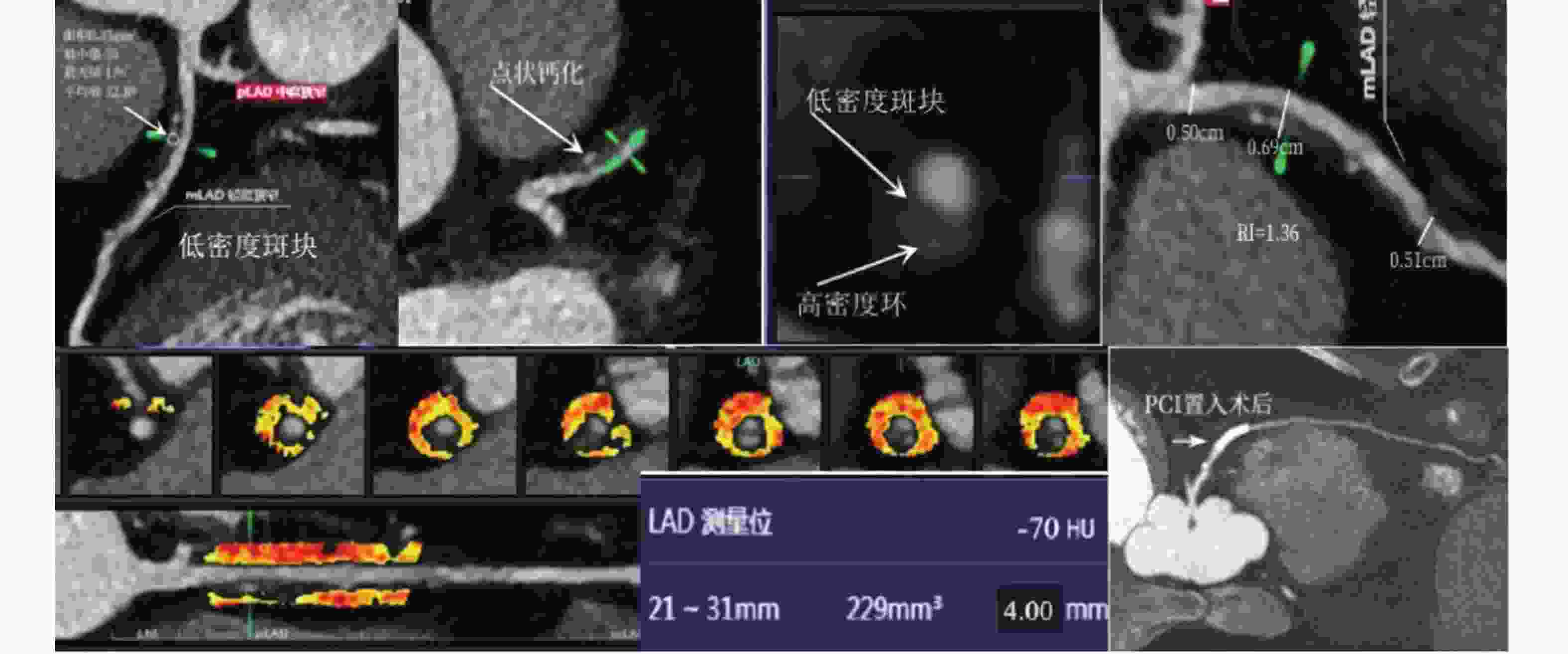
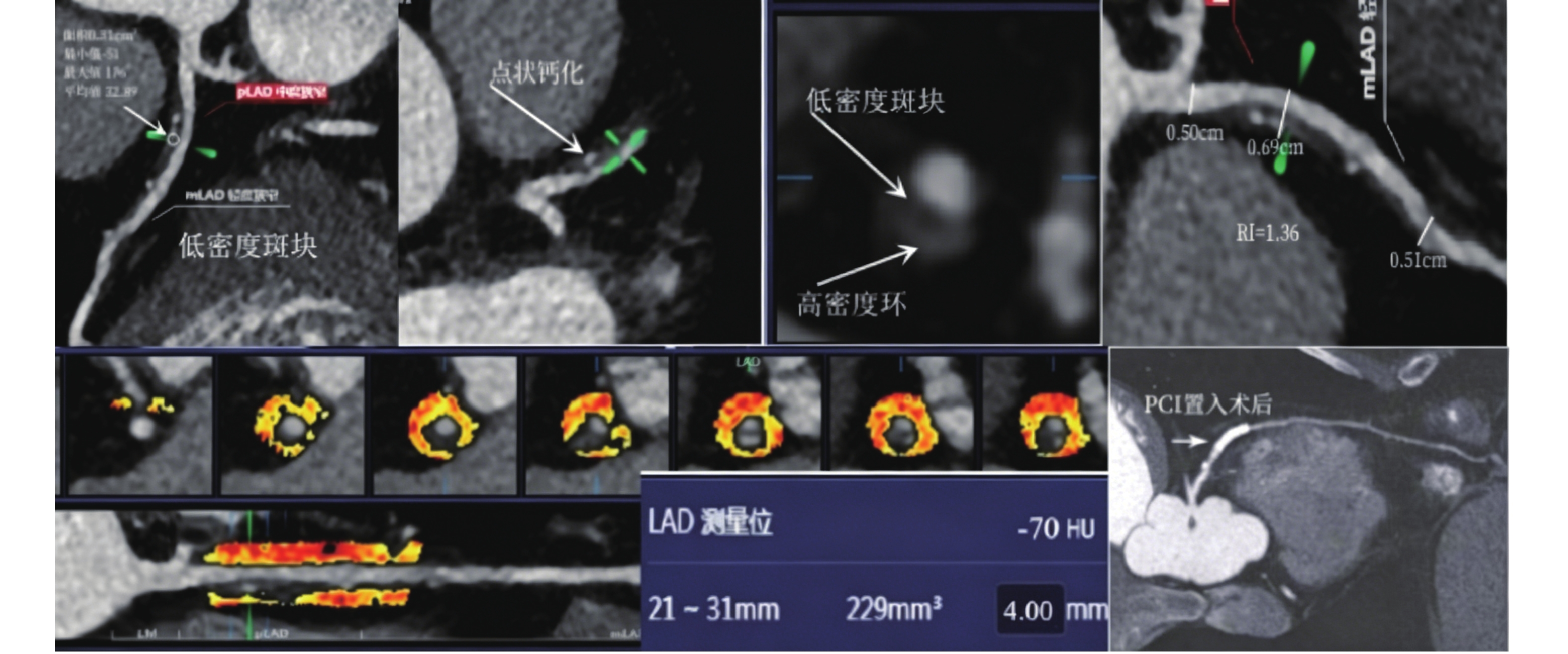
图 6 随访病例
注:男性,57岁。CCTA提示冠状动脉LAD低密度斑块、点状钙化、餐巾环征及正性重构四种高危特征(箭头),斑块周围FAI值 -70HU。随访1年8个月后出现急性心肌梗死(ACS),并行PCI术后复查。
Figure 6. Follow-up cases
表 1 高危斑块与非高危斑块FAI(Hu)比较($ \bar x \pm s $)
Table 1. Comparison of FAI(Hu) between high-risk plaques and non-high-risk plaques ($ \bar x \pm s $)
指标 FAI(Hu) 非高危斑块(n = 110) −79.25 ± 8.55 高危斑块(n = 289) −74.89 ± 6.86 t 5.289 P <0.001* *P < 0.05。  下载: 导出CSV
表 2 不同高危特征数量的高危斑块FAI(Hu)比较($ \bar x \pm s $)
Table 2. Comparison of FAI(Hu)of high-risk plaques with different numbers of high-risk features ($ \bar x \pm s $)
特征数量 FAI 2项(n=184) −75.93 ± 6.59 3项(n=69) −73.36 ± 7.62∆ 4项(n=36) −72.50 ± 5.59∆ F 6.218 P 0.002* *P < 0.05;与2项特征组比较,∆P < 0.05。
下载: 导出CSV
表 3 2种高危特征的斑块FAI(Hu)比较($ \bar x \pm s $)
Table 3. Comparison of FAI (HU) among plaques with two high-risk features($ \bar x \pm s $)
指标 FAI(Hu) 低密度+正性重构(n = 139) −74.98 ± 6.94a 点状钙化+正性重构(n = 35) −79.48 ± 3.93 低密度+点状钙化(n = 10) −76.60 ± 4.79 F 7.215 P 0.001* *P < 0.05;与点状钙化+正性重构组比较,aP < 0.05。
下载: 导出CSV
表 4 3种高危特征的斑块FAI(Hu)比较($ \bar x \pm s $)
Table 4. Comparison of FAI (Hu) among plaques with three high-risk features($ \bar x \pm s $)
指标 FAI(Hu) FAI(Hu) −74.87 ± 5.65 低密度+点状钙化+正性重构(n = 23) −74.87 ± 5.65 餐巾环征+正性重构+低密度(n = 46) −72.61 ± 8.40 t −1.164 P 0.249
下载: 导出CSV
表 5 有无餐巾环征的高危斑块FAI(Hu)比较($ \bar x \pm s $)
Table 5. Comparison of FAI (Hu) of high-risk plaques with and without napkin ring sign($ \bar x \pm s $)
指标 FAI (Hu) 有餐巾环征(n = 82) 72.56 ± 7.25 无餐巾环征(n = 207) −75.81 ± 6.49 t 3.709 P <0.001* *P < 0.05。
下载: 导出CSV
表 6 有无点状钙化的高危斑块FAI(Hu)比较($ \bar x \pm s $)
Table 6. Comparison of FAI (Hu) of high-risk plaques with and without spot calcification($ \bar x \pm s $)
指标 FAI(Hu) 有点状钙化(n = 104) −75.77 ± 5.76 无点状钙化(n = 185) −74.39 ± 7.38 t −1.639 P 0.1
下载: 导出CSV
表 7 不同FAI(Hu)/特征组合斑块风险分层的临床基线及随访结果比较
Table 7. Comparison of clinical baseline and follow-up outcomes of plaque risk stratification with different FAI(Hu)/feature combinations
变量 合计(n=90) 高危组(n = 30) 非高危组(n = 60) t/χ2 P 平均FAI(Hu) −79.79 ± 7.29 −72.24 ± 6.94 −81.61 ± 6.12 6.342 <0.001* MACE事件 8(8.89) 8(26.7) 0(0.00) 17.56 <0.001* 含低密度+正性重构 27(30.0) 27(90.0) 0(0.00) 77.14 <0.001* 含餐巾环征 9(10.0) 9(30.0) 0(0.00) 20.00 <0.001* *P < 0.05。
下载: 导出CSV
-
[1] 李梅, 陈蕊, 周烨, 等. 急性冠脉综合征斑块病理的性别差异[J]. 国际心血管病杂志, 2024, 51(3): 144-147. doi: 10.3969/j.issn.1673-6583.2024.03.005 [2] 中华医学会心血管病学分会, 中国老年医学学会, 中华心血管病杂志编辑委员会. 冠状动脉CT血管成像在慢性冠脉综合征的风险分层和诊疗中应用的专家共识[J]. 中华心血管病杂志, 2025, 53(1): 16-27. doi: 10.3760/cma.j.cn112148-20240919-00552 [3] Kuneman J H, van Rosendael S E, van der Bijl P, et al. Pericoronary adipose tissue attenuation in patients with acute coronary syndrome versus stable coronary artery disease[J]. Circ Cardiovasc Imaging, 2023, 16(2): e014672. [4] Jing M, Xi H, Yang J, et al. Relationship between pericoronary fat-attenuation values quantified by coronary computed tomography angiography and coronary artery disease severity[J]. Clin Radiol, 2024, 79(8): e1021-e1030. doi: 10.1016/j.crad.2024.02.020 [5] Kinoshita D, Suzuki K, Fujimoto D, et al. High-risk plaque features and perivascular inflammation[J]. J Cardiovasc Comput Tomogr, 2025, 19(3): 299-305. doi: 10.1016/j.jcct.2025.01.010 [6] Zhu L, Ha R, Machida H, et al. Image quality of coronary CT angiography at ultra low tube voltage reconstructed with a deep-learning image reconstruction algorithm in patients of different weight[J]. Quant Imaging Med Surg, 2023, 13(6): 3891-3901. doi: 10.21037/qims-22-1141 [7] 许玲, 吴红燕, 徐瑶, 等. 急性冠脉综合征患者炎症指数、血脂指标变化与冠状动脉病变严重程度的相关性[J]. 国际医药卫生导报, 2025, 31(11): 1780-1784. doi: 10.3760/cma.j.cn441417-20241230-11004 [8] Jing M, Xi H, Wang Y, et al. Association between pericoronary fat attenuation index values and plaque composition volume fraction measured by coronary computed tomography angiography[J]. Acad Radiol, 2024, 31(9): 3579-3589. doi: 10.1016/j.acra.2024.05.001 [9] Rotzinger D C, Magnin V, van der Wal A C, et al. Coronary CT angiography for the assessment of atherosclerotic plaque inflammation: Postmortem proof of concept with histological validation[J]. Eur Radiol, 2024, 34(3): 1755-1763. doi: 10.1007/s00330-023-10169-2 [10] Oikonomou E K, Marwan M, Desai M Y, et al. Non-invasive detection of coronary inflammation using computed tomography and prediction of residual cardiovascular risk (the CRISP CT study): A post-hoc analysis of prospective outcome data[J]. Lancet, 2018, 392(10151): 929-939. doi: 10.1016/S0140-6736(18)31114-0 [11] Kinoshita D, Watanabe M, Jang I K. The role of high-risk plaque in refining cardiovascular event prediction[J]. Curr Opin Cardiol, 2025, 40(6): 432-439. doi: 10.1097/HCO.0000000000001249 [12] Kruk M, Wardziak Ł, Kolossvary M, et al. Identification of noncalcified coronary plaque characteristics using machine learning radiomic analysis of non-contrast high-resolution computed tomography[J]. Kardiol Pol, 2023, 81(10): 978-989. doi: 10.33963/v.kp.97206 [13] 孙明菲, 刘婷, 袁雪, 等. 冠脉斑块CT血管造影的定性特征对预测心肌缺血的诊断价值[J]. 中国临床医学影像杂志, 2021, 32(3): 190-194. doi: 10.12117/jccmi.2021.03.008 [14] Kinoshita D, Suzuki K, Yuki H, et al. Coronary plaque phenotype associated with positive remodeling[J]. J Cardiovasc Comput Tomogr, 2024, 18(4): 401-407. doi: 10.1016/j.jcct.2024.04.009 [15] Nurmohamed N S, Gaillard E L, Malkasian S, et al. Lipoprotein(a) and long-term plaque progression, low-density plaque, and pericoronary inflammation[J]. JAMA Cardiol, 2024, 9(9): 826-834. doi: 10.1001/jamacardio.2024.1874 [16] Wang C, Wu X, Liu Y, et al. Association between coronary plaque vulnerability features and multiparametric pericoronary fat indices on coronary computed tomography angiography: A cross-sectional study[J]. Quant Imaging Med Surg, 2025, 15(8): 6897-6909. doi: 10.21037/qims-24-1002 [17] Nie J Y, Chen W X, Wu Q D. Relationship analysis between pericoronary fat attenuation index and parameters of single plaque[J]. J Comput Assist Tomogr, 2024, 48(4): 647-651. doi: 10.1097/RCT.0000000000001589 [18] Kishan Singh, R, Astha, K, Tarvinder Bir, S, et al. Characteristics of coronary artery vulnerable plaque on coronary computed tomography angiography (CCTA) and optical coherence tomography (OCT) - a review article[J]. J Cardiovasc Med Cardiol, 2025, 12(1): 1-6. doi: 10.17352/2455-2976.000218 [19] 王志伟. 冠脉斑块特征及斑块周围FAI在预测急性冠脉综合征的价值[D]. 大连: 大连医科大学, 2023. [20] 陈治宇, 徐晗, 颜欢欢, 等. 基于冠状动脉CT血管成像评估高危斑块特征与血清高敏C反应蛋白水平的关系[J]. 中国中西医结合影像学杂志, 2024, 22(5): 554-558. doi: 10.3969/j.issn.1672-0512.2024.05.012 [21] Busco M, Ricchiuto A, Bianchini F, et al. Optical coherence tomography imaging for the evaluation and treatment of calcified coronary artery disease[J]. G Ital Cardiol (Rome), 2024, 25(6 Suppl 1): 8S-15S. [22] Qin Z, Yu L, Zhang Y, et al. Coronary artery calcification and plaque stability: An optical coherence tomography study[J]. Heliyon, 2023, 9(12): e23191. doi: 10.1016/j.heliyon.2023.e23191 -

 点击查看大图
计量
- 文章访问数: 2
- HTML全文浏览量: 1
- PDF下载量: 0
- 被引次数: 0
