

Histological and Ultrastructural Studies of Lung Tissues in A Piglet Model of Early Onset Scoliosis with Combination of the Thoracic Insufficiency Syndrome
-
摘要:
目的 探究早发性脊柱侧凸(early onset scoliosis,EOS)合并胸廓发育不良综合征(thoracic insufficiency syndrome,TIS)幼猪在生长友好技术治疗前后肺组织学及超微结构变化。 方法 选择6周龄幼猪,随机分为对照组(n = 3)、模型组(n = 5)和治疗组(n = 5),在6 周龄时构建EOS+TIS幼猪模型。在14周龄时解除拴系的同时行矫正治疗。18周龄时对幼猪实施安乐死,取肺组织行HE染色、Masson染色,对3组肺组织的肺实质、肺血管发育进行病理学观察;通过量化放射状肺泡计数(RAC)、肺泡壁厚度(AWT)、肺泡腔面积比(ASDR)、呼吸膜厚度(RFT)、平均胶原纤维量(ACFV)、肺毛细血管密度(PCD)、肺动脉壁厚度比值(WT%)和肺动脉壁横截面积比值(WA%)指标评价支气管肺发育不良、肺纤维化表现;同时在透射电镜下观察幼猪模型肺组织超微结构的改变。 结果 3组幼猪模型病理学观察:与对照组比较,模型组表现出支气管肺发育不良及肺纤维化,治疗组上述表现获得缓解。 3 组幼猪RAC、AWT、ASDR、RFT、ACFV、PCD组间两两比较,差异有统计学意义(P < 0.05);WT%、WA%组间两两比较,差异无统计学意义(P > 0.05)。透射电镜下模型组细胞结构受到破坏、完整性差,治疗组细胞结构得到改善。 结论 EOS+TIS幼猪肺组织表现出支气管肺发育不良及肺纤维化的特征;运用生长友好技术治疗EOS+TIS幼猪,其支气管肺发育不良及肺纤维化得到一定程度的改善。 Abstract:Objective To explore the histological and ultrastructural changes in lung tissues of piglets with the early onset scoliosis (EOS) with the combination of thoracic insufficiency syndrome (TIS) before and after the treatment with the growth-friendly techniques. Methods Six-week-old piglets were selected and randomly divided into the control group (n=3), the model group (n=5) and the treatment group (n=5). An EOS+TIS piglet model was constructed at 6 weeks of age. At 14 weeks of age, orthodontic treatment was performed at the same time as the tethering was released. The piglets were euthanized at 18 weeks of age, and the lung tissues were taken for HE staining and Masson staining. Pathological observations were conducted on lung parenchyma and pulmonary vascular development in the three groups. Bronchopulmonary dysplasia and pulmonary fibrosis were evaluated quantitatively with the use of following indicators: radial alveolar count (RAC), alveolar wall thickness (AWT), alveolar septal density ratio (ASDR), respiratory film thickness (RFT), average collagen fiber volume (ACFV), pulmonary capillary density (PCD), pulmonary artery wall thickness ratio (WT%), and pulmonary artery wall cross-sectional area ratio (WA%). At the same time, the ultrastructural changes in the lung tissue of piglets model were observed under the transmission electron microscope. Results Compared with the control group, pathological observation of the three piglet groups showed that the model group exhibited the bronchopulmonary dysplasia and pulmonary fibrosis, while the treatment group showed the alleviation of these manifestations.The differences in these groups of young pigs RAC, AWT, ASDR, RFT, ACFV and PCD were statistically significant (P < 0.05). The differences between the WT% and WA% groups were not statistically significant(P > 0.05). Under the transmission electron microscopy, the cell structure of the model group was damaged and had the poor integrity, while the cell structure of the treatment group was improved. Conclusion The lung tissue of EOS+TIS piglets shows the characteristics of bronchopulmonary dysplasia and pulmonary fibrosis; using growth-friendly technology to treat EOS+TIS piglets, its bronchopulmonary dysplasia and pulmonary fibrosis have been improved to a certain extent. -
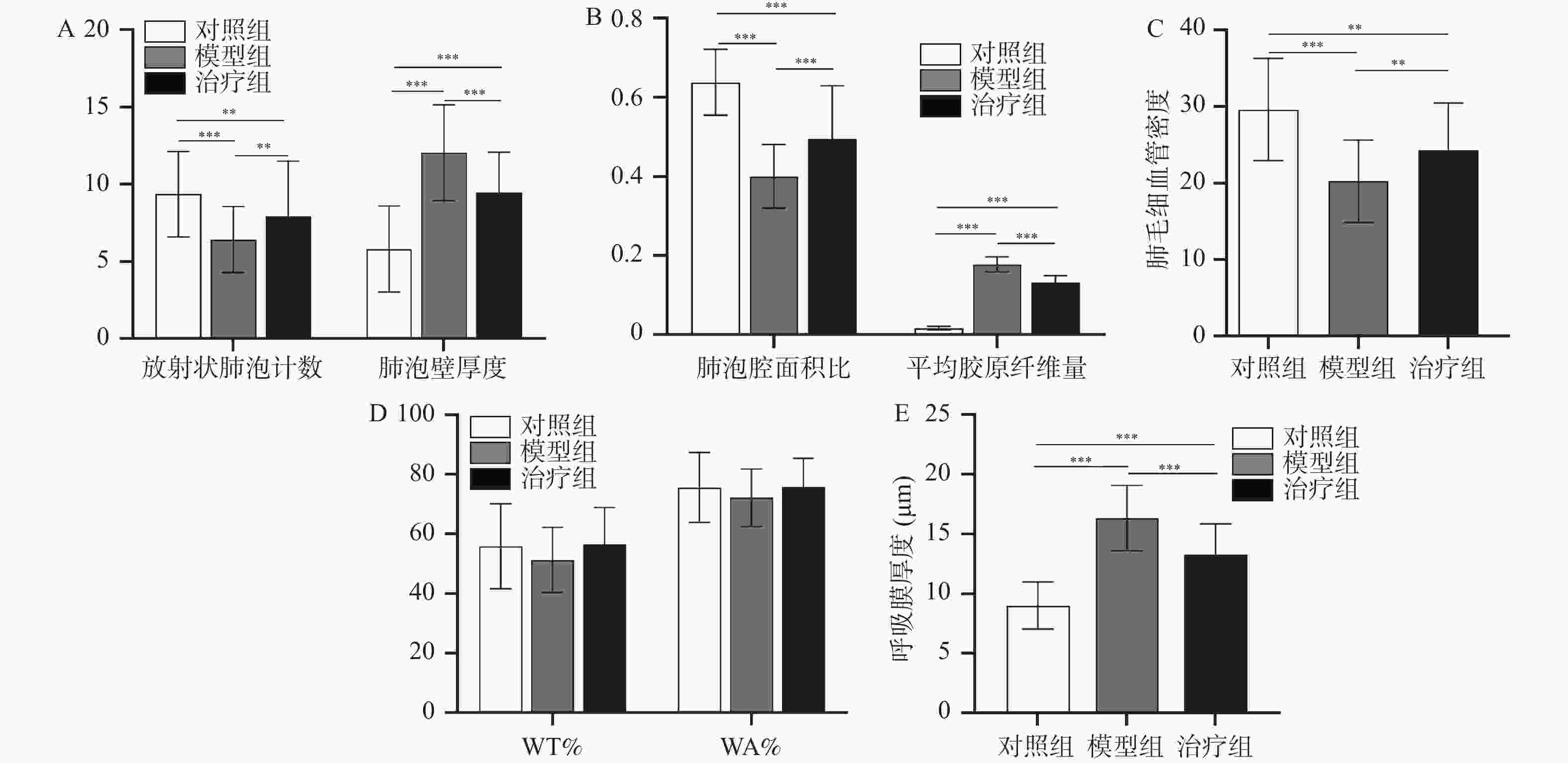
图 1 3组幼猪肺组织HE和Masson染色统计结果对比
A:3组幼猪放射状肺泡计数、肺泡壁厚度统计图;B:3组幼猪肺泡腔面积比、平均胶原纤维量统计图;C:3组幼猪肺毛细血管密度统计图;D:3组幼猪WT%、WA%统计图;E:3组幼猪呼吸膜厚度统计图;*P < 0.05;**P < 0.01;***P < 0.001。
Figure 1. Comparison of HE and Masson staining results in 3 groups of piglet lung tissues
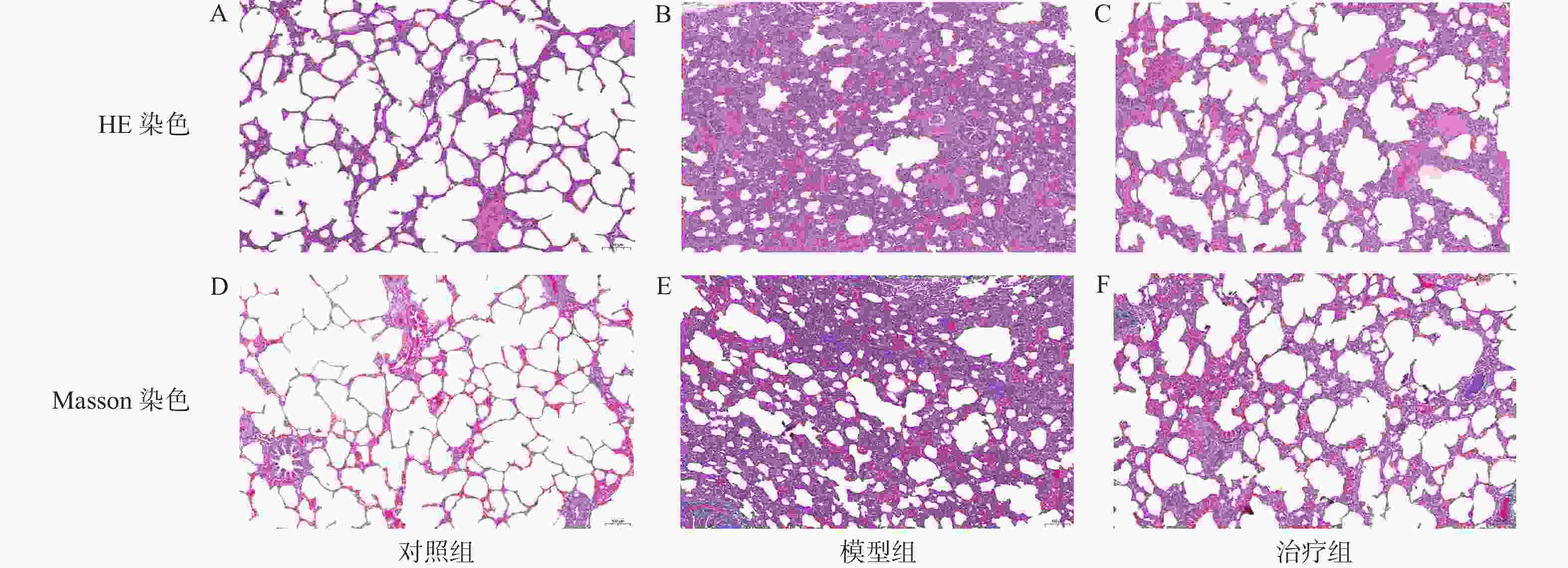
图 2 3组幼猪肺组织HE和Masson染色结果比较(胶原纤维呈蓝色,肌纤维、纤维素和红细胞呈红色)(标尺:100 μm)
A:对照组肺HE染色;B:模型组肺HE染色;C:治疗组肺HE染色;D:对照组肺Masson染色;E:模型组肺Masson染色;F:治疗组肺Masson染色。
Figure 2. Comparison of HE and Masson staining results of lung tissue of three groups of piglets (collagen fibers are blue,muscle fibers,cellulose and red blood cells are red) (scale bar: 100 μm)
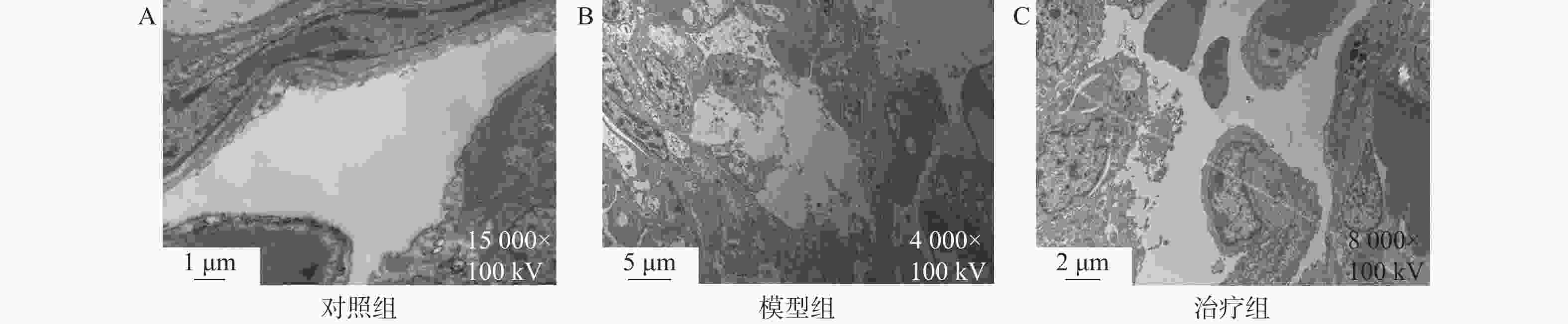
图 3 3组幼猪肺组织透射电镜观察结果
A:对照组电镜下肺组织;B:模型组电镜下肺组织;C:治疗组电镜下肺组织。
Figure 3. Transmission electron microscope observation results of lung tissue of three groups of piglets
表 1 3组幼猪左侧肺组织HE和Masson染色的测量结果($ \bar x \pm s $)
Table 1. Measurement results of HE and Masson staining of left lung tissue of three groups of piglets($ \bar x \pm s $)
对照组(n=3) 模型组(n=5) 治疗组(n=5) F P 放射状肺泡计数(个) 9.35±2.77 6.41±2.14* 7.92±3.56*# 19.92 <0.001※ 肺泡腔面积比(%) 63.82±8.34 40.06±8.05* 49.49±13.47*# 95.62 <0.001※ 呼吸膜厚度(μm) 9.01±1.97 16.35±2.72* 13.29±2.56*# 160.78 <0.001※ 肺泡壁厚度(μm) 5.81±2.78 12.04±3.10* 9.44±2.63*# 89.81 <0.001※ 平均胶原纤维量 0.02±0.01 0.18±0.02* 0.13±0.02*# 1921.33 <0.001※ 毛细血管密度(‰) 29.62±6.66 20.24±5.39 24.36±6.90*# 11.66 <0.001※ 肺动脉壁厚度比值(WT%) 55.93±14.29 51.31±10.95 56.56±12.39 1.89 0.156 肺动脉壁横截面积比值(WA%) 75.66±11.68 72.22±9.65 75.81±9.64 1.38 0.257 与对照组比较,*P < 0.05;与模型组比较,#P < 0.05;※P < 0.001。  下载: 导出CSV
-
[1] Campbell R M,Smith M D,Mayes T C,et al. The characteristics of thoracic insufficiency syndrome associated with fused ribs and congenital scoliosis[J]. J Bone Joint Surg Am,2003,85(3):399-408. doi: 10.2106/00004623-200303000-00001 [2] Yang S,Andras L M ,Redding G J,et al. Early-onset scoliosis: A review of history,current treatment,and future directions[J]. Pediatrics,2016,137(1): e20150709. [3] Mayer O H. Management of thoracic insufficiency syndrome[J]. Curr Opin Pediatr,2009,21(3):333-343 doi: 10.1097/MOP.0b013e328329a500 [4] Cunin V. Early-onset scoliosis: Current treatment[J].Orthop Traumatol Surg Res,2015,101(1 Suppl): S109-118. [5] Zhang Y,Shi Z,Li W ,et al. A porcine model of early-onset scoliosis combined with thoracic insufficiency syndrome: Construction and transcriptome analysis[J].Gene,2023,858(23): 147202. [6] Cooney T P,Thurlbeck W M. The radial alveolar count method of emery and mithal: A reappraisal 2--intrauterine and early postnatal lung growth[J].Thorax,1982,37(8): 580-583. [7] Olson J C,Kurek K C,Mehta H P ,et al. Expansion thoracoplasty affects lung growth and morphology in a rabbit model: A pilot study[J]. Clin Orthop Relat Res,2011,469(5): 1375-1382. [8] Olson J C,Glotzbecker M P,Takahashi A,et al. Expansion thoracoplasty in rabbit model: Effect of timing on preserving pulmonary growth and correcting spine deformity[J]. Spine (Phila Pa 1976),2018,43(15): E877-E884. [9] Olson J C,Takahashi A,Glotzbecker M P,et al. Extent of spine deformity predicts lung growth and function in rabbit model of early onset scoliosis[J]. PLoS One,2015,10(8):e0136941. doi: 10.1371/journal.pone.0136941 [10] Mehta H P,Snyder B D,Callender N N,et al. The reciprocal relationship between thoracic and spinal deformity and its effect on pulmonary function in a rabbit model: A pilot study[J]. Spine (Phila Pa 1976),2006,31(23): 2654-2664. [11] Mehta H P,Snyder B D,Baldassarri S R,et al. Expansion thoracoplasty improves respiratory function in a rabbit model of postnatal pulmonary hypoplasia: A pilot study[J]. Spine (Phila Pa 1976),2010,35(2): 153-161. [12] Campbell R M,Smith M D,Hell-Vocke A K. Expansion thoracoplasty: The surgical technique of opening-wedge thoracostomy. Surgical technique[J]. J Bone Joint Surg Am,2004,86(A Suppl 1): 51-64. [13] Campbell R M,Smith M D,Mayes T C,et al. The effect of opening wedge thoracostomy on thoracic insufficiency syndrome associated with fused ribs and congenital scoliosis[J]. J Bone Joint Surg Am,2004,86(8):1659-1674. doi: 10.2106/00004623-200408000-00009 [14] Tepper R S,Morgan W J,Cota K,et al. Physiologic growth and development of the lung during the first year of life[J]. Am Rev Respir Dis,1986,134(3):513-519. [15] Harding C O,Green C G,Perloff W H,et al. Respiratory complications in children with spondyloepiphyseal dysplasia congenita[J]. Pediatr Pulmonol,1990,9(1):49-54. doi: 10.1002/ppul.1950090112 [16] Dimeglio A. Growth of the spine before age 5 years[J]. Pediatr Orthop,1993,1(2):102-107. [17] Abman S H. Bronchopulmonary dysplasia: "A vascular hypothesis"[J]. Am J Respir Crit Care Med,2001,164(10 Pt 1): 1755-1756. [18] Gaengel K,Genové G,Armulik A,et al. Endothelial-mural cell signaling in vascular development and angiogenesis[J]. Arterioscler Thromb Vasc Biol,2009,29(5):630-638. doi: 10.1161/ATVBAHA.107.161521 [19] Morrisey E E,Hogan B L. Preparing for the first breath: Genetic and cellular mechanisms in lung development[J]. Dev Cell,2010,18(1):8-23. doi: 10.1016/j.devcel.2009.12.010 [20] Nardiello C,Miž í kov á I,Silva D M,et al. Standardisation of oxygen exposure in the development of mouse models for bronchopulmonary dysplasia[J]. Dis Model Mech,2017,10(2):185-196. [21] Lederer D J,Martinez F J. Idiopathic pulmonary fibrosis[J]. N Engl J Med,2018,378(19):1811-1823. doi: 10.1056/NEJMra1705751 [22] Wu D,Birukov K,Endothelial cell mechano-metabolomic coupling to disease states in the lung microvasculature[J]. Front Bioeng Biotechnol,2019,7(5): 172. [23] Wu H,Yu Y,Huang H,et al. Progressive pulmonary fibrosis is caused by elevated mechanical tension on alveolar stem cells[J]. Cell,2020,180(1):107-121. doi: 10.1016/j.cell.2019.11.027 -


 点击查看大图

计量
- 文章访问数: 359
- HTML全文浏览量: 400
- PDF下载量: 6
- 被引次数: 0
