Measurement of Articular Cartilage Thickness of Talus in Normal Adults by MRI
-
摘要:
目的 通过MRI测量正常成人距骨表面软骨厚度,得出正常成人距骨软骨厚度的参数以及与性别、年龄、体重、身高的关系。 方法 选择踝关节无关节炎、无外伤史的成人100名,男、女各50名,18~60岁,行MRI检查,通过MRI测量前、中、后距骨关节面软骨厚度,测量结果采用SPSS25.0统计学软件包进行统计学分析,P < 0.05表示差异有统计学意义。 结果 男性前、中、后距骨关节面软骨厚度分别为(1.00±0.18)mm、(1.40±0.21)mm、(0.87±0.18)mm;女性前、中、后距骨关节面软骨厚度分别为(0.96±0.19)mm、(1.29±0.20)mm、(0.86±0.15)mm;男性和女性距骨前、后关节软骨面厚度,差异无统计学意义(P > 0.05),距骨中关节面软骨厚度,差异有统计学意义(P < 0.05),男性与女性年龄,差异无统计学意义(P > 0.05)。 结论 男性距骨中关节面平均软骨厚度大于女性;前后关节面软骨厚度无差异;男、女性身高、体重、年龄与前、中、后关节面软骨厚度均无相关性;在距骨软骨损伤时对需要修复的软骨量提供依据,在距骨解剖假体设计时加入相应软骨厚度。 Abstract:Objective To measure the thickness of cartilage on the surface of the talus of normal adults by MRI and to obtain the parameters of the thickness of the cartilage of the talus of normal adults and the relationship with gender, age, weight and height. Methods 100 adults, 50 males and 50 females, aged 18 ~ 60, without arthritis and trauma history of ankle joint in the outpatient department were selected for MRI examination.The thickness of articular cartilage of anterior, middle and posterior talus was measured by MRI. The measurement results were measured by SPSS 25. 0 statistical software package for statistical analysis. Results The cartilage thicknesses of the anterior, middle, and posterior talus articular surface in men were (1.00±0.18) mm, (1.40±0.21) mm, and (0.87±0.18) mm respectively; the thicknesses of cartilage in the anterior, middle, and posterior talar articular surfaces of women were (0.96±0.18) mm, 0.19) mm, (1.29 ± 0.20) mm, (0.86 ± 0.15) mm respectively; male and female anterior and posterior articular cartilage thicknesses of the talus were not statistically different (P > 0.05), there was significant difference in the thickness of articular cartilage in talus (P < 0.05), and there was no statistically significant difference in age between men and women (P > 0.05). Conclusion The average cartilage thickness of the articular surface of the talus in men is greater than that in women and there is no difference in the thickness of cartilage on the front and rear articular surfaces; There is no correlation between the height, weight, and age of men’s and women’s articular cartilage thickness. The amount of cartilage provides the basis, and the corresponding cartilage thickness is added to the design of the talar anatomical prosthesis. -
Key words:
- MRI /
- Talus /
- Cartilage thickness /
- Method of measurement
-
随着影像学学科的飞速发展,通过对影像技术的灵活运用可以对骨骼表面软骨进行更好的观察和测量,其中超声与MRI可对软骨进行测量[1-2]。二者无辐射,安全无害,但大量影像循证医学研究表明MRI在真实软骨测量准确性方面好于超声,是间接观察软骨的最佳选择[3]。随着MRI检查手段和系统的不断升级,可以很好的利用其对关节软骨损伤和病变检测,1.5T磁共振T1WI TSE成像技术可以测量出真实的软骨厚度[4]。对于踝关节损伤患者,除了考虑骨折和韧带损伤,软骨的损伤也逐渐得到骨科医生的关注,许多研究表明软骨厚度通常小于2 mm,然而软骨厚度也会因地方、性别、和个体的不同而表现出很大的差异[5]。因此对距骨软骨厚度测量将帮助骨科医生预测需要修复的软骨量,并以合理的成本进行治疗,而不会过度侵犯患者,在距骨解剖假体的设计过程中也要考虑软骨存在[6-8]。Giannicola等[9]研究发现软骨厚度分布与骨骼长短、粗细均不相关,即我们不能简单从骨骼CT和X光片显像中预测出软骨厚度。当前国内尚未有对距骨关节面软骨厚度测量的相关文献报道,因此应用MRI测量成人正常距骨表面软骨厚度,构建国内成人正常距骨表面软骨厚度参数势在必行。
1. 资料与方法
1.1 一般资料
选取2021年01月至2021年11月在云南省中医医院MRI室行检查的踝关节无关节炎、无外伤史的成人100例,其中男性、女性各50例,年龄18~60岁,中位年龄37岁,签署知情同意告知书,对踝关节进行扫描,如实记录体重、身高与年龄。已通过医院伦理委员会审批。
1.2 仪器与方法
使用荷兰飞利浦1.5T磁共振仪。扫描线圈:SENSE-FLEX-M-COIL,C1-COIL,C3-COIL,T/R HEAD COIL KNEE/FOOT COIL。扫描序列:冠状位T1WI TSE ,矢状位T2WI TSE,矩阵512×512,FOV 16 cm,层厚为3 mm,层距为1 mm。扫描体位:取仰卧位,足部先进,沙袋固定足部于中立位。
1.3 测量方法
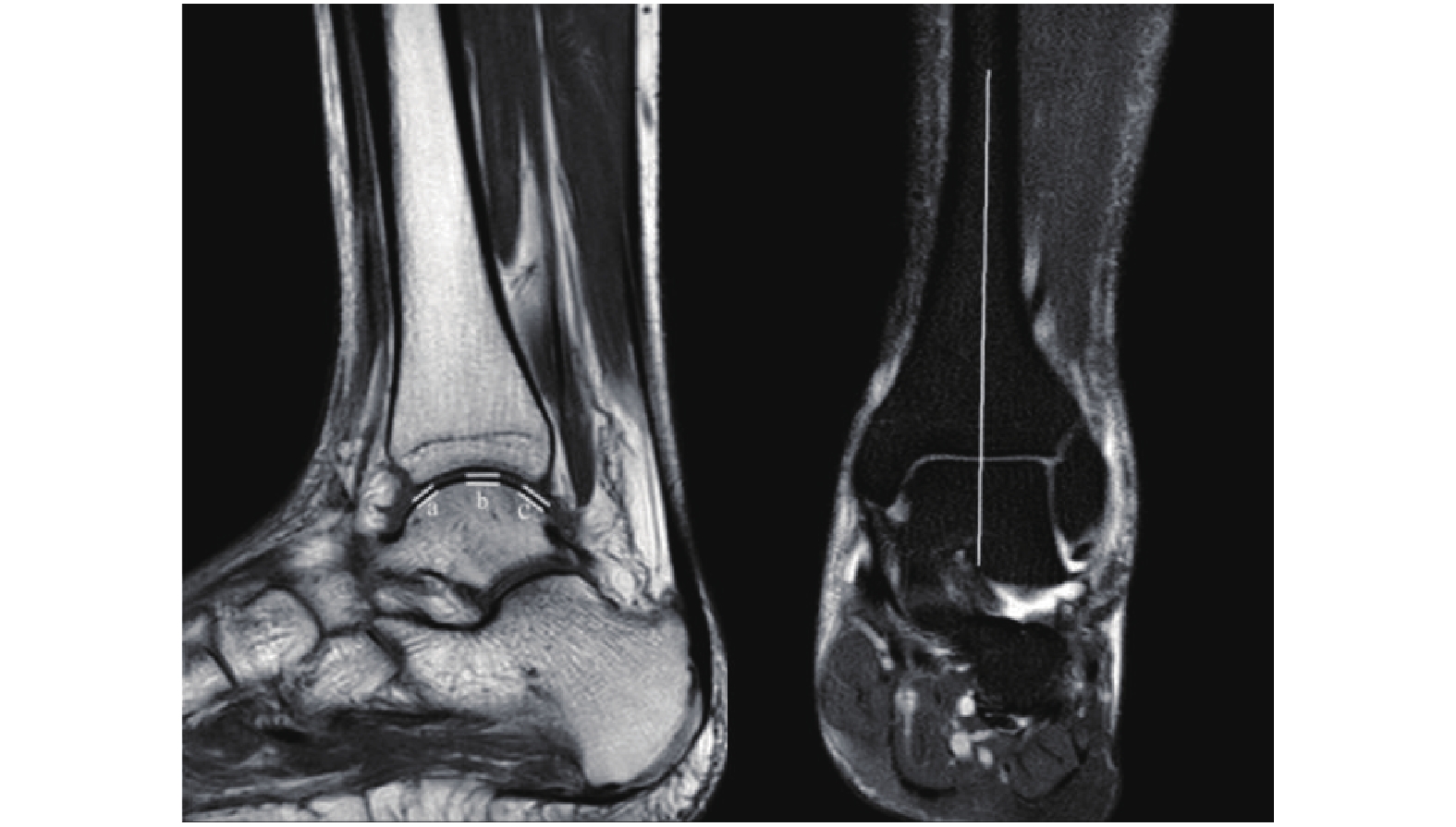
选择在冠状位通过距骨关节面中垂线的矢状位图像作为测量图像,以胫距关节面前、中、后3个点作为测量点,各测量点垂直于相应的关节面,测量软骨厚度为关节软骨低信号的垂直高度,得出软骨厚度a、b、c(图1)。所有图片均放大3倍,由3名骨科医师独立分别完成3个测量点测量,每个点取3者所得平均值作为最终结果,测量精度为0.01 mm。
软骨测量点的选择:选择在冠状位通过距骨关节面中垂线的矢状位图像作为测量图像,以胫距关节面前、中、后3个点作为测量点,各测量点垂直于相应的关节面,测量软骨厚度为关节软骨低信号的垂直高度,得出软骨厚度a、b、c。
1.4 统计学处理
采用SPSS25.0统计学软件包进行统计学分析,基本描述正态分布计量资料采用均数据±标准差表示,正态分布计量资料两组间比较采用两独立样本t检验,相关性分析采用Pearson相关性分析,P < 0.05表示差异有统计学意义。
2. 结果
2.1 性别与前、中、后距骨关节面软骨厚度的关系
男性前、中、后距骨关节面软骨厚度分别为(1.00±0.18)mm、(1.40±0.21)mm、(0.87±0.18)mm;女性前、中、后距骨关节面软骨厚度分别为(0.96±0.19)mm、(1.29±0.20)mm、(0.86±0.15)mm;男性和女性距骨前、后关节软骨面厚度差异无统计学意义(P > 0.05),距骨中关节面软骨厚度,差异有统计学意义(P < 0.05),见表1。
表 1 男、女性各指标比较Table 1. Comparison of male and female indicators指标 男性 女性 t P a(mm) 1.00 ± 0.18 0.96 ± 0.19 1.342 0.183 b(mm) 1.40 ± 0.21 1.29 ± 0.20 2.715 0.008* c(mm) 0.87 ± 0.18 0.86 ± 0.15 0.089 0.930 年龄(岁) 37.00 ± 11.96 41.42 ± 14.10 −1.680 0.096 身高(m) 1.70 ± 0.06 1.64 ± 0.06 5.194 < 0.001* 体重(Kg) 67.61 ± 6.29 58.04 ± 4.47 8.707 < 0.001* a:前关节面软骨厚度;b:中关节面软骨厚度;c:后关节面软骨厚度。*P < 0.05。 2.2 身高、体重、年龄与距骨前、中、后关节面软骨厚度的关系
男性与女性年龄差异无统计学意义(P > 0.05),男性身高、体重均大于女性,差异有统计学意义(P < 0.05),考虑为男女差异。经Pearson相关性分析男、女性身高、体重、年龄与距骨前、中、后关节面软骨厚度均无相关性(P > 0.05),见表2、表3、表4。
表 2 身高与各指标相关性分析Table 2. Correlation analysis between height and various indexes指标 r P a(mm) 0.11 0.297 b(mm) 0.05 0.600 c(mm) −0.03 0.764 a:前关节面软骨厚度;b:中关节面软骨厚度;c:后关节面软骨厚度。 表 3 体重与各指标相关性分析Table 3. Correlation analysis between body weight and various indexes指标 r P a(mm) 0.08 0.429 b(mm) 0.17 0.099 c(mm) 0.06 0.588 a:前关节面软骨厚度;b:中关节面软骨厚度;c:后关节面软骨厚度。 表 4 年龄与各指标相关性分析Table 4. Correlation analysis between age and various indicators指标 r P a(mm) −0.14 0.182 b(mm) −0.09 0.389 c(mm) −0.15 0.131 a:前关节面软骨厚度;b:中关节面软骨厚度;c:后关节面软骨厚度。 3. 讨论
在磁共振成像中使用各同向性体素进行厚度测量,来自各同向性体素的MRI数据结果表明,在关节间隙宽度和软骨厚度的测量值的位置准确测量软骨厚度是体素大小的2倍或更大是科学合理的,因此本研究所有测量图像均放大3倍;通过与3个模型、5个正常尸体髋部标本和9个骨关节炎患者的实验结果进行比较,验证了该模拟方法的有效性。MRI提供了软骨和软骨下骨厚度的良好表征,支持其在骨软骨结构和改变的研究和临床诊断中的应用[10-11],也正是如此为本研究MRI测量软骨厚度提供了研究基础。
虽然超声与MRI可对软骨进行测量,二者无辐射,安全无害,超声可以作为评估股骨中、后股骨区域相对软骨厚度的可行临床工具。然而,在软骨厚度 < 2 mm时,超声在相关测量值之间的绝对一致性较差,由于更多的基于MRI测量,超声测量的绝对有效性受到同行质疑[12]。而CT需要通过造影技术对软骨进行测量,有创且辐射大,不适于对人体进行研究,但动物研究可考虑CT造影技术;目前来说,相对于超声、CT测量软骨厚度,MRI测量更为准确、安全、敏感,因此本研究选择了MRI测量。
近年来关节软骨损伤越来越受到骨科医生所关注,距骨软骨面损伤是踝关节最常见的软骨损伤[13],主要表现为踝部肿痛、关节异物感、活动受限,多见于青壮年男性。X线片是踝关节扭伤后需拍摄的常规影像学检查,以前是距骨软骨面损伤的主要诊断依据,但X线片会忽略50%以上的软骨病变。MRI敏感度高,可清晰显示距骨软骨病变、骨髓水肿范围,是软骨损伤最佳无创检查[14];关节软骨损伤的患者日益受到重视,准确的诊断软骨损伤显得更为重要,而X线片误诊率较高,并不利于疾病的诊疗,因此目前MRI检查更有意义。当然,本研究只是起了一个抛砖引玉的作用,而且由于各方面的原因存在一定的局限性,本研究纳入的样本量有限,研究的还不够深入,由于选择健康志愿者进行检查成本高,实施难度大,本研究对象根据病情需要选择踝关节无关节炎、无外伤史的门诊就诊成人,节约成本,也能代表正常距骨软骨厚度,下一步将对运动前后、踝关节损伤后距骨软骨厚度的变化进行探索,希望更多的学者加入进一步更加全面的研究。
通过MRI对距骨表面软骨厚度测量研究发现,成人后距骨表面软骨厚度趋于稳定,与身高、体重、年龄均无相关性,负重区距骨软骨厚度明显大于非负重区,且负重区软骨厚度男性大于女性。展望未来,对于这一现象相信随着影像学学科的不断前进,笔者能更好的对不同区域的软骨进行更深入的研究。
-
表 1 男、女性各指标比较
Table 1. Comparison of male and female indicators
指标 男性 女性 t P a(mm) 1.00 ± 0.18 0.96 ± 0.19 1.342 0.183 b(mm) 1.40 ± 0.21 1.29 ± 0.20 2.715 0.008* c(mm) 0.87 ± 0.18 0.86 ± 0.15 0.089 0.930 年龄(岁) 37.00 ± 11.96 41.42 ± 14.10 −1.680 0.096 身高(m) 1.70 ± 0.06 1.64 ± 0.06 5.194 < 0.001* 体重(Kg) 67.61 ± 6.29 58.04 ± 4.47 8.707 < 0.001* a:前关节面软骨厚度;b:中关节面软骨厚度;c:后关节面软骨厚度。*P < 0.05。 表 2 身高与各指标相关性分析
Table 2. Correlation analysis between height and various indexes
指标 r P a(mm) 0.11 0.297 b(mm) 0.05 0.600 c(mm) −0.03 0.764 a:前关节面软骨厚度;b:中关节面软骨厚度;c:后关节面软骨厚度。 表 3 体重与各指标相关性分析
Table 3. Correlation analysis between body weight and various indexes
指标 r P a(mm) 0.08 0.429 b(mm) 0.17 0.099 c(mm) 0.06 0.588 a:前关节面软骨厚度;b:中关节面软骨厚度;c:后关节面软骨厚度。 表 4 年龄与各指标相关性分析
Table 4. Correlation analysis between age and various indicators
指标 r P a(mm) −0.14 0.182 b(mm) −0.09 0.389 c(mm) −0.15 0.131 a:前关节面软骨厚度;b:中关节面软骨厚度;c:后关节面软骨厚度。 -
[1] Steppacher S D,Hanke M S,Zurmühle C A,et al. Ultrasonic cartilage thickness measurement is accurate,reproducible,and reliable-validation study using contrast-enhanced micro-CT[J]. J Orthop Surg Res,2019,14(1):67. doi: 10.1186/s13018-019-1099-8 [2] Wang N,Badar F,Xia Y. Experimental Influences in the accurate measurement of cartilage thickness in MRI[J]. Cartilage,2019,10(3):278-287. doi: 10.1177/1947603517749917 [3] Song K,Pietrosimone B G,Nissman D B,et al. Ultrasonographic measures of talar cartilage thickness associate with magnetic resonance-based measures of talar cartilage volume[J]. Ultrasound Med Biol,2020,46(3):575-581. [4] 俞佳凤,孙岩,张静,等. 喙突下撞击综合征的磁共振成像测量研究[J]. 实用放射学杂志,2020,36(8):1281-1285. doi: 10.3969/j.issn.1002-1671.2020.08.026 [5] Maier J,Black M,Bonaretti S,et al. Comparison of different approaches for measuring tibial cartilage thickness[J]. J Integr Bioinform,2017,14(2):20170015. [6] Giannicola G,Spinello P,Scacchi M,et al. Cartilage thickness of distal humerus and its relationships with bone dimensions:Magnetic resonance imaging bilateral study in healthy elbows[J]. J Shoulder Elbow Surg,2017,26(5):e128-e136. doi: 10.1016/j.jse.2016.10.012 [7] Giannicola G,Scacchi M,Sedati P,et al. Anatomical variations of the trochlear notch angle:MRI analysis of 78 elbows[J]. Musculoskelet Surg,2016,100(Suppl 1):89-95. [8] 吕嘉玲,林伟强,张隐笛,等. 基质诱导的自体软骨细胞移植术的磁共振成像评估及其与软骨下骨的相关性[J]. 实用放射学杂志,2020,36(5):776-779. doi: 10.3969/j.issn.1002-1671.2020.05.022 [9] Giannicola G,Sedati P,Polimanti D,et al. Contribution of cartilage to size and shape of radial head circumference:Magnetic resonance imaging analysis of 78 elbows[J]. J Shoulder Elbow Surg,2016,25(1):120-126. doi: 10.1016/j.jse.2015.07.003 [10] 袁文昭,邓德茂,段高雄,等. MRI量化评分在腕关节类风湿关节炎治疗中的应用价值[J]. 实用放射学杂志,2018,34(9):1410-1413,1457. doi: 10.3969/j.issn.1002-1671.2018.09.025 [11] Cheng Y,Guo C,Wang Y,et al. Accuracy limits for the thickness measurement of the hip joint cartilage in 3-D MR images:Simulation and validation[J]. IEEE Trans Biomed Eng,2013,60(2):517-533. doi: 10.1109/TBME.2012.2230002 [12] Schmitz R J,Wang H M,Polprasert D R,et al. Evaluation of knee cartilage thickness:A comparison between ultrasound and magnetic resonance imaging methods[J]. Knee.,2017,24(2):217-223. doi: 10.1016/j.knee.2016.10.004 [13] 刘三彪,张文涛,唐陆波,等. 距骨骨软骨损伤修复材料研究进展[J]. 足踝外科电子杂志,2021,8(2):56-62. [14] 陈言智,张洪涛,程宇,等. 距骨骨软骨损伤的诊疗现状[J]. 足踝外科电子杂志,2021,8(2):52-55,62. -


 下载:
下载:
 下载:
下载:

