Effects of Exercise Rehabilitation on Cardiac Fibrosis and Function in Rats with Myocardial Infarction
-
摘要:
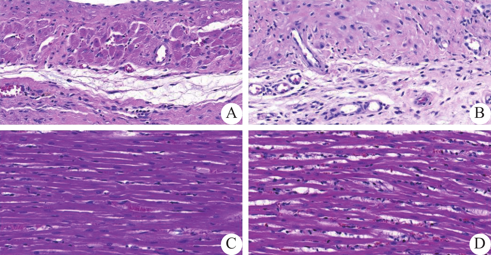
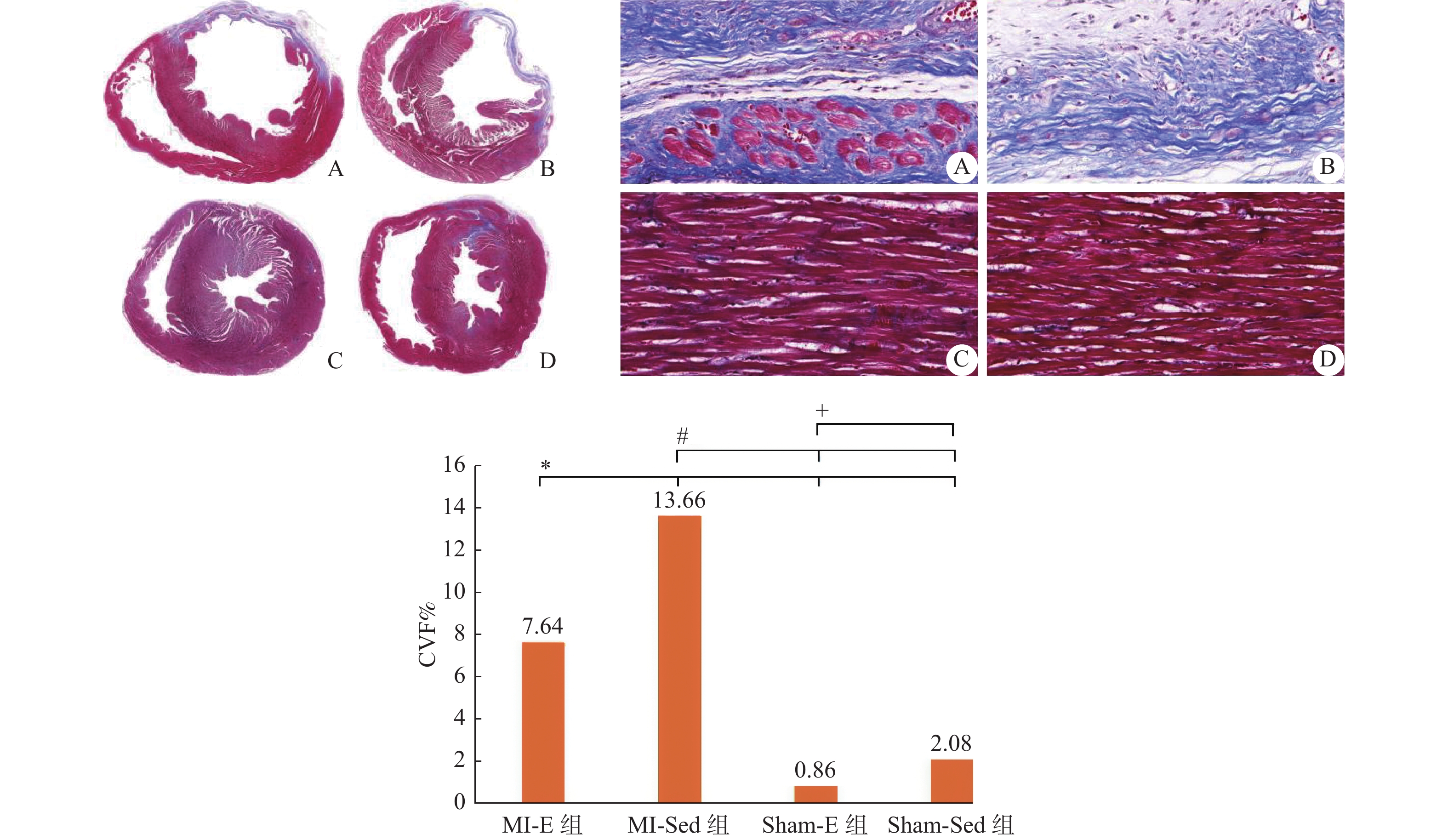
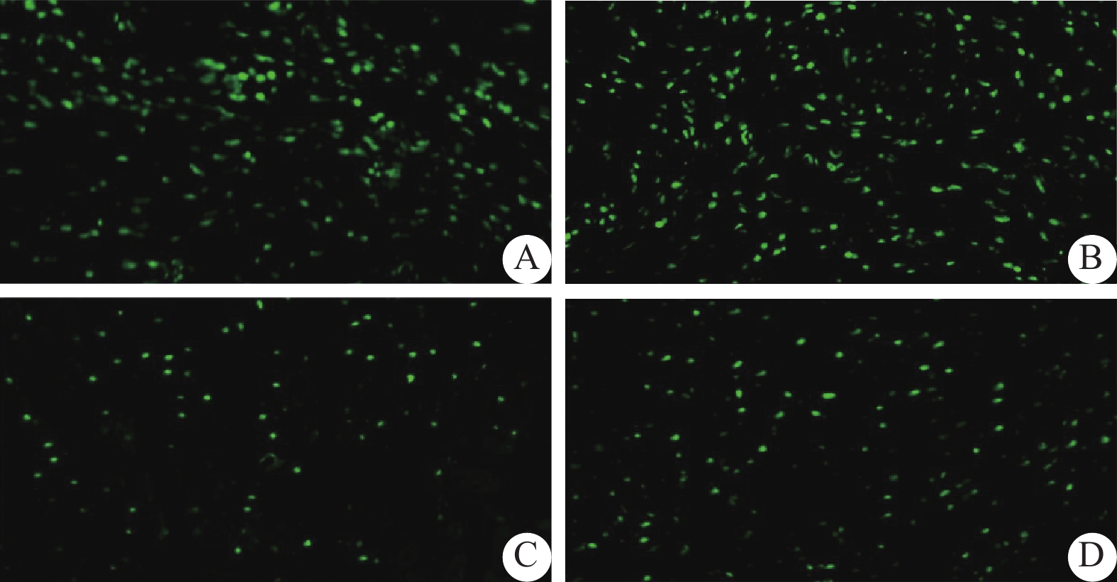
目的 探究运动康复对心肌梗死大鼠心脏纤维化及心功能的影响。 方法 24只雄性SD大鼠随机分为4组:心肌梗死运动组(MI-E组)、心肌梗死不运动组(MI-Sed组)、假手术运动组(Sham-E组)、假手术不运动组(Sham-Sed组),每组5~7只。采用结扎左冠状动脉前降支(LAD)制备心肌梗死模型,而假手术组只穿针不结扎。术后1周开始运动训练,包括适应性运动1周和正式运动4周,而不运动组全程不参加运动训练。运动结束后行超声心动图检查,HE、Masson和Tunel染色,以及透射电镜检测。 结果 与Sham组比较,MI组左心室室壁变薄及收缩功能降低,以MI-Sed组明显(P < 0.05);而MI-E组与MI-Sed组比较,LVESD、LVEF和FS有所改善(P < 0.05);Sham-E组与Sham-Sed组比较差异无统计学意义(P > 0.05)。HE染色显示与Sham组比较,MI组心肌细胞呈现不同程度溶解,伴炎性细胞浸润,成纤维细胞增生,而MI-E组损伤较轻。Masson染色显示MI组较Sham组心肌细胞排列紊乱,心肌纤维化明显增加,而MI-E组与MI-Sed组、Sham-E组与Sham-Sed组相比较,胶原容积分数(CVF%)降低(P < 0.05)。Tunel染色显示MI组较Sham组心肌细胞凋亡数增多,而MI-E组与MI-Sed组、Sham-E组与Sham-Sed组相比较,心肌细胞凋亡指数降低(P < 0.05)。透射电镜显示MI组较Sham组心肌细胞及线粒体损伤严重,肌纤维疏松紊乱,而MI-E组较MI-Sed组心肌细胞损伤较轻,线粒体结构较完整,自噬小体形成较多。 结论 运动康复可减少MI大鼠的心肌细胞损伤和细胞凋亡,减少心肌梗死面积,改善心肌纤维化及心功能;运动可适度诱导自噬水平发挥心脏保护作用。 Abstract:Objective To explore the effect of exercise rehabilitation on cardiac fibrosis and function in rats with myocardial infarction. Methods Twenty-four male SD rats were randomly divided into four groups: MI-E group, MI-Sed group, Sham-E group and Sham-Sed group, with five to seven rats in each group. A myocardial infarction model was prepared using ligation of the left anterior descending coronary artery (LAD), while the sham surgery group underwent needle insertion without ligation. One week after surgery, exercise training was initiated, including one week of adaptive exercise and four weeks of formal exercise. The non-exercise group did not participate in any exercise training throughout the study. After completion of the exercise regimen, echocardiography, HE staining, Masson staining, Tenel staining, and transmission electron microscopy were performed for evaluation. Results Compared with sham group, left ventricular wall thinning and systolic function were decreased in MI groups, especially in MI-Sed group (P < 0.05), while LVESD, LVEF and FS were improved in MI-E group compared with MI-Sed group (P < 0.05); there was no significant difference between Sham-E group and Sham-Sed group (P > 0.05). Compared with the sham group, HE staining showed that cardiomyocyte exhibited different degrees of lysis with inflammatory cell infiltration and fibroblast proliferation in the MI group while the damage was milder in the MI-E group. Masson staining showed that cardiomyocyte was disorganized and myocardial fibrosis was significantly increased in the MI group compared with the sham group, while collagen volume fraction (CVF%) was decreased in the MI-E group compared with the MI-Sed group and the Sham-E group compared with the Sham-Sed group (P < 0.05). Tunel staining showed that compared with the Sham group, the MI group had increased myocardial cell apoptosis, while the MI-E group had a lower myocardial cell apoptosis index compared to the MI-Sed group, Sham-E group, and Sham-Sed group (P < 0.05). Transmission electron microscopy showed that compared with the Sham group, the MI group had severe damage to myocardial cells and mitochondria, as well as loose and disordered muscle fibers, while the MI-E group had milder myocardial cell injury, more intact mitochondrial structure, and more formation of autophagosomes. Conclusion Exercise rehabilitation can reduce cardiomyocyte damage and apoptosis, reduce myocardial infarct area, improve myocardial fibrosis and cardiac function in MI rats. Exercise can moderately induce autophagy level to play a cardioprotective role. -
Key words:
- Myocardial infarction /
- Exercise rehabilitation /
- Autophagy /
- Myocardial fibrosis
-
肺癌是威胁人类健康最常见的恶性肿瘤之一,2022年全球新发肺癌人数和死亡人数高达250万例和180万例,中国新发肺癌106.06万例、死亡73.33万例,均居所有肿瘤第一位[1]。衰弱是一组由于机体的生理储备下降或多系统失调,导致机体易损性增加、抗应激能力减弱的综合征,可增加个体不良结局的风险[2−3]。肿瘤患者由于其疾病本身、心理以及治疗方式都可能是挑战患者生理储备的重要压力源,均可增加其衰弱的发生。肺癌患者症状困扰(37.7%~99.2%)和心理障碍(97.98%)发生率高且较严重[4−5],加之抗肿瘤治疗相关副反应的困扰,是衰弱的高发人群,相关研究显示肺癌患者衰弱发生高达28%~61%(平均发生率为45%)[6],是导致患者不良症状反应、治疗严重并发症及病死率上升,加重患者经济负担,降低生活质量的重要原因[7]。放射治疗是肺癌患者的主要治疗手段之一。目前国内外学者已将衰弱应用到肺癌放疗患者治疗相关并发症及康复结局的预测中,并成为肺癌放疗患者康复结局的重要预测因子[8−9],但目前肺癌衰弱现状及其影响因素的探讨多集中在手术、化疗患者,关于肺癌患者衰弱发生率与放射治疗的相关性还存在争议[10−12],缺乏客观、综合的衰弱评估,更缺乏对肺癌放疗患者衰弱发生现状及影响因素的研究。因此,本研究旨在运用主观和客观相结合的方式,调查肺癌放疗患者衰弱的发生现状,并分析其影响因素,以期为开展肺癌放疗患者衰弱管理提供参考依据。
1. 资料与方法
1.1 研究对象
本研究为横断面调查研究,采用便利抽样调查2023年1月至12月在云南省某三级甲等肿瘤专科医院放疗肺癌患者。纳入标准:(1)病理学或细胞学诊断为肺癌需接受放射治疗患者[13];(2)年龄>18岁,自愿签署知情同意书;(3)意识清楚,无认知障碍,能正常交流沟通;(4)正在接受放疗且总放疗次数 ≥ 20次。排除标准:(1)肺癌术后时间 < 3个月,放疗期间合并化疗的患者;(2)患有其他严重躯体、精神疾病者;(3)严重心、肝、肾功能衰竭及骨髓造血功能不全者。根据Kendall样本量估计方法,可取自变量个数的5~10倍[14],本研究共涉及变量25个,估计需要样本量为125~250例,共收集241例患者,满足样本量需求。本研究获云南省肿瘤医院伦理委员会批准(SLKYLX2023-031)。
1.2 研究工具
1.2.1 一般资料
调查问卷包括肺癌放疗患者年龄、性别、职业、肺癌细胞分型类型、放疗次数、合并症、血红蛋白计数、白蛋白计数等。
1.2.2 衰弱表型量表
Fried等[15]于2001年编制,多用于住院患者的衰弱测量,内容包括5项指标,不明原因体质量减轻、握力低下、体力活动减少、行走速度降、自述疲乏,总分为5分,0分为无衰弱,1~2分为衰弱前期,3分为衰弱期,本研究中将衰弱前期和无衰弱状态统称为非衰弱状态。
1.2.3 安德森症状评估量表
该量表由美国Texas大学和Anderson癌症中心编制[16],包含症状严重度和症状困扰度两部分,共13个条目,评估过去24 h癌症患者出现的伴随症状和症状困扰度。中文版安德森症状测评量表内部一致性信度在0.82~0.94之问,该量表广泛适用于不同类型和治疗的癌症患者[17]。
1.2.4 医院焦虑抑郁量表
该量表包含有焦虑和抑郁两个亚量表,各7个条目,共14个条目组成,两个量表的Cronbach's α系数分别为0.806与0.791[18]。
1.2.5 营养风险筛查2002
用于评估研究对象是否存在营养不良风险,评估内容包括:疾病严重程度评分、营养受损评分、年龄评分[19]。
1.2.6 Barthel指数
用于评估研究对象的日常活动能力,共10个条目,根据患者完成每项内容需协助的程度计分,总分100分[20]。
1.3 数据收集
由经过统一培训的调查员在放射治疗科病房招募符合纳入、排除标准的肺癌放疗患者,向患者介绍研究内容、目的,同意后签署知情同意书,现场发放纸质版问卷填写,无法自行填写者,由调查者采用问答形式协助填写。
1.4 统计学处理
采用SPSS23.0软件进行数据录入与分析。符合正态分布的计量资料采用均数±标准差($\bar x \pm s $)表示,不符合正态分布计量资料采用中位数(四分位数)M(P25,P75)表示,用Wilcoxon秩和检验;计数资料以百分数(n)%表示,组间比较采用χ2检验,等级资料采用非参数检验;采用二元Logistic探讨影响因素,以P < 0.05表示差异有统计学意义。
2. 结果
2.1 人口学资料
本研究共纳入241名肺癌放疗患者,其中男性187例(77.59%),女性54例(22.41%),平均年龄(58.95±9.16)岁,年龄范围(29~85)岁;鳞癌59例(24.48%),腺癌112例(46.47%),大细胞肺癌17例(7.05%),小细胞肺癌53例(21.99%);II期23例(9.54%),III期138例(57.26%),IV期80例(33.20%);局部转移159例(65.98%),远处转移82例(34.02%)。
2.2 肺癌放疗患者衰弱表型量表、医院焦虑抑郁量表、安德森症状评估量表评分情况
肺癌放疗患者衰弱评分,焦虑评分、抑郁评分、安德森症状评估量表评分见表1。
表 1 肺癌放疗患者衰弱表型、焦虑、抑郁、安德森症状评估量表得分[M(P25,P75),分]Table 1. Scores of frailty phenotype,anxiety,depression,and Anderson symptom assessment scale for lung cancer patients undergoing radiotherapy[M(P25,P75),points]变量 得分 衰弱表型量表评分 3.00(1.00,4.00) 医院焦虑抑郁量表 焦虑 8.00(5.00,11.00) 抑郁 7.00(3.50,10.00) 安德森症状评估量表评分 50.18(17.00,74.50) 2.3 不同社会人口特征肺癌放疗患者衰弱状况的单因素分析
年龄、放疗次数、疾病分期、转移情况、是否口服多种药物、白细胞计数、血红蛋白计数、营养风险评分等因素在肺癌放疗患者衰弱中差异具有统计学意义(P < 0.05),见表2。
表 2 不同社会人口特征肺癌放疗患者衰弱状况的单因素分析[n(%)](1)Table 2. Univariate analysis of frailty status in lung cancer radiotherapy patients with different sociodemographic characteristics[n(%)](1)项目 衰弱组 非衰弱组 χ2 P 年龄(岁) 19.527 < 0.001* < 60 57(42.86) 77(71.30) ≥ 60 76(57.14) 31(28.70) 性别 男 105(78.95) 82(75.93) 0.313 0.576 女 28(21.05) 26(24.07) 文化程度 小学及以下 50(37.59) 42(38.89) 1.401 0.705 初中 41(30.83) 37(34.26) 高中或中专 26(19.55) 15(14.89) 大专及以上 16(12.03) 14(12.96) 婚姻状况 已婚 115(86.47) 99(91.67) 1.620 0.203 其他 18(13.53) 9(8.33) 职业 农民 74(55.64) 69(63.89) 4.873 0.181 工人 11(8.27) 12(11.11) 职员 10(7.52) 9(8.33) 无业或退休 38(28.57) 18(16.67) 居住情况 3.861 0.145 与配偶同住 100(75.19) 86(79.63) 与子女同住 28(21.05) 14(12.96) 独居或养老院 5(3.76) 8(7.41) 家庭人均月收入(元) ≤ 2000 56(42.11) 56(51.85) 5.311 0.150 2001 ~4000 35(26.32) 28(25.93) 4001 ~6000 24(18.05) 9(8.33) > 6000 18(13.53) 15(13.89) 医疗费用支付方式 1.905 0.168 职工医保 52(39.10) 33(30.56) 居民医保 81(60.90) 75(69.44) 病程(月) 5.311 0.150 ≤ 6 13(9.77) 22(20.37) 7~12 43(32.33) 36(33.33) 13~24 40(30.08) 30(27.78) > 24 37(27.82) 20(18.52) 肺癌细胞分型 1.781 0.633 腺癌 31(23.31) 28(25.93) 鳞癌 61(45.86) 51(47.22) 大细胞肺癌 8(6.02) 9(8.33) 小细胞肺癌 33(24.81) 20(18.52) 临床分期 12.592 0.002* II期 10(7.52) 13(12.04) III期 66(49.62) 72(66.67) IV期 57(42.86) 23(21.30) *P < 0.05。 表 2 不同社会人口特征肺癌放疗患者衰弱状况的单因素分析[n(%)](2)Table 2. Univariate analysis of frailty status in lung cancer radiotherapy patients with different sociodemographic characteristics[n(%)](2)项目 衰弱组 非衰弱组 χ2 P 转移情况 14.125 < 0.001* 局部转移 74(55.64) 85(78.70) 远处转移 59(44.36) 23(21.30) 曾接受何种治疗 手术 11(8.27) 10(9.26) 0.451 0.930 化疗 88(66.17) 69(63.30) 手术+化疗 20(15.04) 19(17.59) 其他治疗 14(10.53) 10(9.26) 是否口服多种药物 22.897 < 0.001* 无口服药物 82(61.65) 96(88.89) 口服一种及以上药物 51(38.35) 12(11.11) 是否存在多种慢性疾病 0.909 0.363 无合并症 106(79.70) 91(84.26) 存在一种及以上合并症 27(20.30) 17(15.74) 血小板计数(109/L) 0.876 0.349 < 100 16(12.03) 9(8.33) ≥ 100 117(87.97) 99(91.67) 白细胞计数(109/L) 4,329 0.037* < 4 43(32.33) 22(20.37) ≥ 4 90(67.67) 86(79.63) 白蛋白计数(g/L) 3.195 0.074 < 35 26(19.55) 12(11.11) ≥ 35 107(80.45) 96(88.89) 血红蛋白计数(g/L) 16.856 < 0.001* < 120 65(48.87) 25(23.15) ≥ 120 68(51.13) 83(76.85) 营养风险评分(分) 43.268 < 0.001* < 3 47(35.34) 84(77.78) ≥ 3 86(64.66) 24(22.22) 放疗次数(次) 62.879 < 0.001* ≤ 10 36(27.07) 84(77.78) 11~20 46(34.59) 16(14.81) > 20 51(38.35) 8(7.41) Barthel指数(分) 135.235 < 0.001* 100(无需依赖) 23(17.29) 93(86.11) 61~99(轻度依赖) 110(82.71) 15(13.89) *P < 0.05。 2.4 不同医院焦虑抑郁量表、安德森症状评估量表评分肺癌放疗患者衰弱状况的单因素分析
衰弱组焦虑、抑郁、安德森症状评估量表评分均高于非衰弱组,焦虑、抑郁量、安德森症状评估量评分是肺癌放疗患者衰弱发生的影响因素,差异具有统计学意义(P < 0.001),见表3。
表 3 不同医院焦虑抑郁量表、安德森症状评估量表评分肺癌放疗患者衰弱状况的单因素分析[M(P25,P75)]Table 3. Univariate analysis of frailty status in lung cancer radiotherapy patients using anxiety and depression scales and Anderson symptom assessment scales from different hospitals[M(P25,P75)]变量 得分 Z P 衰弱组(n = 133) 非衰弱组(n = 108) 焦虑量表评分 9.00(5.00,11.00) 7.00(4.00,9.00) −4.349 < 0.001* 抑郁量表评分 9.00(5.00,11.00) 4.00(3.00,8.00) −6.567 < 0.001* 安德森症状评估量表评分 72.93(47.00,98.00) 23.08(7.00,35.00) −10.58 < 0.001* *P < 0.05。 2.5 肺癌放疗患者衰弱的多因素分析
以是否发生衰弱为因变量(否 = 0,是 = 1),将单因素分析中差异具有统计学意义的项目作为自变量进行Logistic回归分析,变量进入方程水准为0.05,剔除水准为0.10。结果显示,年龄( < 60岁 = 0, ≥ 60 = 1)、放疗次数(≤ 10次 = 0,10~20次 = 1,> 20次 = 2)、血红蛋白计数( < 120 = 0, ≥ 120 = 1)、Barthel指数评分(100分无依赖 = 0,61~99分有依赖 = 1)、安德森症状评估量量表、焦虑评分是肺癌放疗患者衰弱发生的影响因素,见表4。
表 4 肺癌放疗患者衰弱发生多因素分析Table 4. Multivariate analysis of frailty in lung cancer patients undergoing radiotherapy自变量 β SE Waldχ P OR 95%CI 常量 −3.455 1.340 6.609 0.010* 年龄(对照组: < 60岁) 2.291 0.664 11.892 0.001* 9.883 2.688~36.334 放疗次数(对照组:≤10次) 11~20 次 1.656 0.683 5.873 0.015* 5.240 1.373~20.001 >20次 3.095 0.829 13.931 < 0.001* 22.098 4.349~89.634 血红蛋白(对照组: < 120 g/L) −1.723 0.628 7.518 0.006* 0.178 0.052~0.612 安德森症状评估量表评分 0.072 0.015 21.806 < 0.001* 1.074 1.043~1.107 焦虑评分 −0.393 0.141 7.742 0.005* 0.675 0.512~0.890 Barthel指数评分(对照组:100分无依赖) 3.142 0.679 21.398 < 0.001* 23.151 6.115~87.647 *P < 0.05。 3. 讨论
3.1 肺癌放疗患者衰弱现状
研究结果显示肺癌放疗患者衰弱发生率为55.19%,与意大利的一项系统评价[6]研究结果相近(肺癌患者衰弱发生率28%~61%,总发生率45%),但明显高于美国学者Raghavan等[9]的一项回顾性研究结果(衰弱发生率35%),原因可能是其研究中选取的140例接受SBRT治疗的非小细胞肺癌患者均为早期,而本研究中研究对象III期、IV期患者占(90.46%),III期、IV期患者由于癌细胞对机体重要器官的损伤以及预后差等原因,患者面临更严重的症状及心理困扰,更容易发生衰弱[21];此外,云南省是中国乃至全世界肺癌高发区域,加之肺癌发病隐匿和云南省属于中国西南边境少数民族聚居地区,超过50%的患者确诊时已处于晚期,患者面临的疾病和症状负担更严重[22],导致患者衰弱发生率更高。同时两项研究中患者的治疗方式也存在较大差别,SBRT相较于传统调强放疗,放疗次数更少副反应更低[23]。英国学者Imam等[24]已证实通过实施医院衰弱风险评分分级可预测患者住院时间、再入院率、死亡率和某些特定条件治疗并发症的关系,可为实施衰弱干预提供信息决策。提示临床医护人员应早期识别患者衰弱现状、评估其严重程度,并根据评估结果制定精准化、个性化的衰弱管理策略,帮助患者延缓或逆转衰弱发生,进一步提高患者生活质量。
3.2 肺癌放疗患者衰弱的影响因素
3.2.1 年龄因素
Logistic回归分析结果显示与60岁以下的肺癌放疗患者相比,60岁及以上肺癌放疗患者发生衰弱的风险更高(OR:9.883;95%CI:2.688~36.334,P = 0.001),年龄 ≥ 60岁的肺癌放疗患者衰弱发生风险是年龄 < 60岁患者的9.883倍。中国大陆学者Hou等[25]在1项横断面研究中结果表明年龄是肺癌患者衰弱的独立预测因子,与本研究结果一致。其原因可能随着年龄增长机体各项机能指标逐渐下降,如基础代谢率下降、钙质流失、肌肉力量下降等导致患者生理功能下降有关;此外运动能力也会影响患者衰弱的发生[26],随着年龄增长机体各项机能指标逐渐下降,人体的运动能力跟运动量也在逐步下降,而本研究在调查过程中也发现大多数 ≥ 60岁的患者都表示“自己有高血压糖尿病,自从罹患肺癌后明显感觉呼吸不够用也不运动了”“自从患病后自己也不知道能做什么运动?”,患者的运动量较生病前明显下降。因此,提示临床医护人员在肺癌放疗患者衰弱管理过程中既要重视高龄患者衰弱的早发现、早诊断,也要根据患者的实际情况制定针对性功能锻炼,是帮助患者预防和缓解衰弱的有效手段。
3.2.2 放疗次数
Logistic回归分析结果显示与放疗次数 ≤ 10次相比,10次 > 放疗次数 ≤ 20、放疗次数 > 20次的肺癌放疗患者衰弱发生风险更高(OR:5.240;95%CI:1.373~20.001,P = 0.015;OR:22.098;95%CI:4.349~89.634,P = 0.015),10次 > 放疗次数 ≤ 20、放疗次数 > 20次的衰弱发生风险是放疗次数 ≤ 10的5.240倍和22.09倍。目前关于肺癌患者衰弱发生率与放射治疗次数累积的相关性还存在争议[10−12],本研究结果表明随着放疗次数增加患者衰弱风险不断上升。放射治疗作为肺癌患者主要治疗手段之一,在放疗过程中受肿瘤解剖位置的限制,其照射野区除肿瘤组织外还涉及人体的正常组织器官,在治疗肿瘤的同时也会产生放射治疗相关副反应,根据副反应发生时间分为早期效应和晚期效应,早期效应可在放疗7~10次出现,且随放疗次数增加而累积[27]。疲乏、放射性皮肤损伤、骨髓抑制等是患者常见的放疗早期效应[28−29],而疲乏是衰弱的重要评价指标,本研究中患者疲乏204例发生率(84.65%),研究结果符合患者放疗期间早期效应发生规律,结果具有可靠性。因此,医护人员应加强对多次、高剂量照射肺癌患者衰弱的评估,及时减轻患者放疗相关副反应症状困扰,减轻衰弱发生。
3.2.3 血红蛋白计数
Logistic回归分析结果显示与血红蛋白 < 120 g/L的肺癌放疗患者相比,血红蛋白 ≥ 120 g/L的肺癌放疗患者发生衰弱风险更低(OR:0.178;95%CI: 0.052~0.612, P = 0.004)。血红蛋白 ≥ 120 g/L的肺癌放疗患者衰弱发生风险是血红蛋白 < 120 g/L的肺癌放疗患者的0.178倍,中国大陆学者Ruan等[30]研究中表明贫血和低血红蛋白浓度与衰弱显著相关,与本研究结果一致。骨髓抑制作为肺癌放疗患者常见的放疗副反应,由放射性导致损伤骨髓的造血功能,使血液中的红细胞数量减少或寿命缩短,促红细胞生长素减少[31],头晕、乏力、注意力不集中等是患者的常见症状,而这些症状都是患者衰弱的重要表现。本研究血红蛋白计数 < 120 g/L患者占(37.34%),提示临床医护人员需动态监测患者的血象变化,及早发现可引起患者衰弱的敏感指标。
3.2.4 安德森症状评估量表评分、焦虑评分
Logistic回归分析安德森症状评估量表、焦虑量表评分越高肺癌放疗患者发生衰弱风险越高(OR:1.074;95%CI:1.043~1.107,P < 0.001;OR:0.675;95%CI:0.512~0.890,P = 0.005),Chen等[7]研究结果表明安德森症状评估量表得分是肺癌患者衰弱的危险因素,与本研究结果一致。肺癌患者症状困扰和发生率高且严重,疼痛、呼吸困难、咳嗽、疲乏、焦虑等是患者的常见症状[4],这些不良症状反应及其导致的一般活动能力下降引起患者发生衰弱[32]。本研究中III、IV期患者占(90.46%),患者症状困扰发生率和严重程度更高,同时放射治疗在肿瘤治疗过程中,也会产生放射治疗相关副反应,如放射治疗皮肤损伤、疲乏、放射性食管炎等[28−29],相关症状严重程度可随放疗次数增加而累积。相关研究显示多学科合作下的症状管理是缓解患者衰弱的有效手段[33],提示临床医护人员对于肺癌放疗患者要动态监测患者的症状和心理状态变化轨迹,帮助患者减轻症状困扰和紧张焦虑,早期识别、早期干预高危因素。
3.2.5 Barthel指数
Logistic回归分析结果显示与Barthel指数为100分无依赖的肺癌放疗患者相比,Barthel < 100分有依赖(OR:23.151;95%CI:6.115~87.647, P < 0.001)患者发生衰弱风险更高,Barthel < 100分有依赖的肺癌放疗患者衰弱发生风险是Barthel指数为100分无依赖的肺癌放疗患者的23.15倍。中国大陆学者Wan等[34]证实Barthel指数与患者患衰弱成负相关,与本研究结果一致。Barthel指数评估内容包括患者进食、洗澡、修饰、穿衣、转移、步行、上楼梯和洗澡等,可直观反应患者衰弱的严重程度。晚期肺癌患者由于疾病本身的症状困扰和治疗所致的相关副反应,患者日常生活能力受损严重,Barthel指数评分较低,导致衰弱更为严重。美国学者Gill等[35]研究发现通过改善居家患者身体能力的潜在障碍,可以减缓患者身体功能衰弱进展。临床医护人员应与康复医师紧密合作,根据患者Barthel指数评分有目的、有计划地为患者制定功能康复方案帮助患者缓解或延缓衰弱的发生。
研究结果显示肺癌放疗患者衰弱发生率为55.19%,年龄、放疗次数、血红蛋白计数、Barthel指数、安德森症状评估量表评分、焦虑评分是肺癌放疗患者衰弱的影响因素。因此,在肺癌放疗患者治疗过程中应重视衰弱的评估及管理,早期识别患者衰弱状态和衰弱的高危因素,以多学科合作、循证为基础有目的、有计划地为患者制定干预措施,预防或控制患者衰弱状况,减轻患者治疗相关并发症和再次入院,进一步提高患者生活质量。近年来,随着国家“优势医疗资源扩容下沉”等政策实施,2023年云南省各地州已成立10余家“肿瘤放射治疗科”,本研究结果对帮助云南省肿瘤放射治疗科临床护士提高肺癌放疗衰弱管理意识具有重要意义。但本研究仅选取云南省一家医院肺癌放疗患者调查,样本量较小,限制研究结果的普及性,今后需开展多中心大样本的纵向研究,深入探讨肺癌放疗患者衰弱的发展轨迹及人群异质性。
-
表 1 4组大鼠超声心动图指标(
$ \bar x \pm s $ )Table 1. Parameters of echocardiography in four groups of rats (
$ \bar x \pm s $ )指标 MI-E组(n = 6) MI-Sed组(n = 7) Sham-E组(n = 6) Sham-Sed组(n = 5) F P AWTd (mm) 1.48 ± 0.30 1.23 ± 0.15 2.05 ± 0.34*# 1.82 ± 0.26*# 11.613 < 0.001a AWTs (mm) 1.94 ± 0.30 1.57 ± 0.19 2.64 ± 0.49*# 2.46 ± 0.25*# 14.398 < 0.001a PWTd (mm) 2.16 ± 0.24 1.87 ± 0.27 2.51 ± 0.51# 2.37 ± 0.25# 4.256 0.018a PWTs (mm) 3.02 ± 0.52 2.55 ± 0.34 3.38 ± 0.56# 3.29 ± 0.11# 4.865 0.011a LVEDD (mm) 6.55 ± 0.84 6.88 ± 0.95 6.02 ± 0.31# 5.85 ± 0.53# 2.574 0.083 LVESD (mm) 4.53 ± 0.67 5.52 ± 0.51* 3.50 ± 0.60*# 3.42 ± 0.24*# 14.726 < 0.001a LVEF (%) 58.99 ± 7.60 45.55 ± 3.41* 70.96 ± 7.54*# 70.17 ± 7.08*# 21.368 < 0.001a FS 30.82 ± 6.60 20.00 ± 2.52* 42.10 ± 8.10*# 41.12 ± 6.84*# 17.583 < 0.001a 组间比较,aP < 0.05;与MI-E组比较,*P < 0.05;与MI-Sed组比较,#P < 0.05。 表 2 4组大鼠细胞凋亡指数[(
$ \bar x \pm s $ ),%]Table 2. Apoptosis index in four groups of rats [(
$ \bar x \pm s $ ),%]组别 n 细胞凋亡指数 MI-E组 6 14.00 ± 1.98 MI-Sed组 7 29.85 ± 4.47* Sham-E组 6 1.89 ± 0.14*# Sham-Sed组 5 4.82 ± 2.07*#+ F 41.39 P < 0.001a 组间比较,aP < 0.05;与MI-E组比较,*P < 0.05;与MI-Sed组比较,#P < 0.05;与Sham-E组比较,+P < 0.05。 -
[1] Virani S S,Alonso A,Benjamin E J,et al. Heart disease and stroke statistics-2020 update: A report from the American Heart Association[J]. Circulation,2020,141(9):e139-e596. [2] 中国心血管健康与疾病报告编写组. 中国心血管健康与疾病报告2021概要[J]. 中国循环杂志,2022,37(6):553-578. [3] 中国心血管疾病患者居家康复专家共识编写组. 中国心血管疾病患者居家康复专家共识[J]. 中国循环杂志,2022,37(2):108-121. [4] Thomas R J,Beatty A L,Beckie T M,et al. Home-based cardiac rehabilitation: A scientific statement from the American Association of Cardiovascular and Pulmonary Rehabilitation,the American Heart Association,and the American College of Cardiology[J]. Circulation,2019,140(1):e69-e89. [5] Li H,Qin S,Liang Q,et al. Exercise training enhances myocardial mitophagy and improves cardiac function via Irisin/FNDC5-PINK1/Parkin pathway in MI mice[J]. Biomedicines,2021,9(6):701. doi: 10.3390/biomedicines9060701 [6] Dikic I and Elazar Z. Mechanism and medical implications of mammalian autophagy[J]. Nat Rev Mol Cell Biol,2018,19(6):349-364. doi: 10.1038/s41580-018-0003-4 [7] Zacchigna S,Paldino A,Falcão-Pires I,et al. Towards standardization of echocardiography for the evaluation of left ventricular function in adult rodents: A position paper of the ESC working group on myocardial function[J]. Cardiovasc Res,2021,117(1):43-59. doi: 10.1093/cvr/cvaa110 [8] Hong G,Rui G,Zhang D,et al. A smartphone-assisted pressure-measuring-based diagnosis system for acute myocardial infarction diagnosis[J]. Int J Nanomedicine,2019,14:2451-2464. doi: 10.2147/IJN.S197541 [9] Lai C C,Tang C Y,Fu S K,et al. Effects of swimming training on myocardial protection in rats[J]. Biomed Rep,2022,16(3):19. doi: 10.3892/br.2022.1502 [10] Xiao L,He H,Ma L,et al. Effects of miR-29a and miR-101a expression on myocardial interstitial collagen generation after aerobic exercise in myocardial-infarcted rats[J]. Arch Med Res,2017,48(1):27-34. doi: 10.1016/j.arcmed.2017.01.006 [11] Liang Q,Cai M,Zhang J,et al. Role of muscle-specific histone methyltransferase (Smyd1) in exercise-induced cardioprotection against pathological remodeling after myocardial infarction[J]. Int J Mol Sci,2020,21(19):7010. doi: 10.3390/ijms21197010 [12] Lee Y,Kang E B,Kwon I,et al. Cardiac kinetophagy coincides with activation of anabolic signaling[J]. Med Sci Sports Exerc,2016,48(2):219-226. doi: 10.1249/MSS.0000000000000774 [13] Dai M,Hillmeister P. Exercise-mediated autophagy in cardiovascular diseases[J]. Acta Physiol (Oxf),2022,236(3):e13890. doi: 10.1111/apha.13890 [14] Li J Y,Pan S S,Wang J Y,et al. Changes in autophagy levels in rat myocardium during exercise preconditioning-initiated cardioprotective effects[J]. Int Heart J,2019,60(2):419-428. doi: 10.1536/ihj.18-310 [15] Cho J M,Park S K,Ghosh R,et al. Late-in-life treadmill training rejuvenates autophagy,protein aggregate clearance,and function in mouse hearts[J]. Aging Cell,2021,20(10):e13467. doi: 10.1111/acel.13467 [16] Jiang L,Shen X,Dun Y,et al. Exercise combined with trimetazidine improves anti-fatal stress capacity through enhancing autophagy and heat shock protein 70 of myocardium in mice[J]. Int J Med Sci,2021,18(7):1680-1686. doi: 10.7150/ijms.53899 -


 下载:
下载:

 下载:
下载:


