




M-CSF Mediates Macrophage Polarization to Regulate Chondrocyte Function in Osteoarthritis via the CSF1R/NF-κB Signaling Pathway
-
摘要:
目的 探讨巨噬细胞集落刺激因子(macrophage colony-stimulating factor,M-CSF)通过调控集落刺激因子1受体(colony stimulating factor 1 receptor,CSF1R)介导骨关节炎(osteoarthritis,OA)中巨噬细胞极化对软骨细胞功能的影响,并阐明其具体作用机制。 方法 采用脂多糖(lipopolysaccharide,LPS)和干扰素-γ(interferon-gamma,IFN-γ)诱导小鼠巨噬细胞RAW 264.7和原代分离的小鼠骨髓来源巨噬细胞(bone marrow-derived macrophages,BMDM)建立OA细胞模型,在RAW 264.7细胞中分别转染sh-NC和sh-M-CSF,并使用CSF1R抑制剂PLX3397进行处理。通过Western blot、免疫荧光和酶联免疫吸附实验(enzyme linked immunosorbent assay,ELISA)检测M-CSF和CSF1R对RAW 264.7细胞极化的影响。利用Transwell共培养系统将小鼠软骨细胞与RAW 264.7或BMDM细胞共培养,利用细胞计数试剂盒、Annexin V-FITC/PI凋亡检测试剂盒、ELISA、Western blot和免疫荧光分析RAW 264.7细胞极化对软骨细胞功能的影响。 结果 经LPS和IFN-γ处理后,RAW 264.7和BMDM细胞发生M1极化并伴随促炎细胞因子分泌增加(P < 0.01),M-CSF和CSF1R表达上调(P < 0.001)。与诱导后的RAW 264.7或BMDM细胞共培养的软骨细胞活性降低(P < 0.001)、细胞凋亡率升高(P < 0.001)、促炎细胞因子水平上升(P < 0.001)。敲低M-CSF或使用PLX3397处理抑制RAW 264.7细胞的M1极化(P < 0.05)以及NF-κB信号通路激活(P < 0.05),并减轻其对软骨细胞的损伤作用(P < 0.001)。敲低M-CSF同时经PLX3397处理组进一步抑制M1极化(P < 0.01),促进软骨细胞的功能恢复(P < 0.001)。 结论 敲低M-CSF通过抑制CSF1R表达和NF-κB通路激活,减弱LPS和IFN-γ诱导的RAW 264.7细胞M1极化,从而缓解软骨细胞损伤。 Abstract:Objective To investigate the effects of macrophage colony-stimulating factor (M-CSF) on chondrocyte function through macrophage polarization mediated by colony-stimulating factor 1 receptor (CSF1R) in osteoarthritis (OA), and to elucidate the underlying mechanism. Methods Lipopolysaccharide (LPS) and interferon-gamma (IFN-γ) were used to induce mouse RAW 264.7 macrophages and primary mouse bone marrow-derived macrophages (BMDMs) to establish an OA cell model. RAW 264.7 cells were transfected with sh-NC or sh-M-CSF and treated with the CSF1R inhibitor PLX3397. Western blotting, immunofluorescence, and enzyme-linked immunosorbent assay (ELISA) were used to assess the effects of M-CSF and CSF1R on RAW 264.7 macrophage polarization. Mouse chondrocytes were co-cultured with RAW 264.7 cells or BMDMs using a Transwell co-culture system. Cell Counting Kit, Annexin V-FITC/PI apoptosis assays, ELISA, Western blotting, and immunofluorescence were used to analyze the effects of RAW 264.7 cell polarization on chondrocyte function. Results Following LPS and IFN-γ treatment , RAW 264.7 and BMDM cells underwent M1 polarization, accompanied by increased secretion of pro-inflammatory cytokines (P < 0.01) and upregulated M-CSF and CSF1R expression (P < 0.001). Chondrocyte viability decreased (P < 0.001), apoptosis increased (P < 0.001), and pro-inflammatory cytokine levels rose (P < 0.001) after co-culture with induced RAW 264.7 cells or BMDMs. M-CSF knockdown or PLX3397 treatment inhibited M1 polarization (P < 0.05) and NF-κB pathway activation (P < 0.05) in RAW 264.7 cells and alleviated their damaging effects on chondrocytes (P < 0.001). Combined M-CSF knockdown and PLX3397 treatment further inhibited M1 polarization (P < 0.01) and promoted functional recovery of chondrocytes (P < 0.001). Conclusion M-CSF knockdown attenuates LPS- and IFN-γ-induced M1 polarization of RAW 264.7 cells by inhibiting CSF1R expression and NF-κB pathway activation, thereby alleviating chondrocyte injury. -
Key words:
- Osteoarthritis /
- Macrophage /
- M-CSF /
- Chondrocyte /
- CSF1R
-
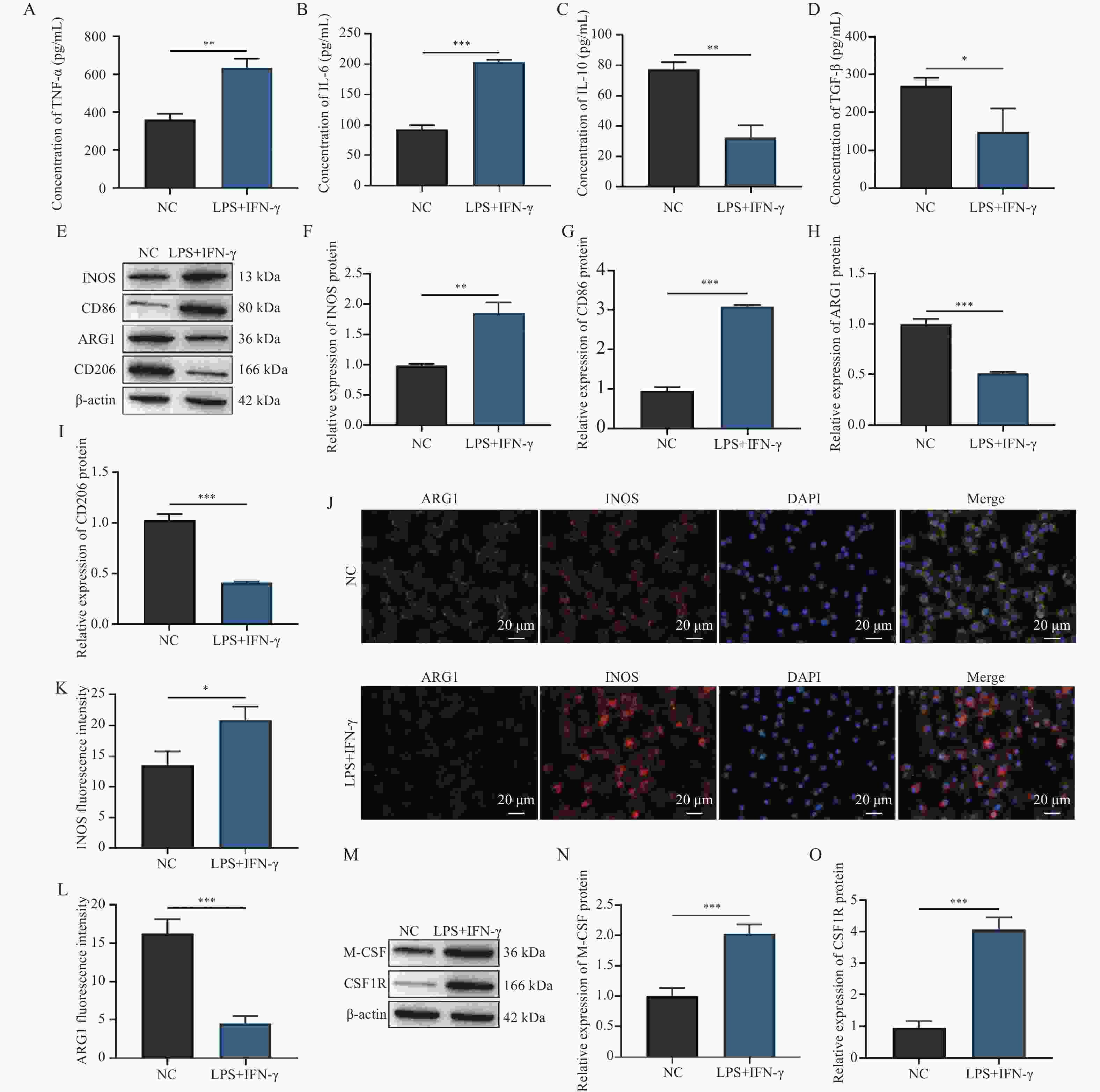
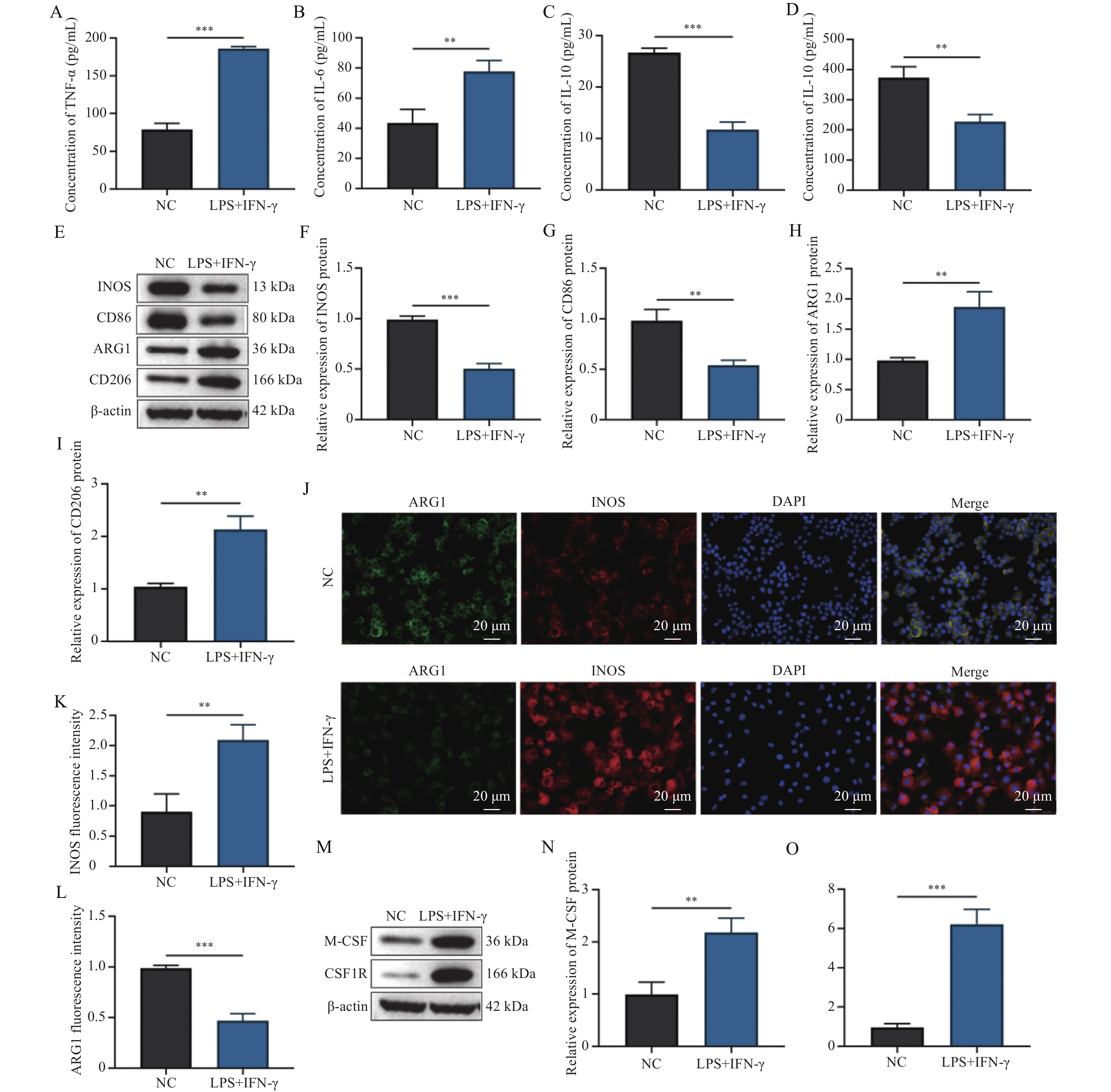
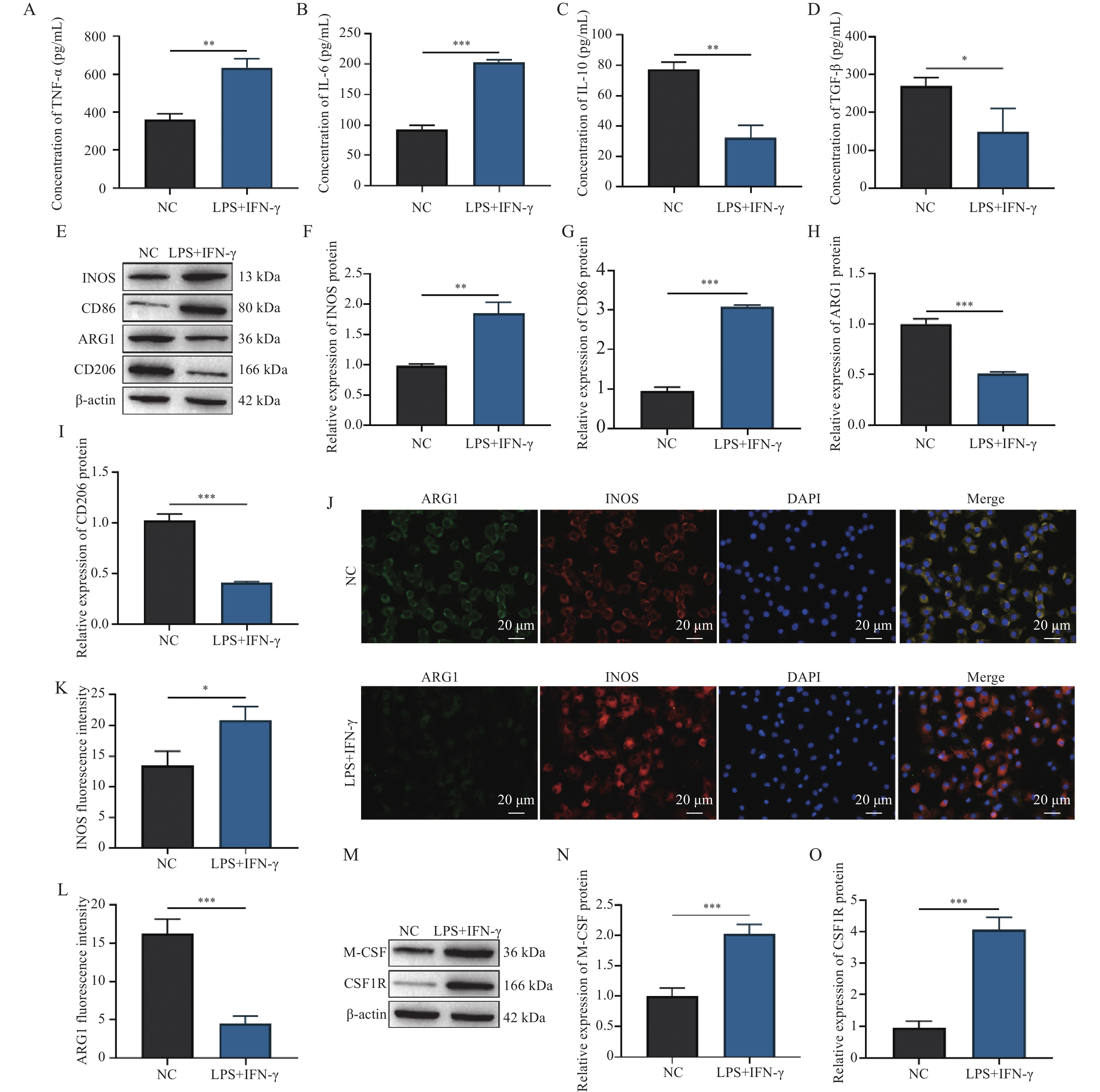
图 1 LPS和IFN-γ诱导RAW 264.7细胞M1极化并上调M-CSF($\bar x \pm s $,n = 3)
A:细胞培养上清液中TNF-α水平;B:细胞培养上清液中IL-6水平;C:细胞培养上清液中IL-10水平;D:细胞培养上清液中TGF-β水平;E~I:Western blot检测RAW 264.7细胞中INOS、CD86、ARG1和CD206蛋白条带及相对表达分析;J~L:ARG1和INOS的IF染色代表性图像及平均荧光强度统计分析;M~O:Western blot检测M-CSF和CSF1R蛋白条带及相对表达分析;*P < 0.05;**P < 0.01;***P < 0.001。
Figure 1. LPS and IFN-γ induce M1 polarization and upregulate M-CSF in RAW 264.7 cells ($\bar x \pm s $,n = 3)
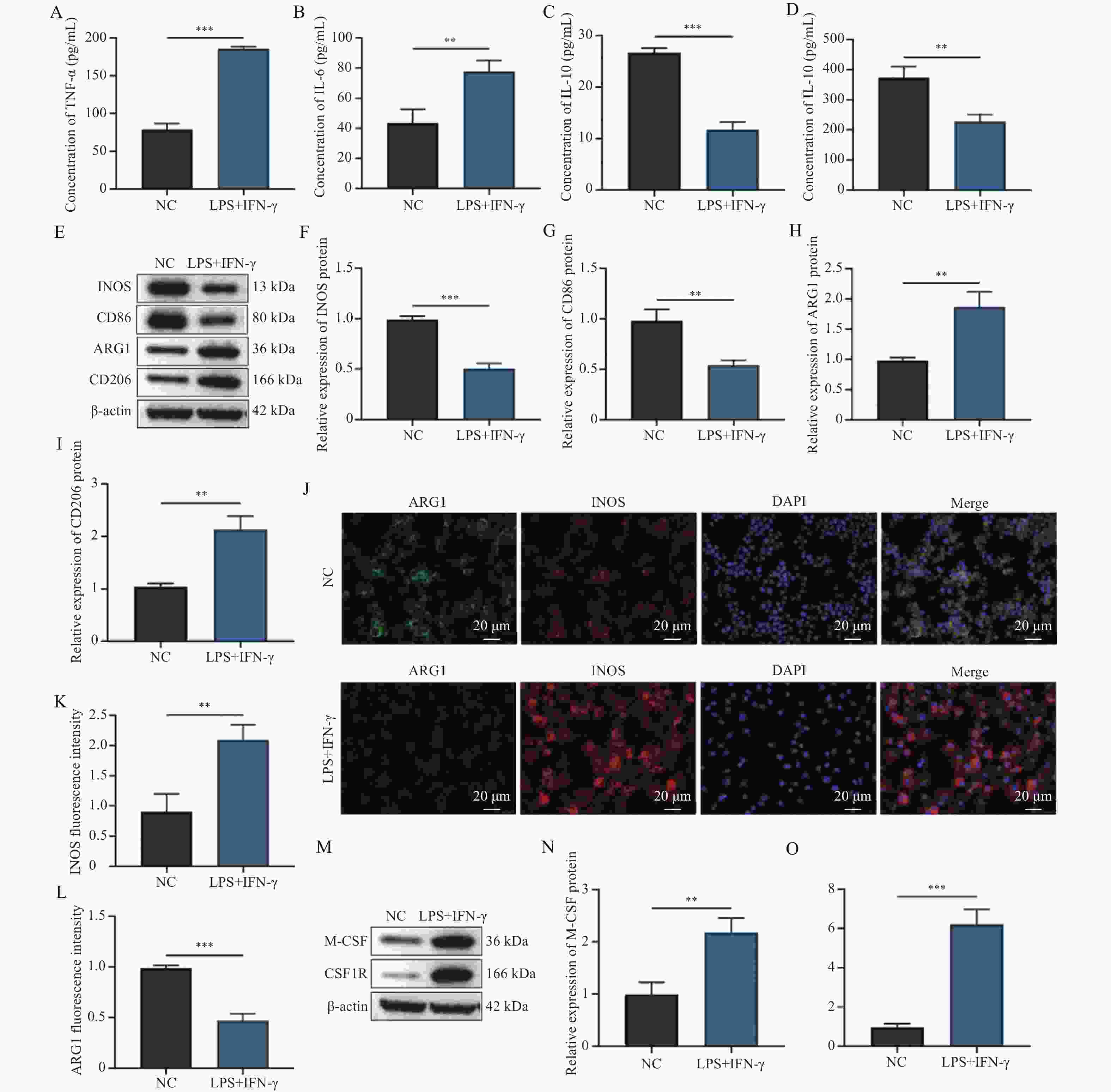
图 2 LPS和IFN-γ诱导BMDM的M1极化并上调M-CSF($\bar x \pm s $,n = 3)
A:细胞培养上清液中TNF-α水平;B:细胞培养上清液中IL-6水平;C:细胞培养上清液中IL-10水平;D:细胞培养上清液中TGF-β水平;E~I:Western blot检测BMDM细胞中INOS、CD86、ARG1和CD206蛋白条带及相对表达分析;J~L:BMDM中ARG1和INOS的IF染色代表性图像及平均荧光强度统计分析;M~O:Western blot检测M-CSF和CSF1R蛋白条带及相对表达分析;**P < 0.01;***P < 0.001。
Figure 2. LPS and IFN-γ induce M1 polarization and upregulate M-CSF in BMDM ($\bar x \pm s $,n = 3)
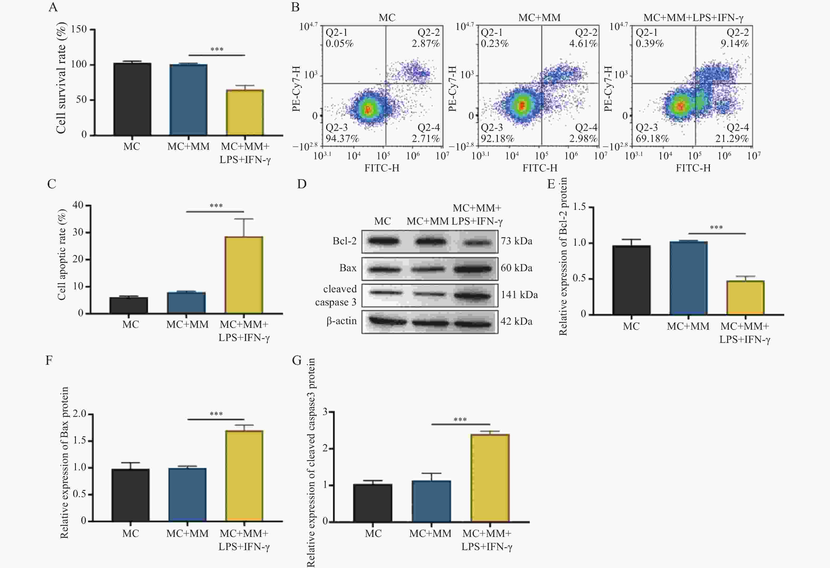
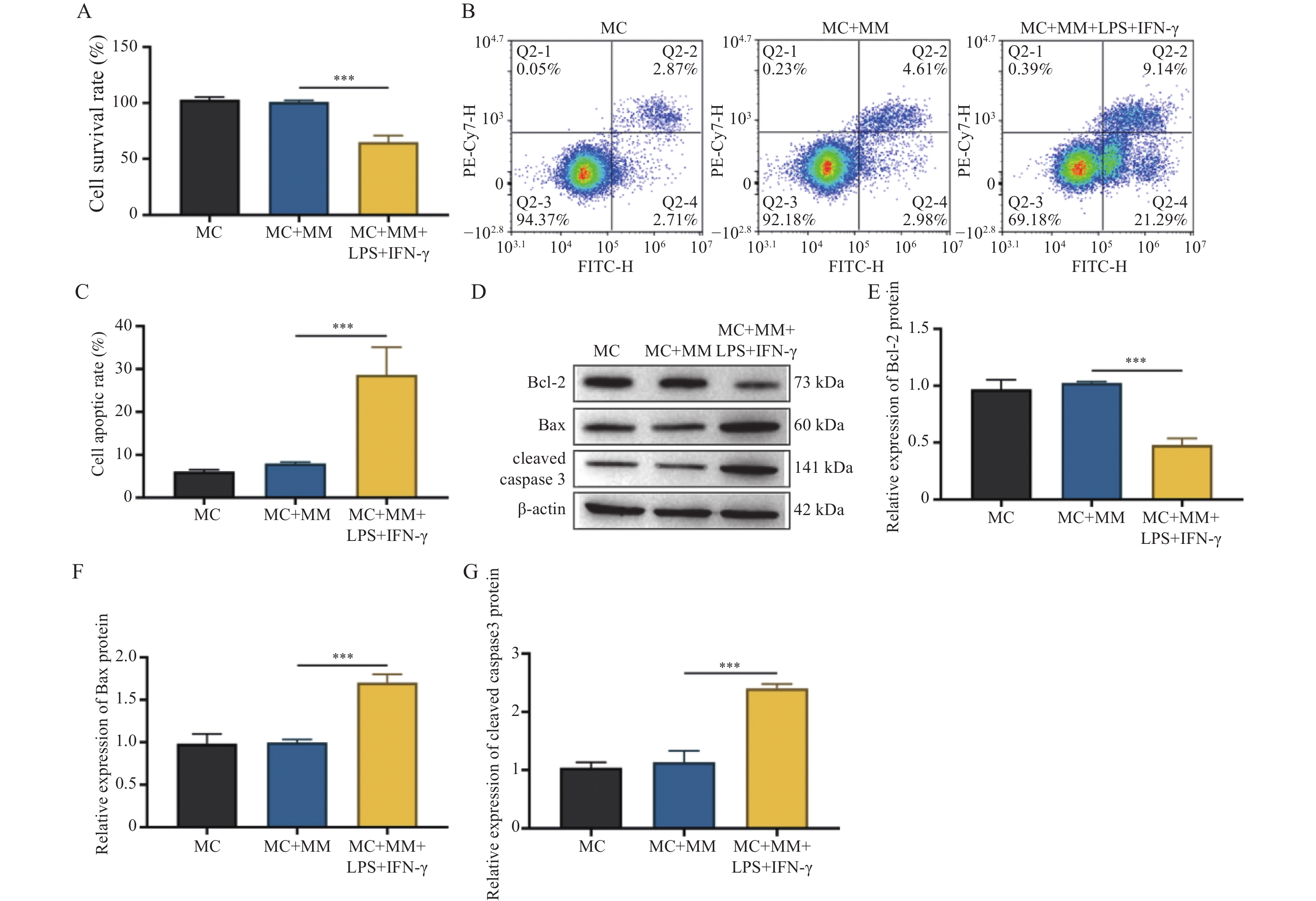
图 3 RAW 264.7细胞M1极化抑制软骨细胞活性促进凋亡($\bar x \pm s $,n = 3)
A:CCK-8检测软骨细胞活力;B~C:Annexin V-FITC/PI细胞凋亡试剂盒检测软骨细胞凋亡率及分析结果;D~G:Western blot检测凋亡相关蛋白Bcl-2、Bax和cleaved caspase 3蛋白条带及相对表达分析;***P < 0.001。
Figure 3. M1-polarized RAW 264.7 cells inhibit chondrocyte viability and promote apoptosis ($\bar x \pm s $,n = 3)
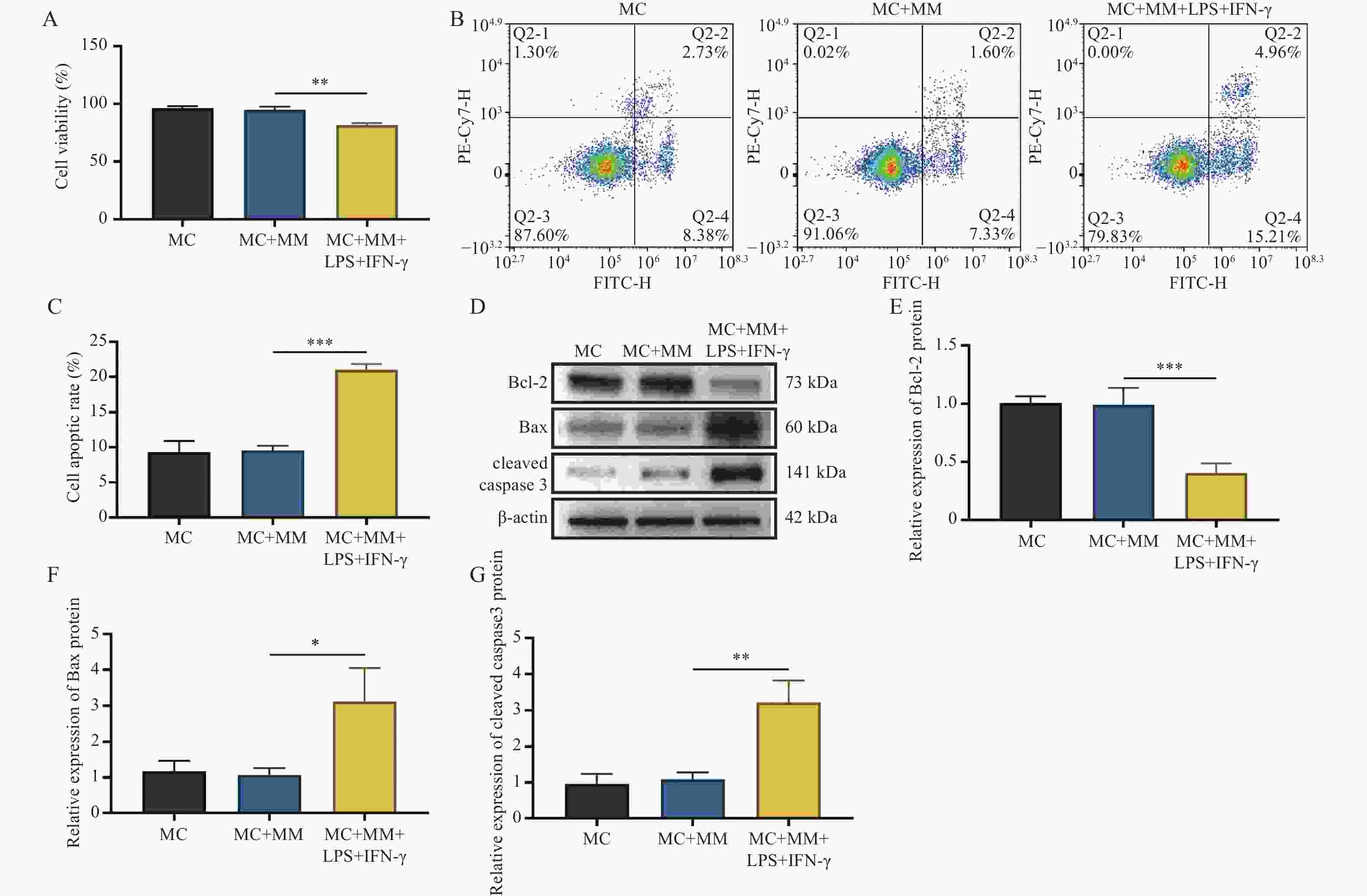
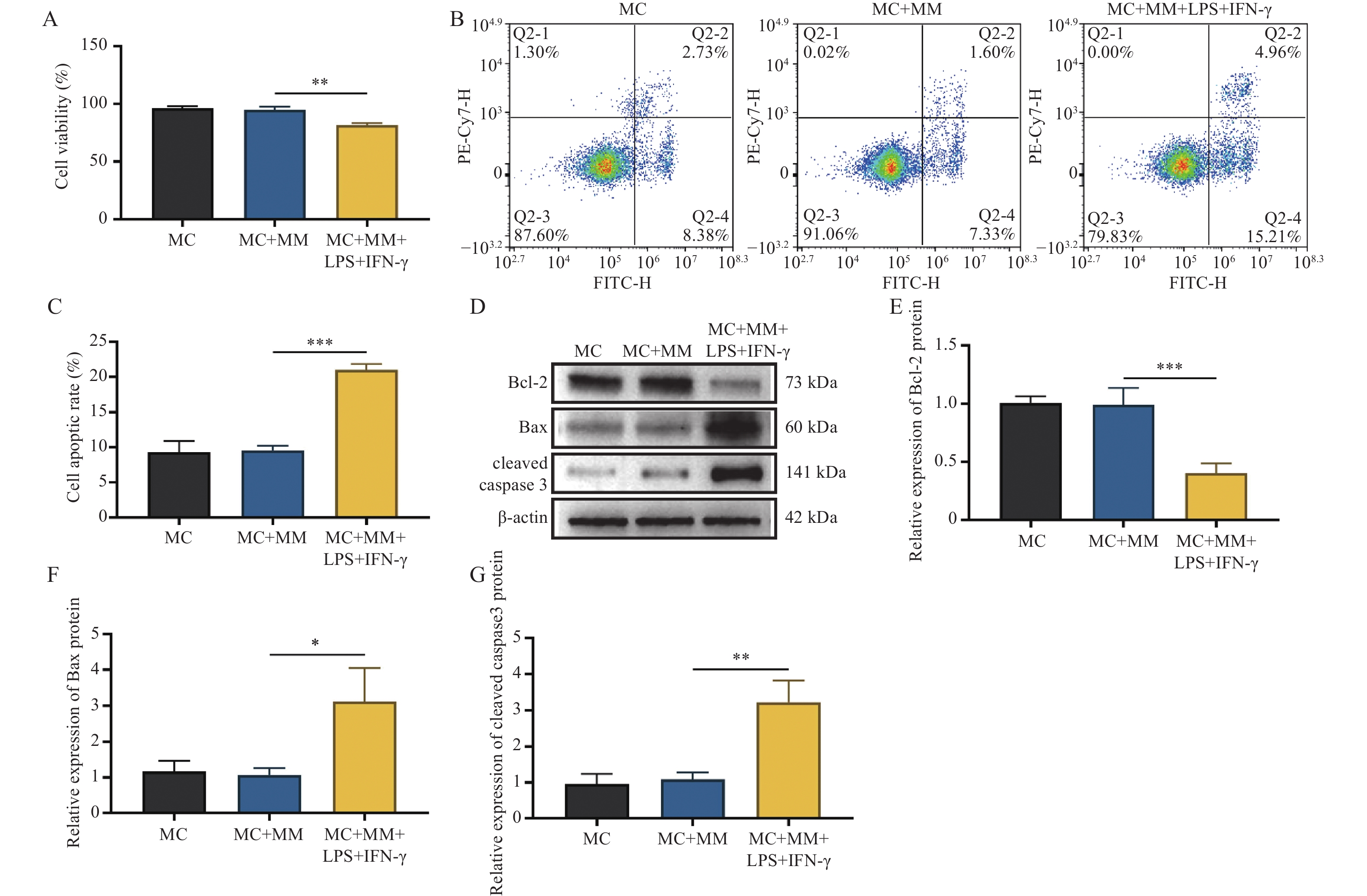
图 4 BMDM细胞M1极化抑制软骨细胞活性促进凋亡($\bar x \pm s $,n = 3)
A:CCK-8检测与BMDM细胞共培养的软骨细胞活力;B~C:Annexin V-FITC/PI细胞凋亡试剂盒检测软骨细胞凋亡率及统计分析;D~G:Western blot检测凋亡相关蛋白Bcl-2、Bax和cleaved caspase 3蛋白条带及相对表达分析;*P < 0.05;**P < 0.01;***P < 0.001。
Figure 4. M1 polarization of BMDMs inhibits chondrocyte viability and promotes apoptosis ($\bar x \pm s $,n = 3)
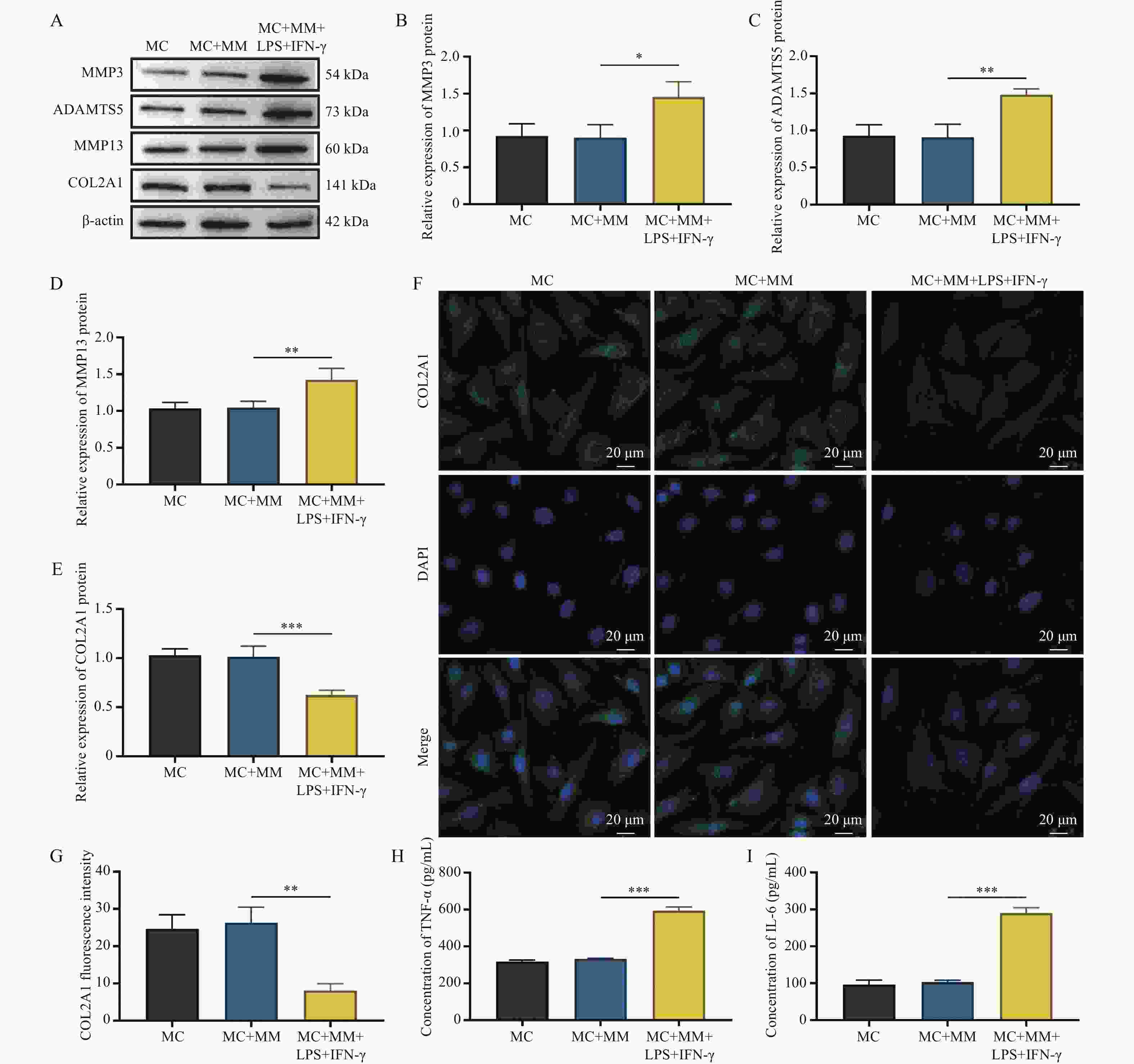
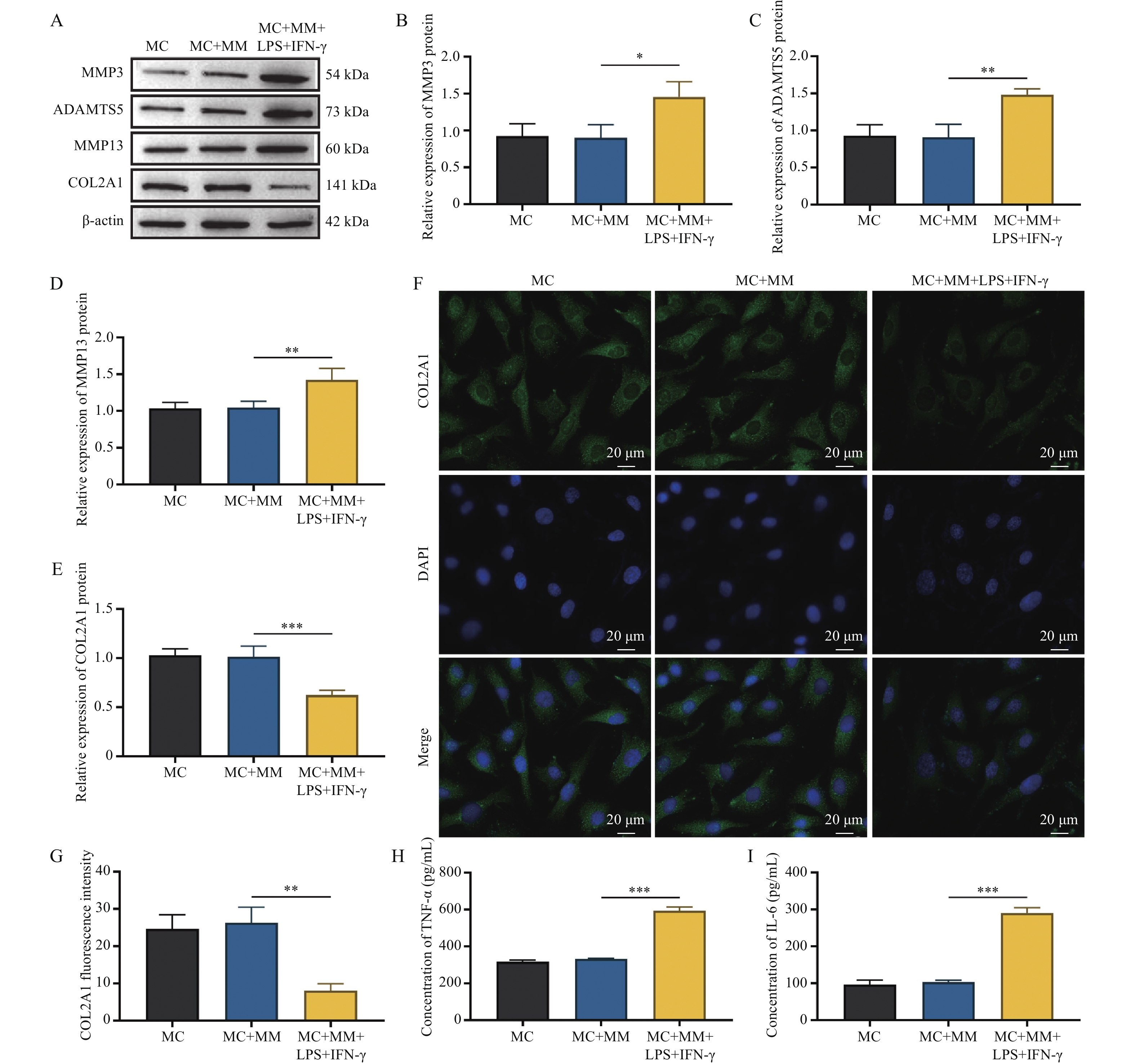
图 5 RAW 264.7细胞M1极化促进软骨细胞变性和炎症反应($\bar x \pm s $,n = 3)
A~E:Western blot检测软骨细胞中MMP3、ADAMTS5、MMP13和COL2A1蛋白条带及相对表达分析;F~G:COL2A1的IF染色代表性图像及平均荧光分析;H~I:ELISA试剂盒检测软骨细胞培养上清液中TNF-α和IL-6水平;*P < 0.05;**P < 0.01;***P < 0.001。
Figure 5. M1 polarization of RAW 264.7 cells promotes chondrocyte degeneration and inflammatory responses ($\bar x \pm s $,n = 3)
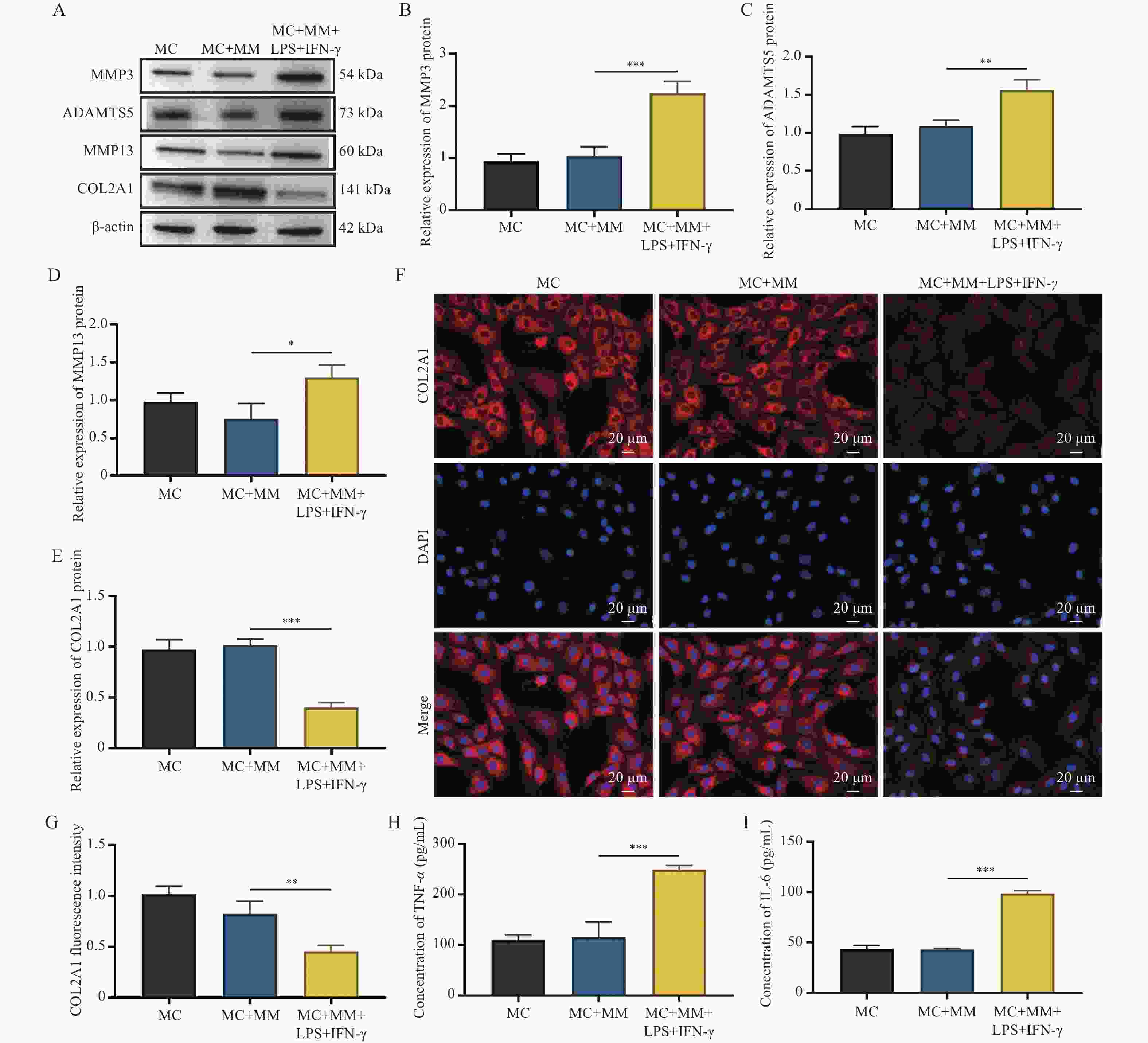
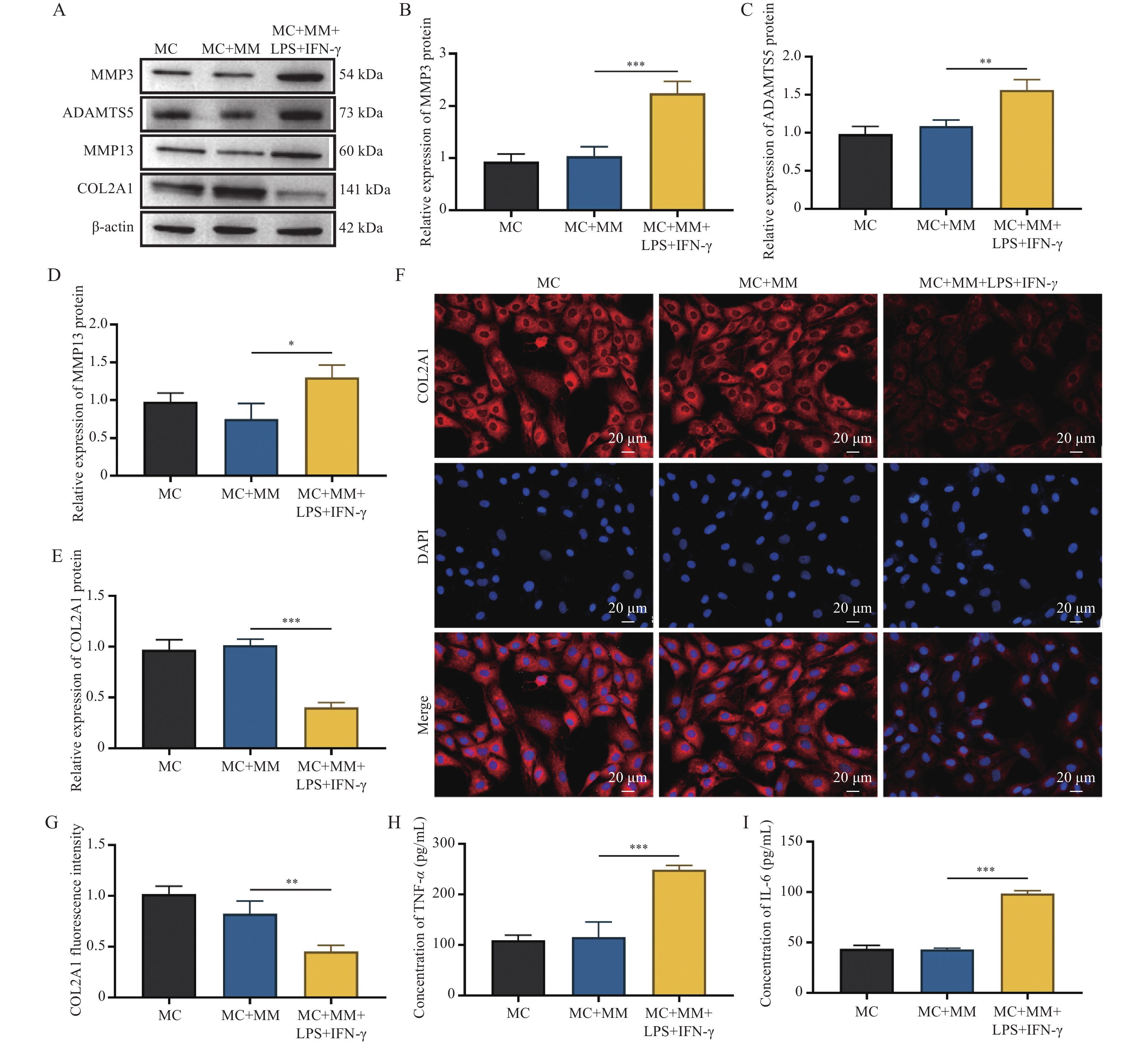
图 6 BMDM细胞M1极化促进软骨细胞变性和炎症反应($\bar x \pm s $,n = 3)
A~E:Western blot检测与BMDM细胞共培养的软骨细胞中MMP3、ADAMTS5、MMP13和COL2A1蛋白条带和相对表达分析;F~G:COL2A1的IF染色代表性图像和平均荧光强度分析;H~I:ELISA试剂盒检测软骨细胞培养上清液中TNF-α和IL-6浓度;*P < 0.05;**P < 0.01;***P < 0.001。
Figure 6. M1 polarization of BMDM cells promotes chondrocyte degeneration and inflammatory responses ($\bar x \pm s $,n = 3)
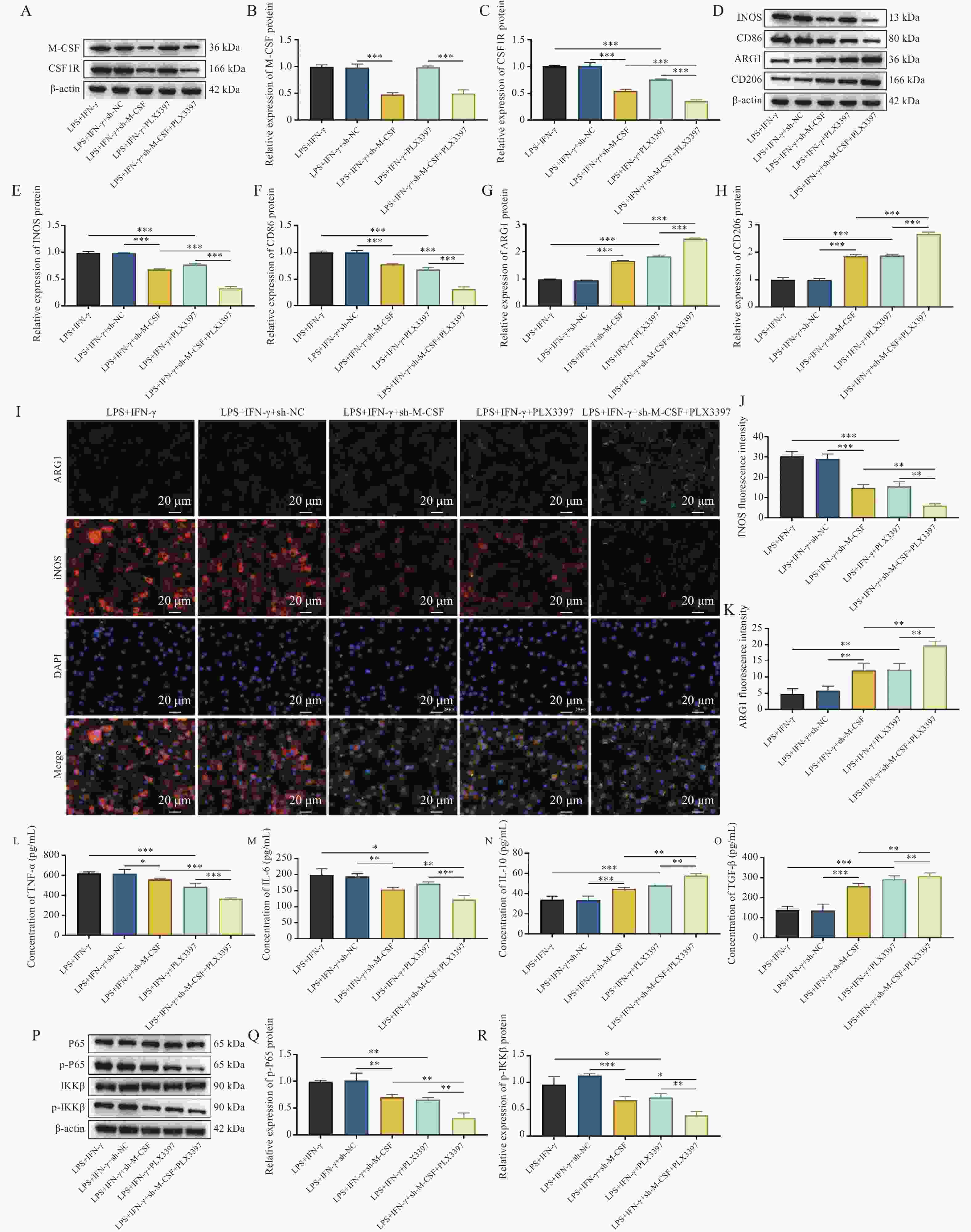
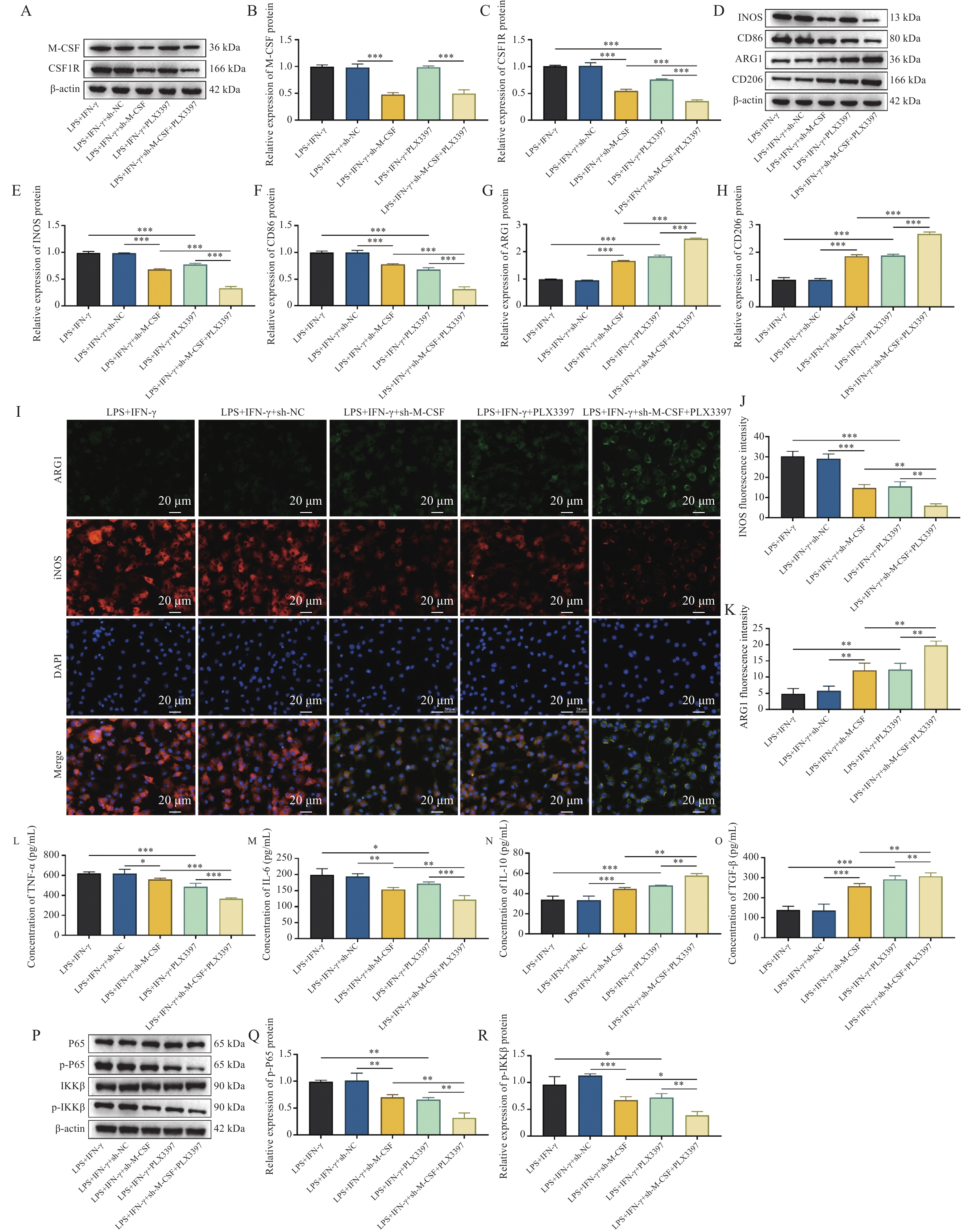
图 7 敲低M-CSF通过下调CSF1R抑制RAW 264.7细胞M1极化($\bar x \pm s $,n = 3)
A~C:Western blot检测敲低M-CSF和PLX3397处理后M-CSF和CSF1R蛋白条带和相对表达分析;D~H:Western blot检测RAW 264.7细胞极化标志物INOS、CD86、ARG1和CD206蛋白条带和相对表达分析;I~K:ARG1和INOS免疫荧光染色代表性图像和平均荧光强度分析;L~O:ELISA检测炎性细胞因子TNF-α、IL-6、IL-10和TGF-β浓度;P~R:Western blot检测P65、p-P65、IKKβ、p-IKKβ蛋白条带和p-P65、p-IKKβ相对表达分析;*P < 0.05;**P < 0.01;***P < 0.001。
Figure 7. Knockdown of M-CSF inhibits M1 polarization in RAW 264.7 cells by downregulating CSF1R ($\bar x \pm s $,n = 3)
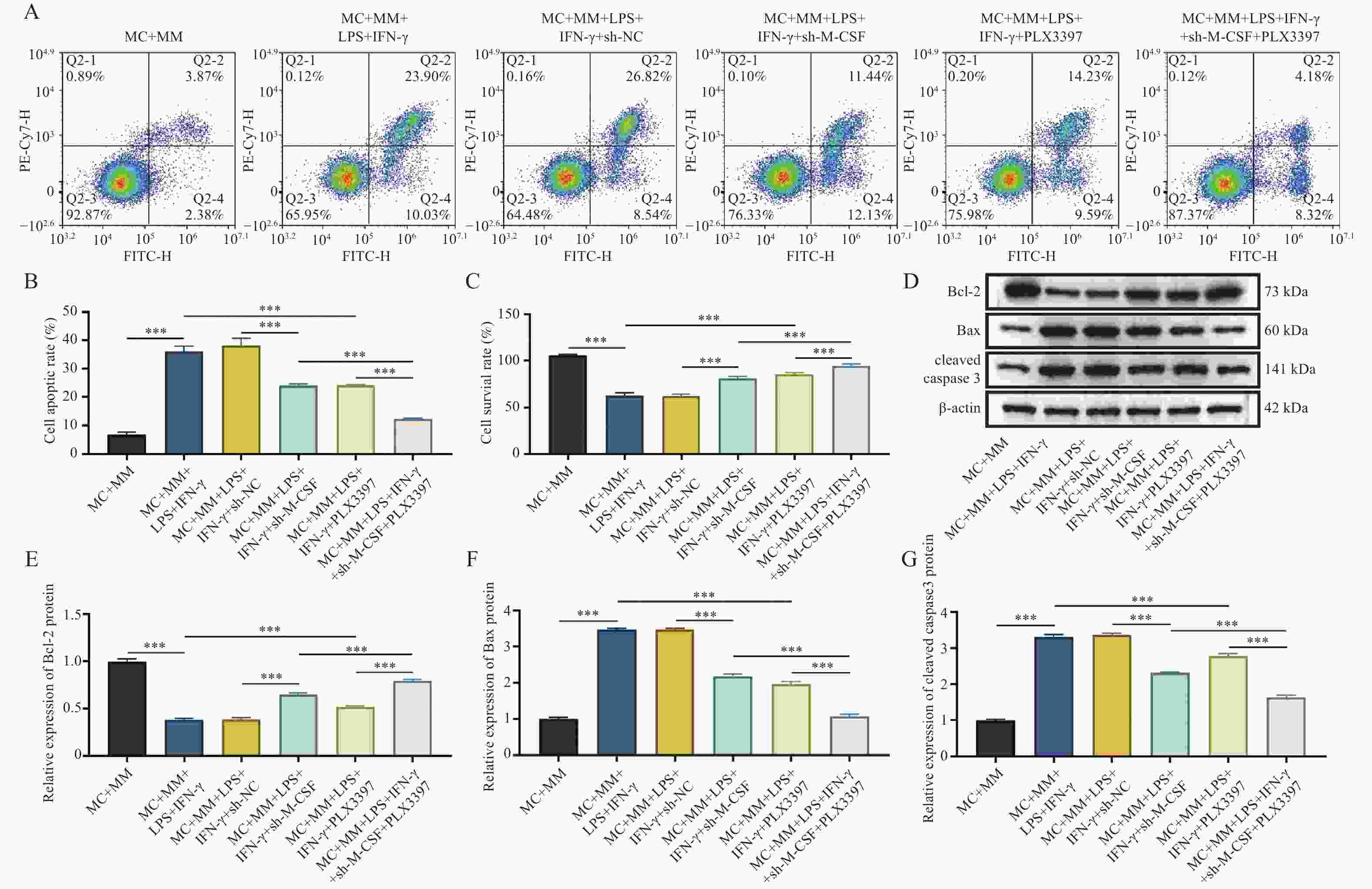
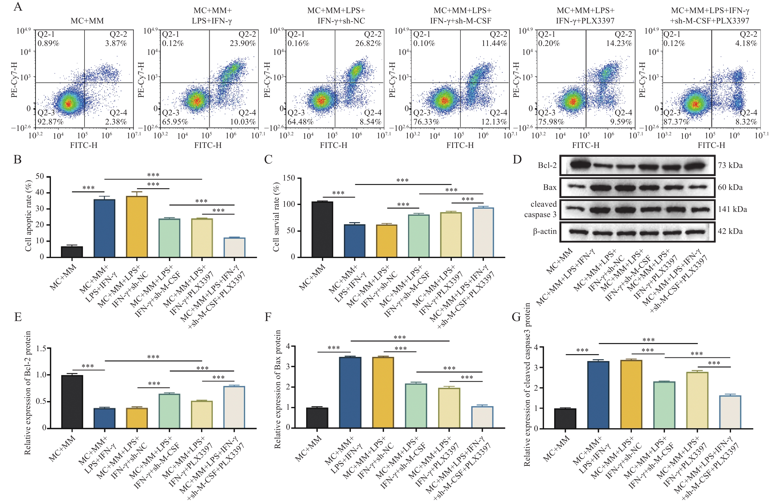
图 8 敲低M-CSF通过下调CSF1R促进软骨细胞增殖并抑制凋亡($\bar x \pm s $,n = 3)
A~B:RAW 264.7细胞中敲低M-CSF和PLX3397处理对软骨细胞凋亡的影响及凋亡率分析;C:CCK-8检测软骨细胞活力;D~G:与敲低M-CSF和PLX3397处理的RAW 264.7细胞共培养软骨细胞中Bcl-2、Bax和cleaved caspase3蛋白条带及相对表达分析;***P < 0.001。
Figure 8. Knockdown of M-CSF promotes chondrocyte proliferation and inhibits apoptosis by downregulating CSF1R ($\bar x \pm s $,n = 3)
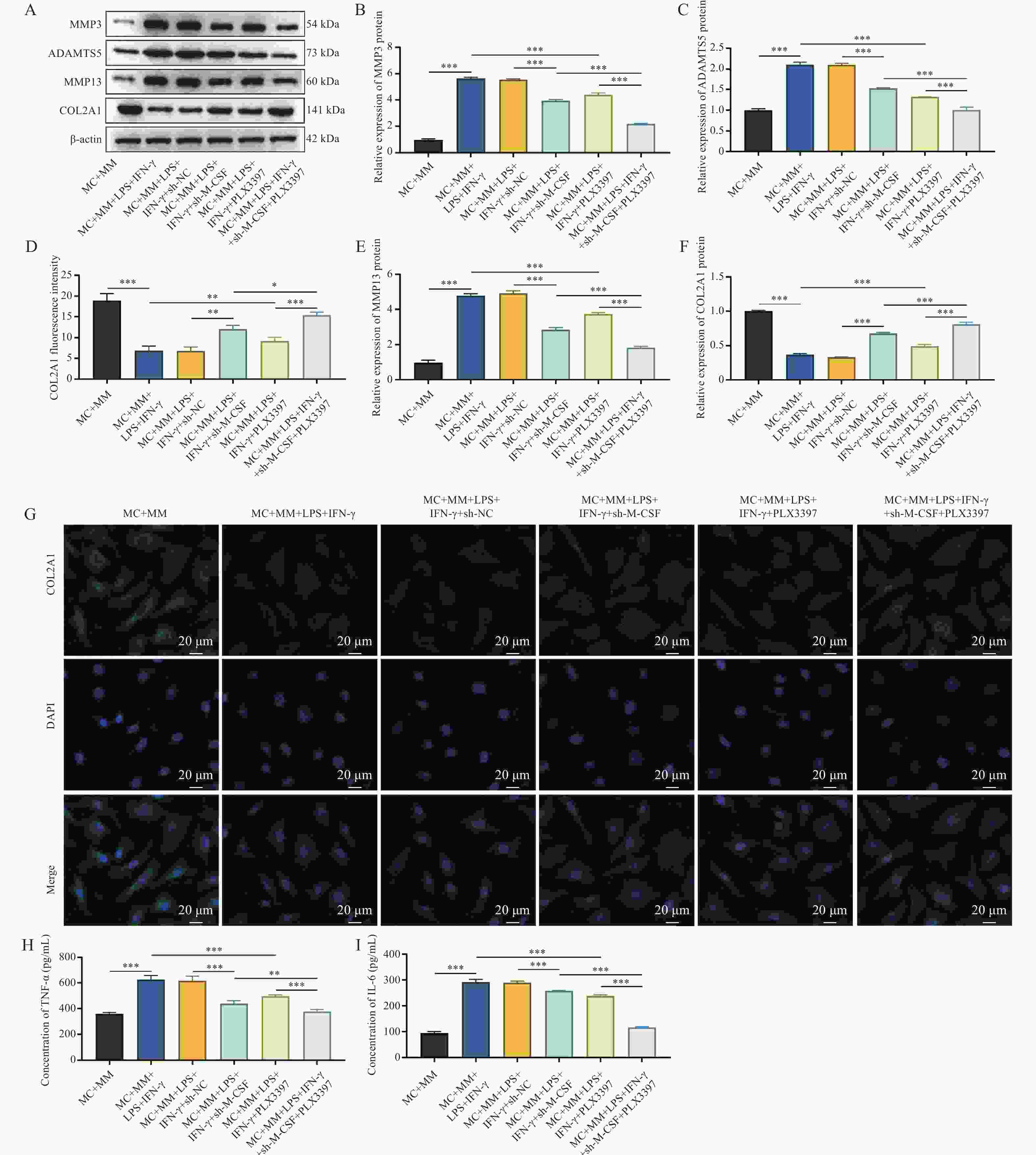
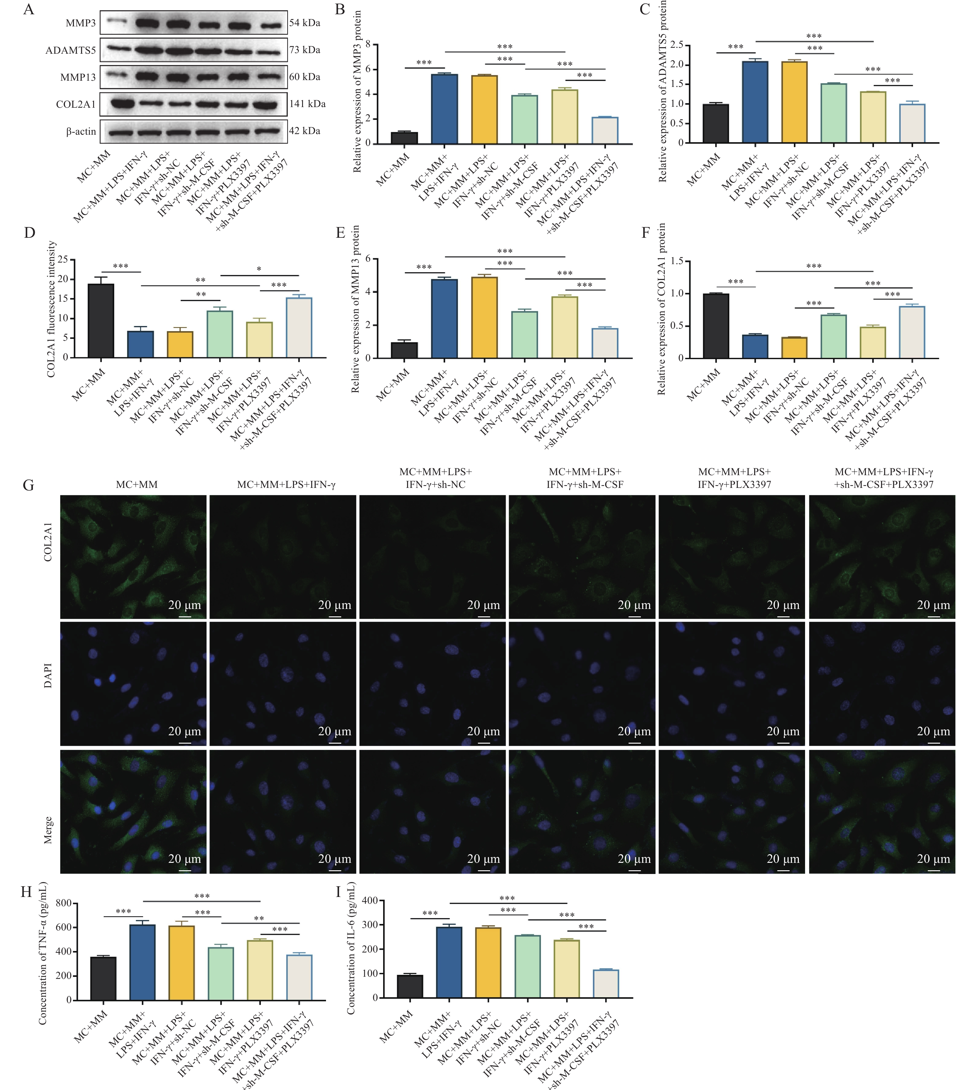
图 9 敲低M-CSF通过下调CSF1R缓解软骨细胞变性($\bar x \pm s $,n = 3)
A~E:Western blot检测与敲低M-CSF和PLX3397处理的RAW 264.7细胞共培养软骨细胞中MMP3、ADAMTS5、MMP13和COL2A1蛋白条带及相对表达分析;F~G:IF检测COL2A1代表性图像及平均荧光强度分析;H~I:ELISA试剂盒检测软骨细胞培养上清液中TNF-α和IL-6水平;*P < 0.05;**P < 0.01;***P < 0.001。
Figure 9. M-CSF knockdown alleviates chondrocyte degeneration by downregulating CSF1R ($\bar x \pm s $,n = 3)
-
[1] Zhang Y, Han Y, Sun Y, et al. Osteoarthritis: Molecular pathogenesis and potential therapeutic options[J]. Signal Transduct Target Ther, 2026, 11: 81. doi: 10.1038/s41392-025-02556-6 [2] Hao T, Pei Z, Hu S, et al. Identification of osteoarthritis-associated chondrocyte subpopulations and key gene-regulating drugs based on multi-omics analysis[J]. Sci Rep, 2025, 15: 12448. doi: 10.1038/s41598-025-90694-w [3] Mei Z, Yilamu K, Ni W, et al. Chondrocyte fatty acid oxidation drives osteoarthritis via SOX9 degradation and epigenetic regulation[J]. Nat Commun, 2025, 16(1): 4892. doi: 10.1038/s41467-025-60037-4 [4] Li Y, Li H, Chen X, et al. CDK5 regulates PPARγ/NF-κB signaling to exacerbate obesity-related osteoarthritis via modulating macrophage polarization and chondrocyte apoptosis[J]. Cell Mol Biol Lett, 2025, 30(1): 135. doi: 10.1186/s11658-025-00822-7 [5] Zhang H, Cai D, Bai X. Macrophages regulate the progression of osteoarthritis[J]. Osteoarthritis Cartilage, 2020, 28(5): 555-561. doi: 10.1016/j.joca.2020.01.007 [6] Wang H, Ning W, Liu Y, et al. Photobiomodulation mitigates chondrocyte catabolism in osteoarthritis by modulating macrophage M1 polarization through the IL-6/JAK/STAT pathway[J]. J Orthop Surg Res, 2025, 20(1): 1082. doi: 10.1186/s13018-025-06506-4 [7] Zhang F, Wu W, Yuan Y, et al. CSF1 regulates inflammation and apoptosis in intervertebral disc degeneration[J]. Sci Rep, 2026, 16(1): 10566. doi: 10.1038/s41598-026-44984-6 [8] Sheng K, Bisson D G, Saran N, et al. The TLR-M-CSF axis is implicated in increased bone turnover and curve progression in adolescent idiopathic scoliosis[J]. Arthritis Res Ther, 2025, 27(1): 68. doi: 10.1186/s13075-025-03535-6 [9] Zheng M, Li Z, Feng Y, et al. The role of CD14 and CSF1R in osteoarthritis and gastritis[J]. Medicine, 2023, 102(43): e35567. doi: 10.1097/MD.0000000000035567 [10] Hu P, Li B, Yin Z, et al. Multi-omics characterization of macrophage polarization-related features in osteoarthritis based on a machine learning computational framework[J]. Heliyon, 2024, 10(9): e30335. doi: 10.1016/j.heliyon.2024.e30335 [11] Salti T, Braunstein I, Haimovich Y, et al. Widespread S-persulfidation in activated macrophages as a protective mechanism against oxidative-inflammatory stress[J]. Redox Biol, 2024, 72: 103125. doi: 10.1016/j.redox.2024.103125 [12] Piccioni G, Maisto N, D’Ettorre A, et al. Switch to phagocytic microglia by CSFR1 inhibition drives amyloid-beta clearance from glutamatergic terminals rescuing LTP in acute hippocampal slices[J]. Transl Psychiatry, 2024, 14: 338. doi: 10.1038/s41398-024-03019-2 [13] Zheng M, Zhu Y, Wei K, et al. Metformin attenuates the inflammatory response via the regulation of synovial M1 macrophage in osteoarthritis[J]. Int J Mol Sci, 2023, 24(6): 5355. doi: 10.3390/ijms24065355 [14] Lopa S, Libonati F, Mareschi K, et al. Using macrophage polarization in human platelet lysate to test the immunomodulatory potential of cells for clinical use[J]. Biomedicines, 2024, 12(4): 833. doi: 10.3390/biomedicines12040833 [15] Fu S, Du H, Ling X, et al. Suppressing chondrocyte cuproptosis by syringaresinol-4-O-β-d-glucoside alleviates gouty arthritis[J]. Front Pharmacol, 2025, 16: 1565422. doi: 10.3389/fphar.2025.1565422 [16] Xu M, Sun X, Ma X, et al. Sodium tanshinone IIA sulfonate alleviates osteoarthritis through targeting SIRT1[J]. Chin Med, 2025, 20(1): 142. doi: 10.1186/s13020-025-01166-2 [17] Li X, Cheng Y, Gu P, et al. Engineered microchannel scaffolds with instructive niches reinforce endogenous bone regeneration by regulating CSF-1/CSF-1R pathway[J]. Adv Mater, 2024, 36(19): e2310876. doi: 10.1002/adma.202310876 [18] Inoue K, Qin Y, Xia Y, et al. Bone marrow Adipoq-lineage progenitors are a major cellular source of M-CSF that dominates bone marrow macrophage development, osteoclastogenesis, and bone mass[J]. eLife, 2023, 12: e82118. doi: 10.7554/eLife.82118 [19] Kim H J, Kang W Y, Seong S J, et al. Follistatin-like 1 promotes osteoclast formation via RANKL-mediated NF-κB activation and M-CSF-induced precursor proliferation[J]. Cell Signal, 2016, 28(9): 1137-1144. doi: 10.1016/j.cellsig.2016.05.018 [20] Kitaura H, Zhou P, Kim H J, et al. M-CSF mediates TNF-induced inflammatory osteolysis[J]. J Clin Invest, 2005, 115(12): 3418-3427. doi: 10.1172/JCI26132 [21] Zeijlon L, Budhwar S, Lindau R, et al. Human amnion epithelial cells induce M2 macrophage polarisation partially via M-CSF secretion but independently of extracellular vesicles in vitro[J]. Front Immunol, 2026, 17: 1723968. doi: 10.3389/fimmu.2026.1723968 [22] Yuan Y, Li Y, Zhao W, et al. WNT4 promotes macrophage polarization via granulosa cell M-CSF and reduces granulosa cell apoptosis in endometriosis[J]. Cytokine, 2023, 172: 156400. doi: 10.1016/j.cyto.2023.156400 [23] Wu J J, Sun Z L, Liu S Y, et al. The ASIC3-M-CSF-M2 macrophage-positive feedback loop modulates fibroblast-to-myofibroblast differentiation in skin fibrosis pathogenesis[J]. Cell Death Dis, 2022, 13: 527. doi: 10.1038/s41419-022-04981-9 [24] Wang Y, Wernersbach I, Strehle J, et al. Early posttraumatic CSF1R inhibition via PLX3397 leads to time- and sex-dependent effects on inflammation and neuronal maintenance after traumatic brain injury in mice[J]. Brain Behav Immun, 2022, 106: 49-66. doi: 10.1016/j.bbi.2022.07.164 [25] Su S, Liu T, Zheng J Y, et al. Huang Lian Jie Du decoction attenuated colitis via suppressing the macrophage Csf1r/Src pathway and modulating gut microbiota[J]. Front Immunol, 2024, 15: 1375781. doi: 10.3389/fimmu.2024.1375781 [26] Li X, Zhuang Z, Hao Y, et al. Paeonol relieves chronic neuropathic pain by reducing communication between schwann cells and macrophages in the dorsal root ganglia after injury[J]. Int J Mol Sci, 2025, 26(9): 3964. doi: 10.3390/ijms26093964 [27] Fujiwara T, Yakoub M A, Chandler A, et al. CSF1/CSF1R signaling inhibitor pexidartinib (PLX3397) reprograms tumor-associated macrophages and stimulates T-cell infiltration in the sarcoma microenvironment[J]. Mol Cancer Ther, 2021, 20(8): 1388-1399. doi: 10.1158/1535-7163.MCT-20-0591 [28] Adams R C, Carter-Cusack D, Llanes G T, et al. CSF1R inhibition promotes neuroinflammation and behavioral deficits during graft-versus-host disease in mice[J]. Blood, 2024, 143(10): 912-929. doi: 10.1182/blood.2023022040 -

 下载:
下载:
 点击查看大图
计量
- 文章访问数: 5
- HTML全文浏览量: 3
- PDF下载量: 0
- 被引次数: 0
