




Diterpenoids Derived from Strophioblachia Fimbricalyx Regulate Cardiac Hypertrophy by Blocking Autophagic Flux
-
摘要:
目的 从宿萼木中筛选抗心肌肥厚活性二萜单体化合物 C080914,探究其抗心肌肥厚作用及分子机制。 方法 分离SD乳鼠原代心肌细胞,分别采用苯肾上腺素(PE,n = 6-10)、血管紧张素 Ⅱ(AngⅡ,n = 6)、异丙肾上腺素(ISO,n = 6)构建体外心肌肥厚模型;CCK-8检测化合物细胞毒性;Western blot 检测心肌肥厚、自噬相关蛋白;F-actin 免疫荧光观察心肌细胞肥大表型(n = 10);mRFP-GFP-LC3双标腺病毒示踪自噬流(n = 5);网络药理学结合分子对接预测并验证候选靶点;采用自噬抑制剂3-MA、激动剂雷帕霉素开展功能干预实验(n = 6);多组间比较采用单因素方差分析,Tukey 法进行两两比较。 结果 C080914 细胞毒性低,CC50>50 μM;可显著抑制 PE、AngⅡ、ISO诱导ANP、MYH7蛋白表达上调(P < 0.05 或P < 0.001),缩小心肌细胞表面积(P < 0.001)。C080914 上调 LC3B、P62,下调 ATG9A 蛋白表达,造成自噬流阻断;分子对接显示 C080914 与 ATG9A 结合活性强,Vina 评分为-7.660 kcal/mol;3-MA 可部分逆转 C080914 抗心肌肥厚效应(P < 0.01);雷帕霉素同样发挥抗肥厚作用,但二者调控自噬模式存在明显差异(P < 0.05)。 结论 宿萼木来源二萜C080914体外可抑制原代心肌细胞病理性肥厚,该效应伴随ATG9A 下调与自噬流阻断,可作为心肌肥厚候选先导分子,ATG9A为潜在研究靶点。 Abstract:Objective To screen the diterpenoid monomer C080914 with anti-myocardial-hypertrophy activity from Strophioblachia fimbricalyx, and to investigate its anti-hypertrophic effect against myocardial hypertrophy as well as the underlying molecular mechanism. Methods Primary cardiomyocytes were isolated from neonatal SD rats. In-vitro myocardial hypertrophy models were induced with phenylephrine (PE, n = 6-10), angiotensin Ⅱ (AngⅡ, n = 6), and isoproterenol (ISO, n = 6), respectively. CCK-8 assay was used to evaluate the cytotoxicity of the compound. Western blot was performed to detect proteins associated with myocardial hypertrophy and autophagy. F-actin immunofluorescence staining was applied to observe the hypertrophic phenotype of cardiomyocytes (n = 10). mRFP-GFP-LC3 dual-labeled adenovirus was utilized to trace autophagic flux (n = 5). Network pharmacology combined with molecular docking was adopted to predict and verify candidate targets. Functional intervention experiments were carried out using the autophagy inhibitor 3-MA and autophagy agonist rapamycin (n = 6). One-way analysis of variance (ANOVA) was used for comparisons among multiple groups, followed by Tukey’ s post-hoc test for pairwise comparisons. P < 0.05 was considered statistically significant. Results C080914 exhibited low cytotoxicity with CC50>50 μM; it significantly suppressed the up-regulation of ANP and MYH7 protein expressions induced by PE, AngⅡ and ISO (P < 0.05 or P < 0.001) and decreased cardiomyocyte surface area (P < 0.001). C080914 up-regulated LC3B and P62 but down-regulated ATG9A protein expression, leading to autophagic flux blockage. Molecular docking demonstrated strong binding affinity between C080914 and ATG9A , with a Vina score of −7.660 kcal/mol. 3-MA partially reversed the anti-hypertrophic effect of C080914 (P < 0.01). Rapamycin also produced anti-hypertrophic effects, yet distinct differences existed in their autophagy-modulating modes. Conclusion The diterpenoid C080914 from Strophioblachia fimbricalyx inhibits pathological hypertrophy of primary cardiomyocytes in vitro, This effect is accompanied by ATG9A down-regulation and autophagic flux blockage. C080914 may be used as a candidate lead molecule against myocardial hypertrophy, with ATG9A as a potential therapeutic target. -
Key words:
- Strophioblachia fimbricalyx /
- Diterpenoids /
- Myocardial hypertrophy /
- Autophagy /
- ATG9A /
- Natural medicines
-
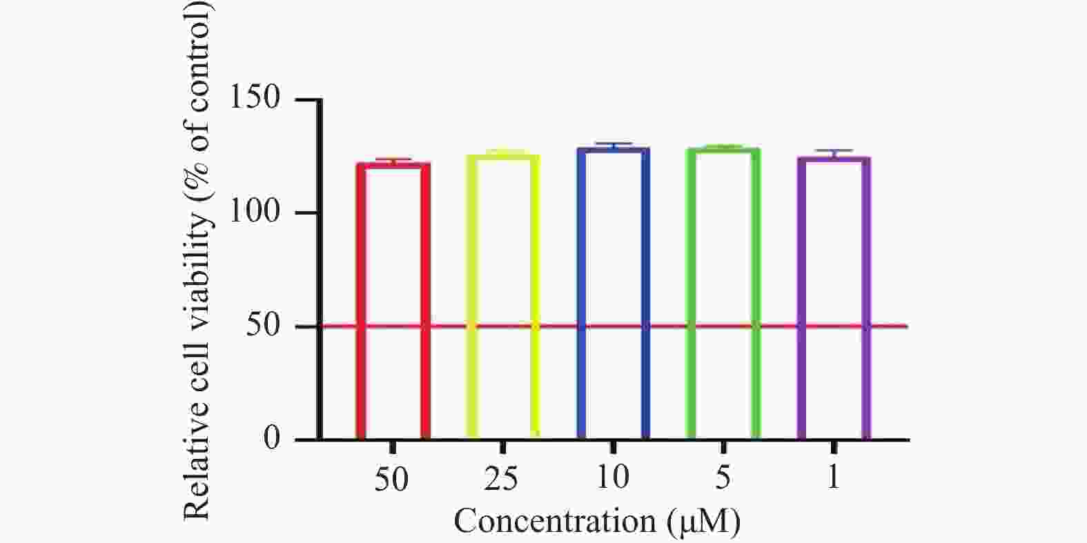
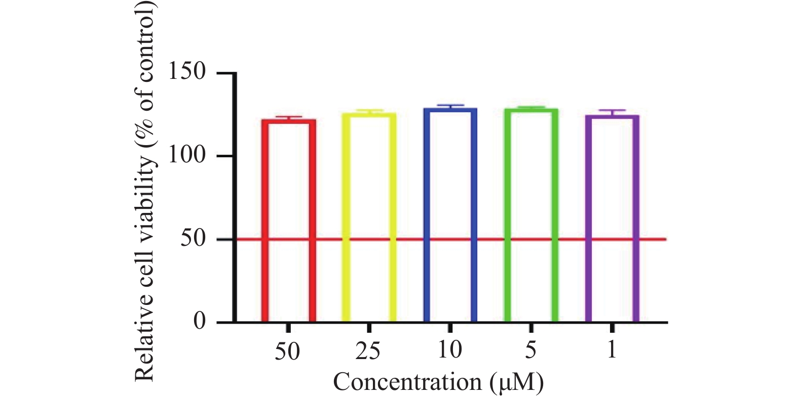
图 1 不同浓度的化合物对原代心肌细胞活性的影响[($ \bar x \pm s $),n = 6]
Figure 1. Effects of different concentrations of the compound on the viability of primary cardiomyocytes[($ \bar x \pm s $),n = 6]
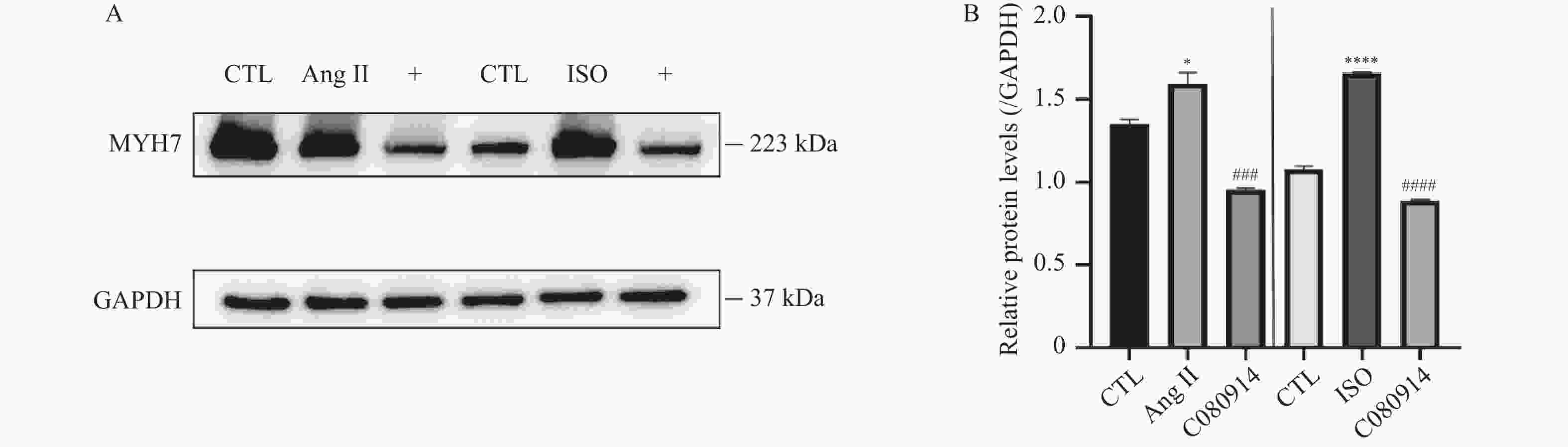
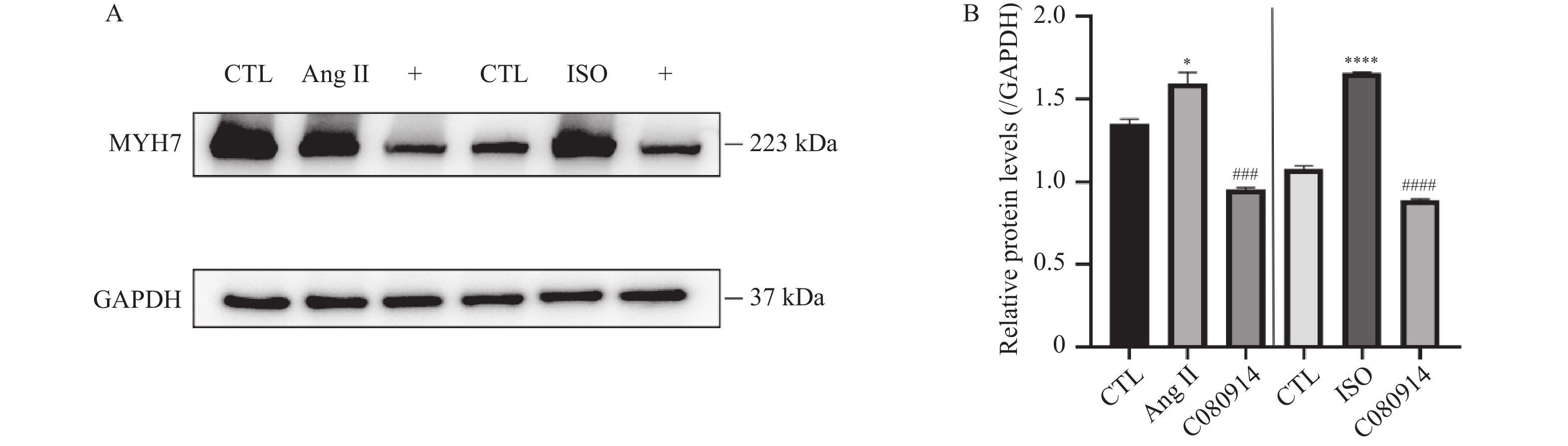
图 2 C080914对ISO、AngⅡ诱导的心肌肥厚的抑制作用[($ \bar x \pm s $),n = 6]
A:AngII/ISO 诱导心肌肥厚模型经C080914处理后,肥厚标志物 MYH7 蛋白的代表性免疫印迹图;B:MYH7蛋白相对定量统计图(目标蛋白灰度值与GAPDH灰度值的比值),竖线左侧为ISO诱导模型,右侧为 AngⅡ 诱导模型,与CTL组相比,*P < 0.05,****P < 0.0001;与AngⅡ、ISO组相比,###P < 0.001,####P < 0.0001。
Figure 2. Inhibitory effects of C080914 on ISO- and AngⅡ-induced myocardial hypertrophy [($ \bar x \pm s $),n = 6]
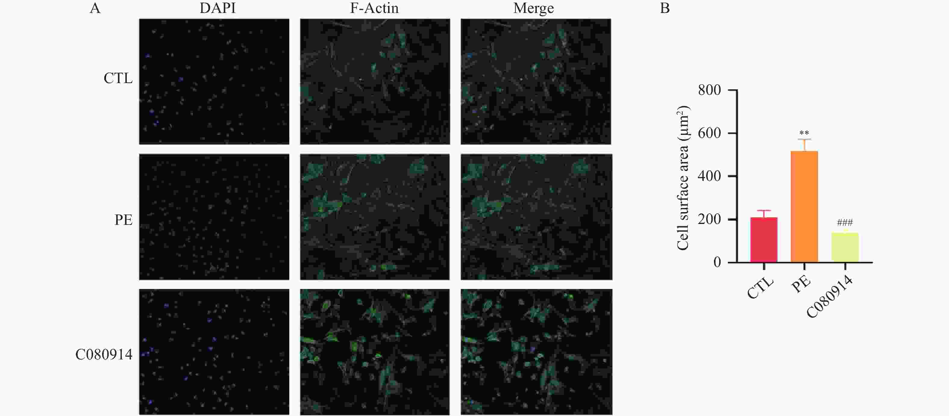
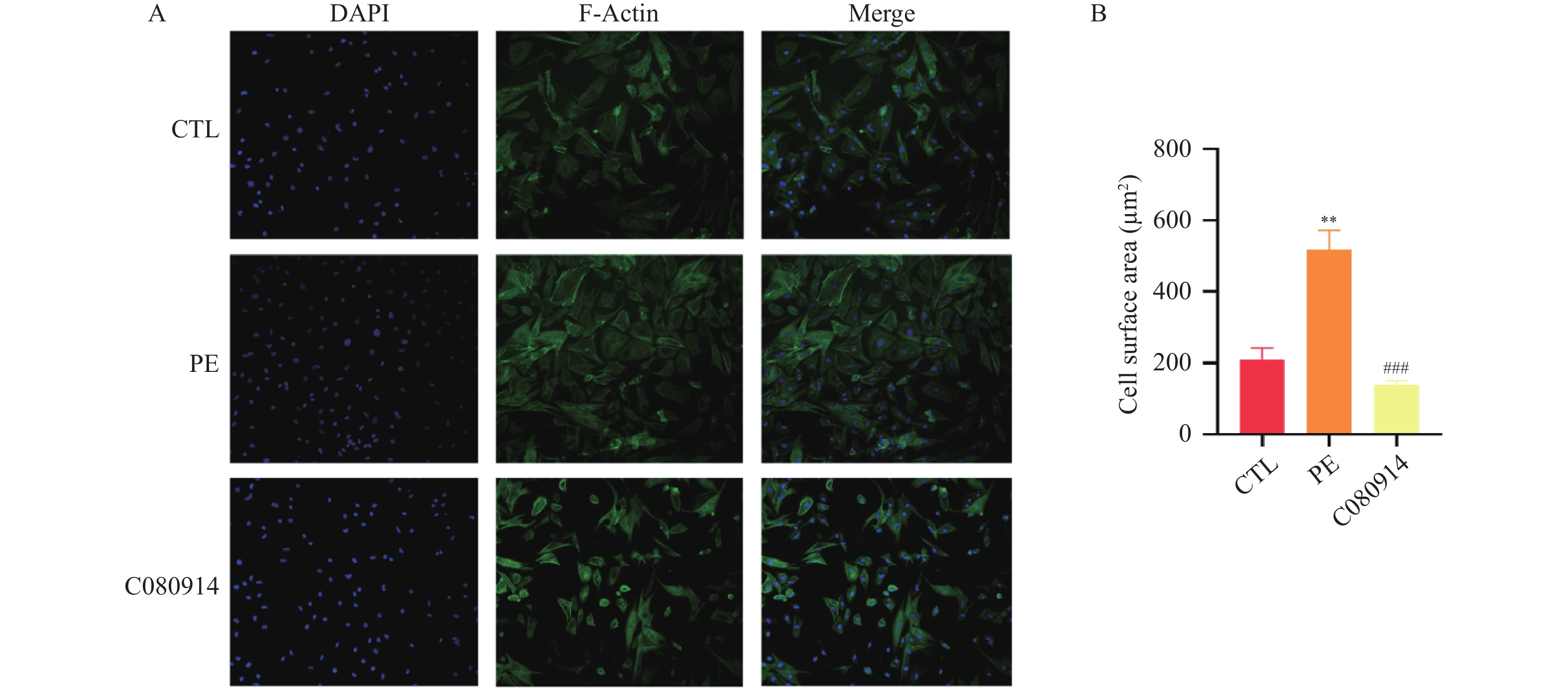
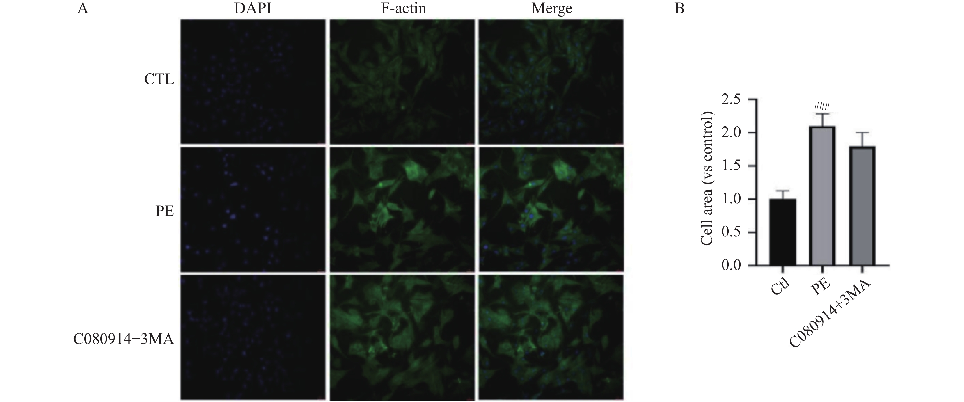
图 3 C080914对心肌细胞肥大及细胞面积的影响[($ \bar x \pm s $),n = 10]
A:心肌细胞表面积免疫荧光代表图(×40);B:运用ImageJ计算细胞表面积;与CTL组相比,**P < 0.01;与PE组相比,###P < 0.001。
Figure 3. Effects of C080914 on cardiomyocyte hypertrophy and cell surface area [($ \bar x \pm s $),n = 10]
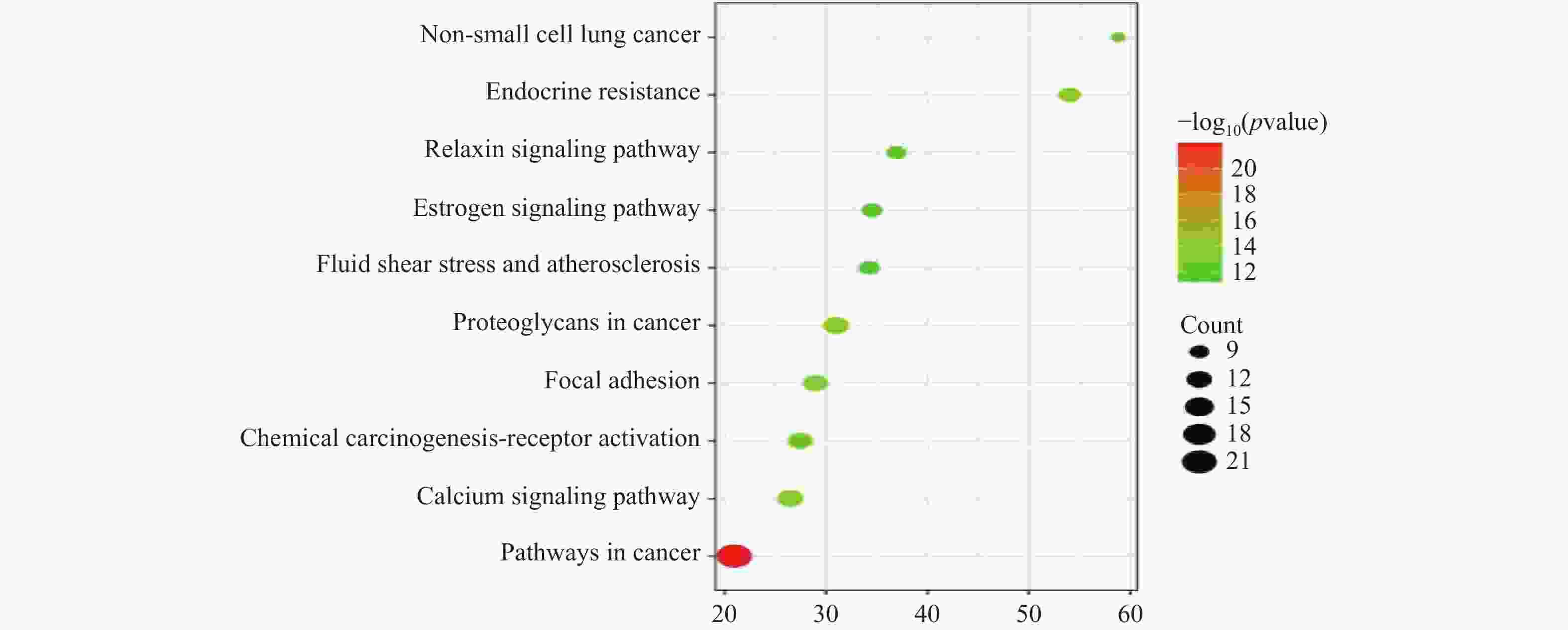
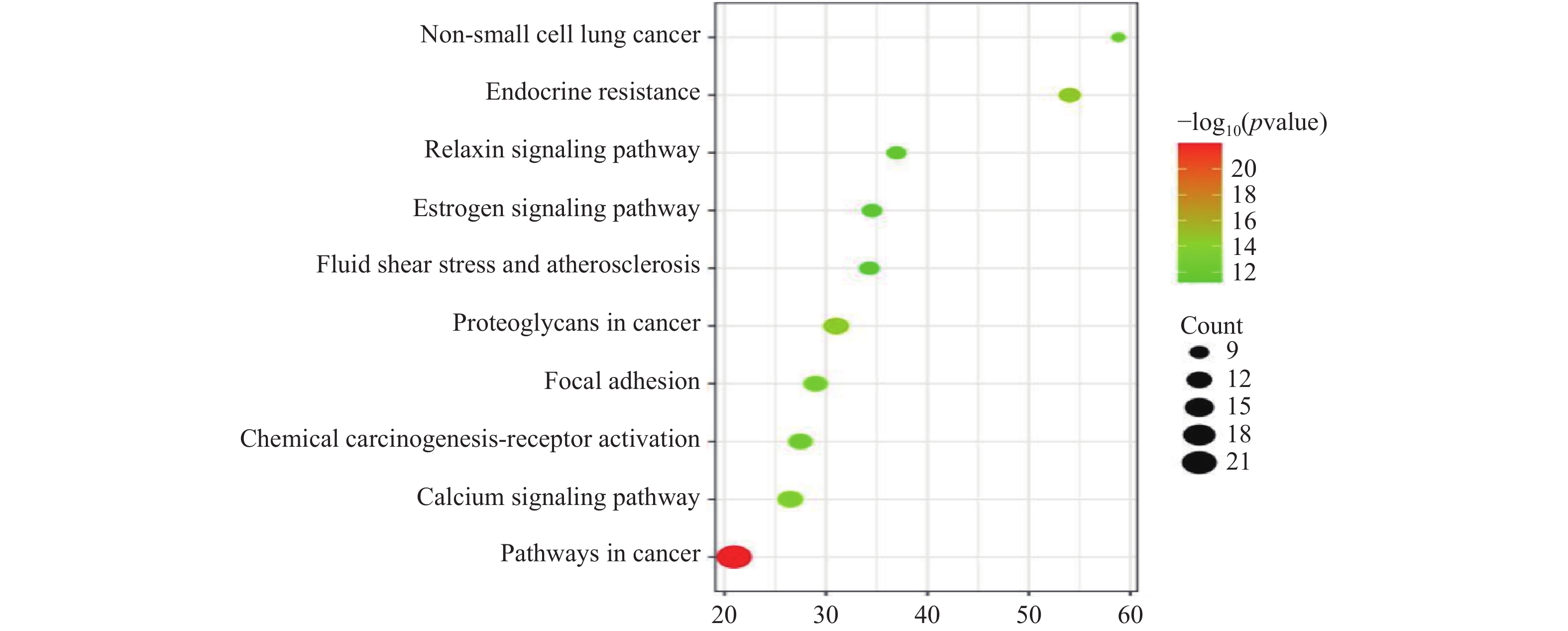
图 5 相关性排名前10信号通路的富集气泡图
Figure 5. Bubble plot of enrichment for the top 10 correlated signaling pathways by correlation ranking
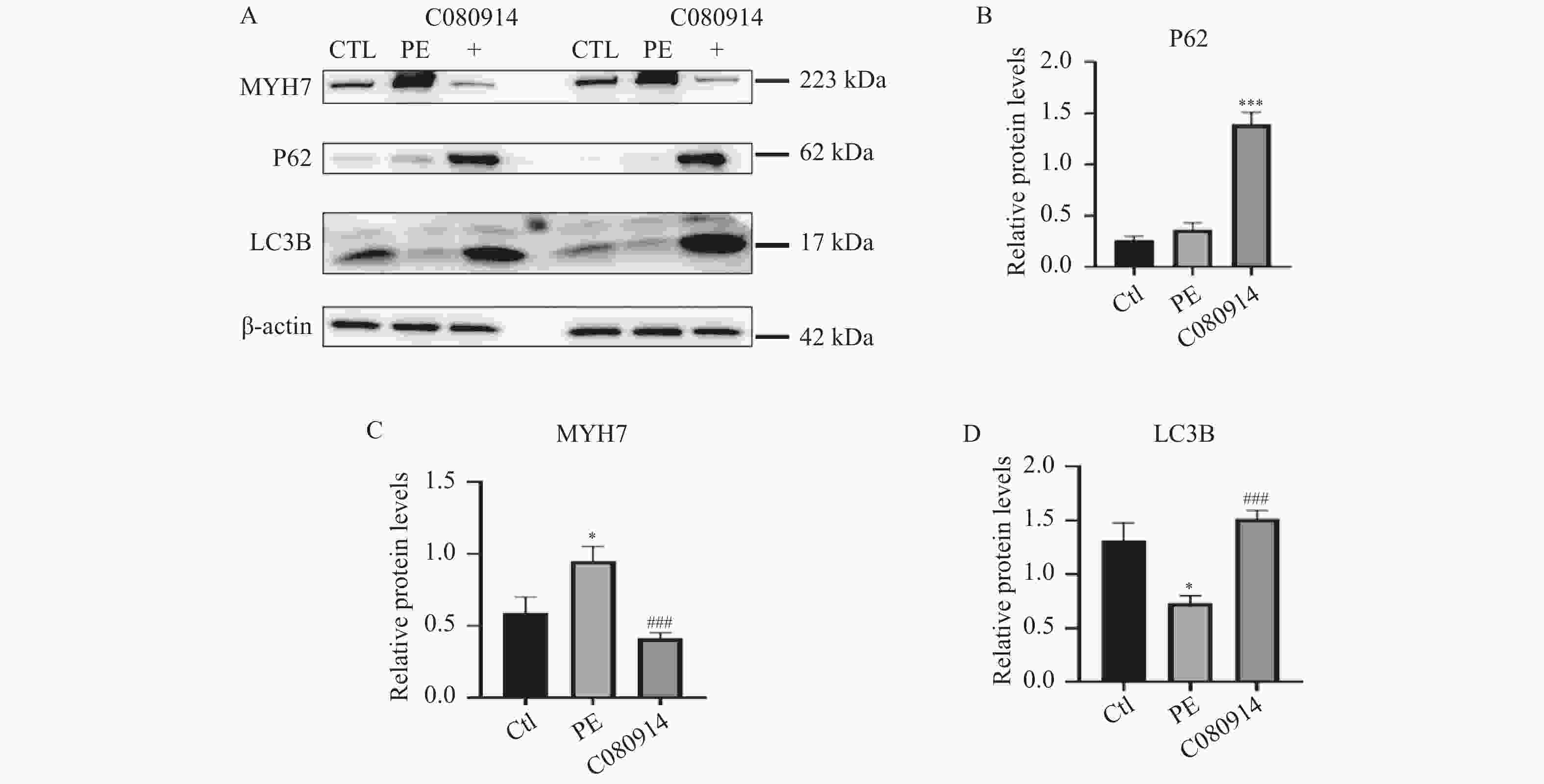
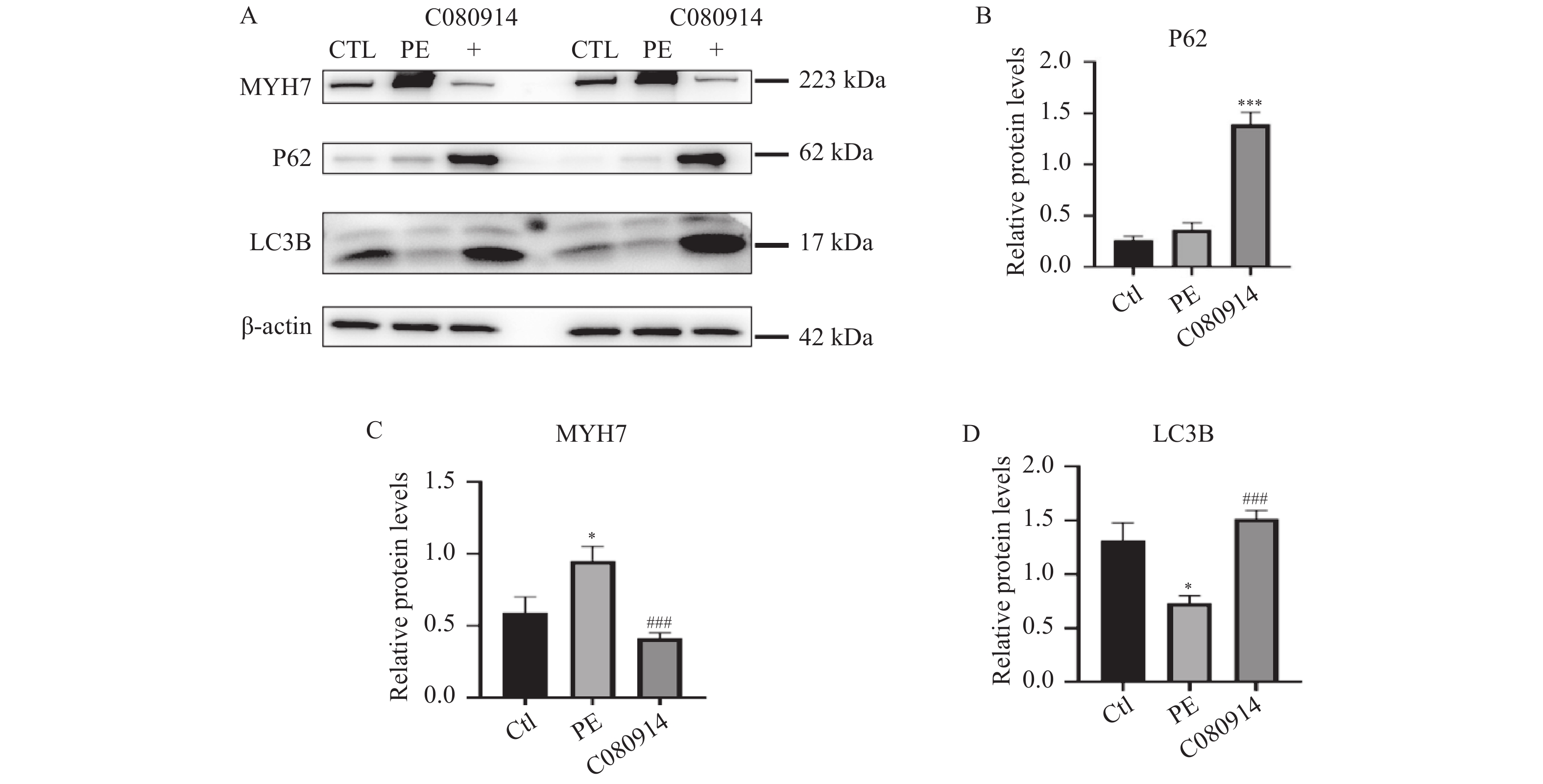
图 6 C080914减轻心肌细胞肥厚并调控自噬相关蛋白表达[($ \bar x \pm s $),n = 6]
A:PE诱导心肌肥厚后加入C080914处理后自噬相关蛋白LC3-Ⅱ、P62蛋白水平的代表性免疫印迹图;B:自噬相关蛋白LC3-Ⅱ相对定量统计图(目标蛋白灰度值与β-actin 灰度值的比值);C:MYH7相对定量统计图(目标蛋白灰度值与β-actin 灰度值的比值);D:自噬相关蛋白LC3-Ⅱ相对定量统计图(目标蛋白灰度值与β-actin灰度值的比值);与CTL组相比,*P < 0.05,***P < 0.001;与AngⅡ、ISO组相比,###P < 0.001。
Figure 6. C080914 alleviates cardiomyocyte hypertrophy and regulates the expression of autophagy-related proteins [($ \bar x \pm s $),n = 6]
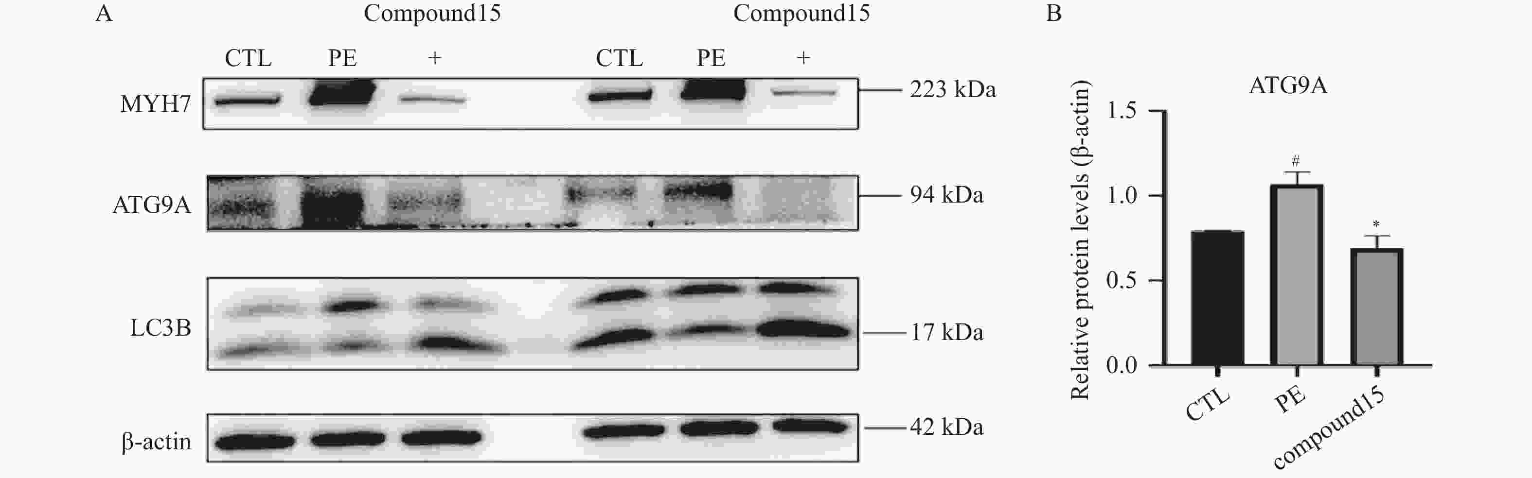
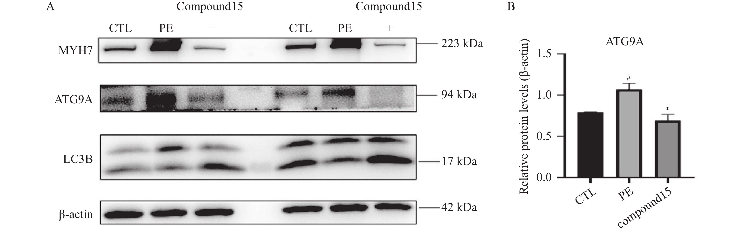
图 7 C080914使ATG9A表达下调[($ \bar x \pm s $),n = 6]
A:MYH7、ATG9A、LC3B 蛋白的电泳条带;B:ATG9A 蛋白相对表达水平定量分析(目标蛋白灰度值与β-actin 灰度值的比值);与 CTL 组比较,#P < 0.05;与 PE 组比较,*P < 0.05。
Figure 7. C080914 downregulates ATG9A expression [($ \bar x \pm s $),n = 6]
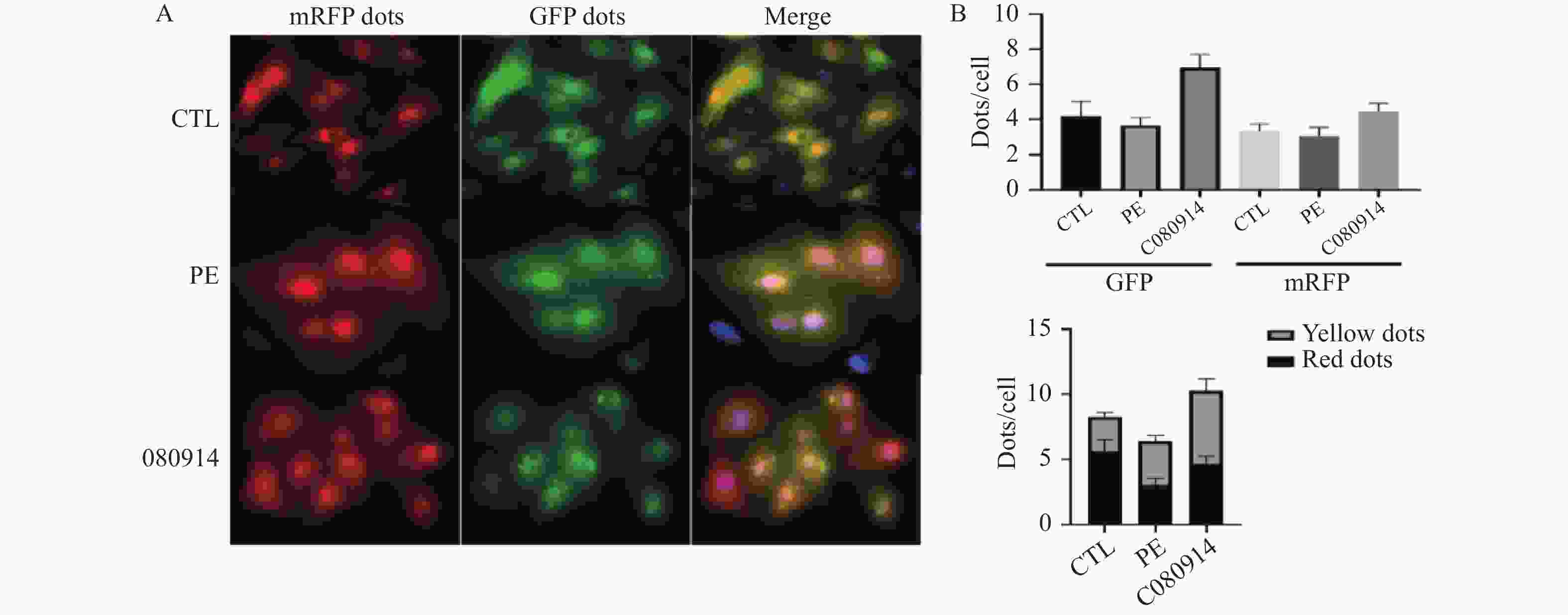
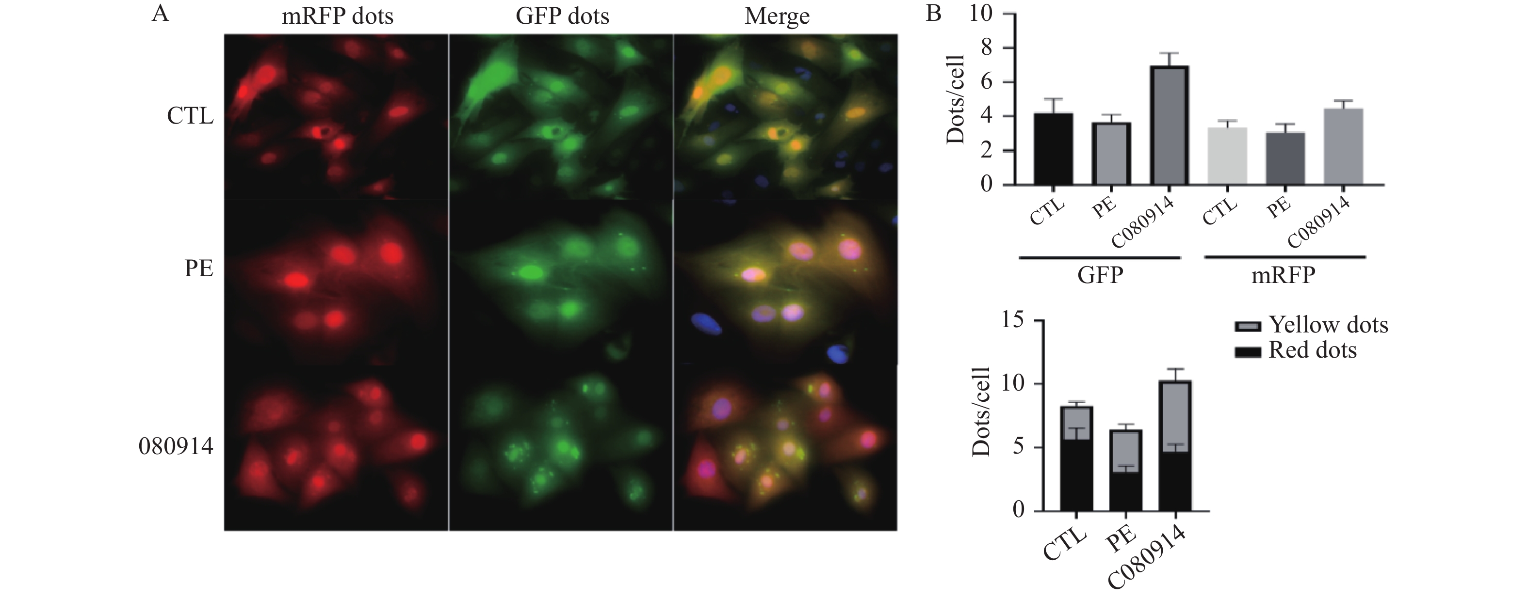
图 8 mRFP-GFP-LC3双标腺病毒检测原代心肌细胞自噬流[($ \bar x \pm s $),n = 5]
A:mRFP-GFP-LC3荧光代表图像(×40); B:各组细胞GFP斑点、mRFP斑点、黄色共定位斑点(未融合自噬体)、红色斑点(自噬溶酶体)定量统计。
Figure 8. Detection of autophagic flux in primary cardiomyocytes using mRFP-GFP-LC3 dual-labeled adenovirus [($ \bar x \pm s $),n = 5]
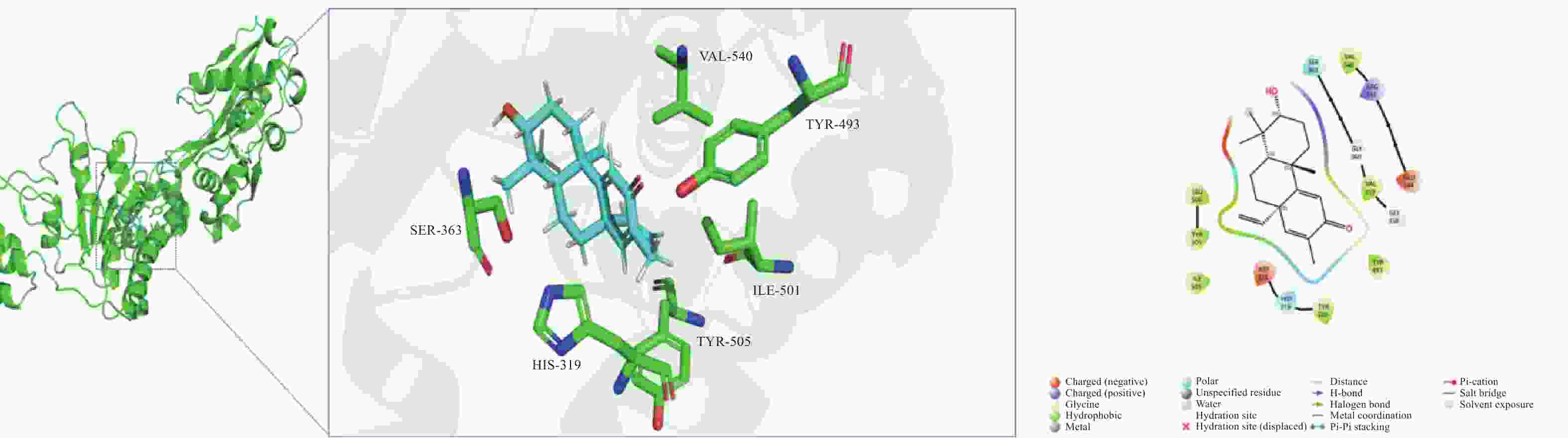
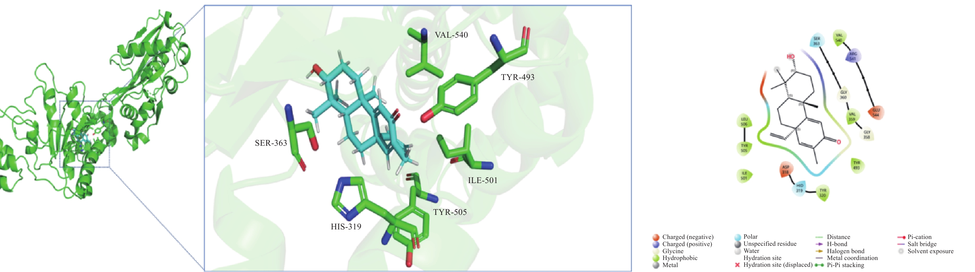
图 9 C080914与ATG9A对接结合相互作用图
注:C080914(青色)- ATG9A(绿色);主要为疏水作用和范德华接触。
Figure 9. Molecular docking interaction between C080914 and ATG9A
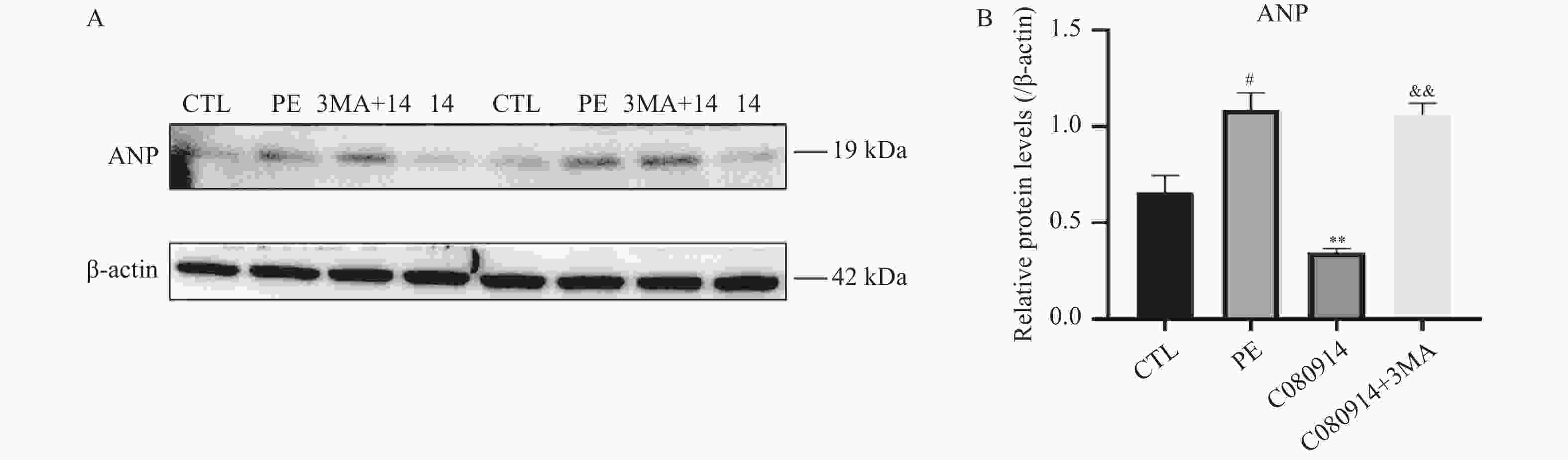
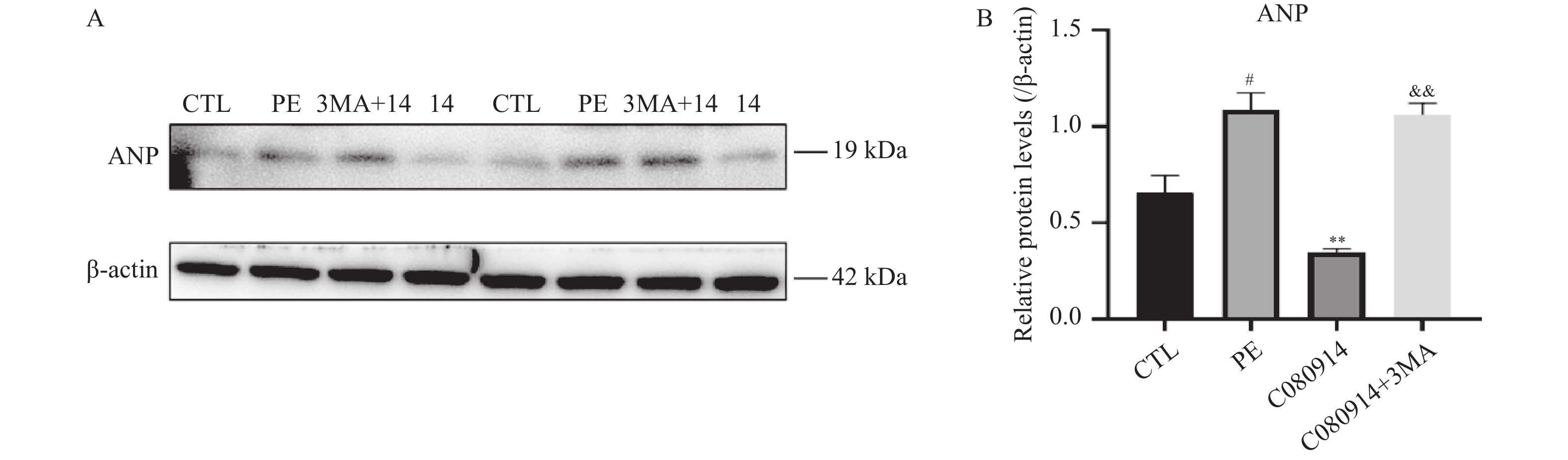
图 10 加入自噬抑制剂3MA后ANP蛋白表达WB图和统计图[($ \bar x \pm s $),n = 6]
A:ANP 蛋白的电泳条带;B:ANP 蛋白相对表达水平定量分析(目标蛋白灰度值与β-actin 灰度值的比值);与PE组比较,**P < 0.01;与CTL组比较,#P < 0.05;与080914组比较,&&P < 0.01。
Figure 10. Western blot and statistical analysis of ANP protein expression after treatment with autophagy inhibitor 3MA [($ \bar x \pm s $),n = 6]
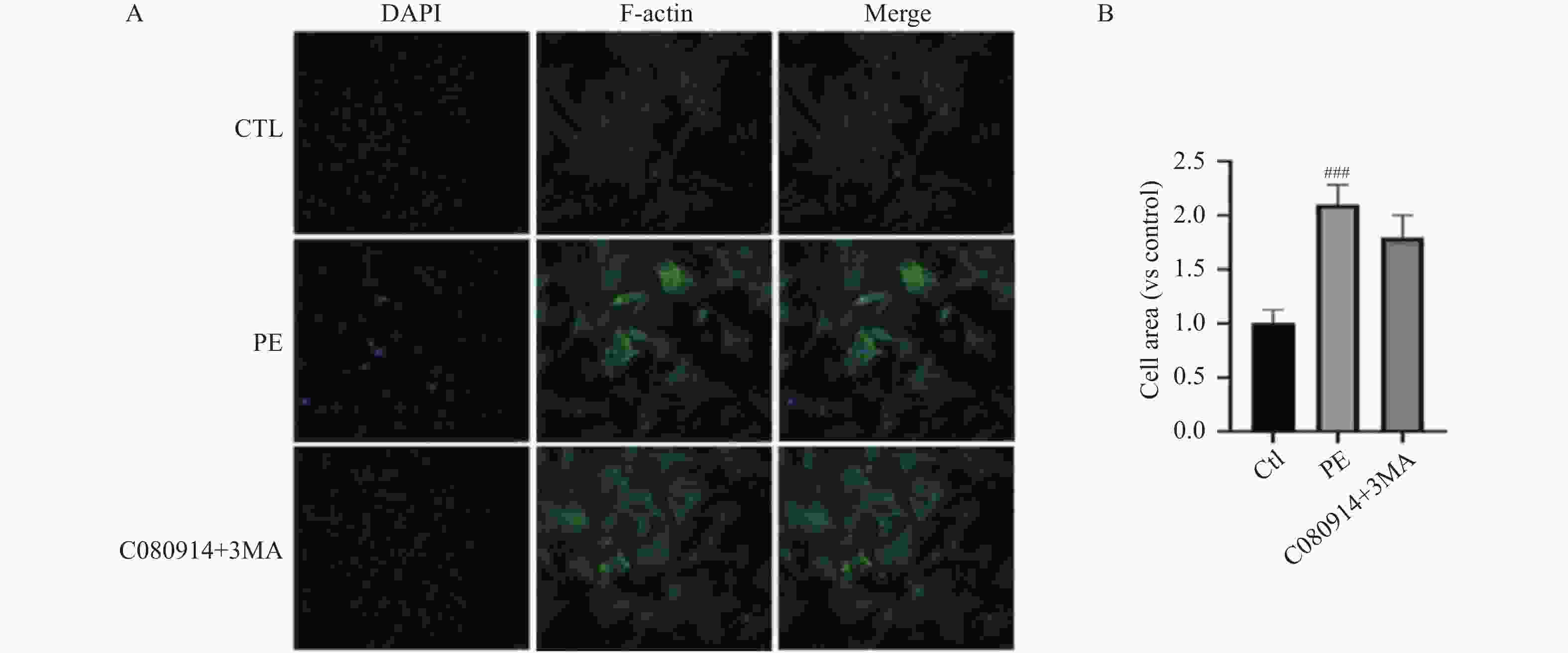
图 11 细胞表面积免疫荧光[($ \bar x \pm s $),n = 10]
A:心肌细胞表面积免疫荧光代表图(×40);B:运用ImageJ计算细胞表面积;与CTL组比较,###P < 0.001。
Figure 11. Immunofluorescence analysis of cell surface area [($ \bar x \pm s $),n = 10]
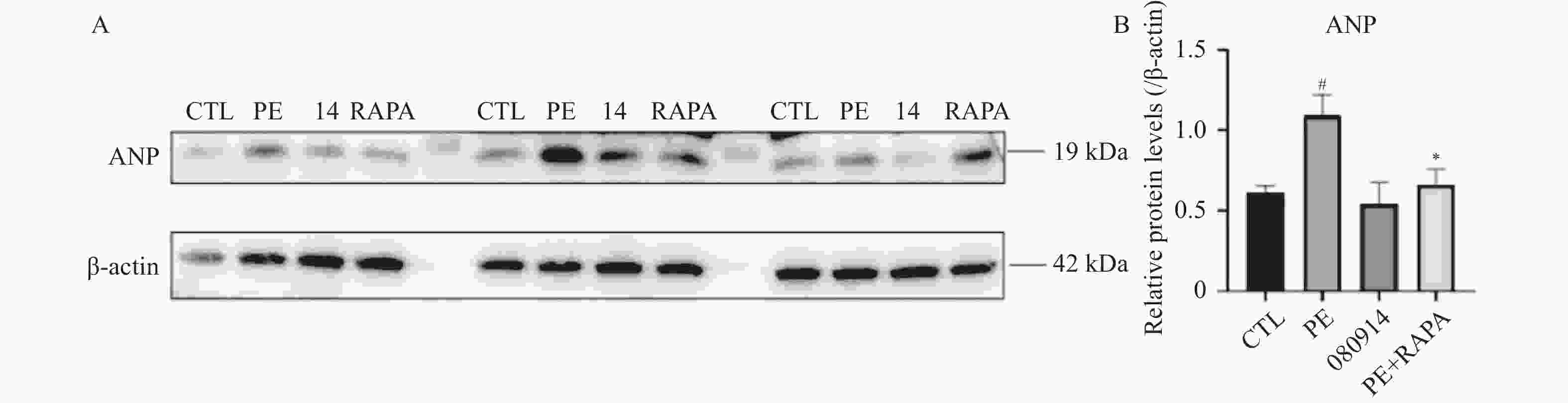
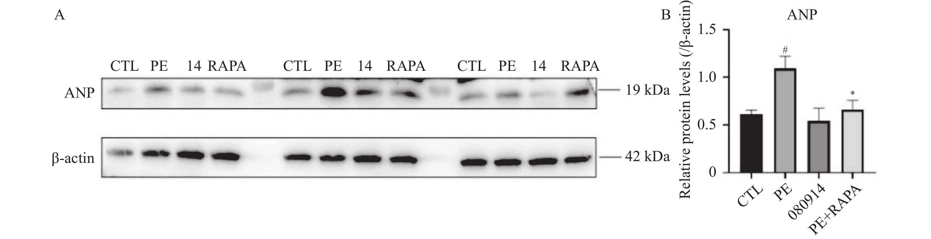
图 12 自噬激动剂Rapa可改善PE诱导的心肌肥厚[($ \bar x \pm s $),n = 6]
A:ANP 蛋白的电泳条带;B:ANP 蛋白相对表达水平定量分析(目标蛋白灰度值与β-actin灰度值的比值);与PE组比较,*P < 0.05;与CTL组比较,#P < 0.05。
Figure 12. Autophagy agonist Rapa ameliorates PE-induced cardiomyocyte hypertrophy [($ \bar x \pm s $),n = 6]
表 1 抗体信息
Table 1. Antibody information
抗体名称 批号 品牌 稀释倍数 NPPA抗体 27426 -1-APproteintech 1∶ 1000 MYH7抗体 22280 -1-APproteintech 1∶ 1000 LC3B抗体 ab192890 Abcam 1∶ 2000 β-actin抗体 ab134964 Abcam 1∶ 5000 GAPDH抗体 5174 Cell Signaling Technology 1∶ 1000 ATG9A抗体 ab228525 Abcam 1∶ 1000 P62抗体 Ab109012 Abcam 1∶ 10000 辣根过氧化物酶(HRP)标记羊抗兔 IgG(H+L)抗体 65- 6120 Invitrogen 1∶ 5000 辣根过氧化物酶(HRP)标记羊抗鼠 IgG(H+L)抗体 62- 6520 Invitrogen 1∶ 5000  下载: 导出CSV
-
[1] Nakamura M, Sadoshima J. Mechanisms of physiological and pathological cardiac hypertrophy[J]. Nat Rev Cardiol, 2018, 15(7): 387-407. doi: 10.1038/s41569-018-0007-y [2] Chen L, Wang Y, Liu Y, et al. Natural products targeting pathological cardiac hypertrophy: Molecular mechanisms and therapeutic potential[J]. Phytomedicine, 202X: ■-■. [3] Li D, Guo Y Y, Cen X F, et al. Lupeol protects against cardiac hypertrophy via TLR4-PI3K-Akt-NF-κB pathways[J]. Acta Pharmacol Sin, 2022, 43(8): 1989-2002. doi: 10.1038/s41401-021-00820-3 [4] Qiu X, Huang Y X, Yuan J, et al. Strophioblachins A-K, structurally intriguing diterpenoids from Strophioblachia fimbricalyx with potential anticardiac hypertrophic inhibitory activity[J]. J Nat Prod, 2023, 86(5): 1211-1221. doi: 10.1021/acs.jnatprod.2c01115 [5] Zhi X, Shi S, Li Y, et al. S100a9 inhibits Atg9a transcription and participates in suppression of autophagy in cardiomyocytes induced by β1-adrenoceptor autoantibodies[J]. Cell Mol Biol Lett, 2023, 28(1): 74. doi: 10.1186/s11658-023-00486-1 [6] Qiu R B, Zhao S T, Xu Z Q, et al. Thymoquinone mitigates cardiac hypertrophy by activating adaptive autophagy via the PPAR-γ/14-3-3γ pathway[J]. Int J Mol Med, 2025, 55(4): 59. doi: 10.3892/ijmm.2025.5500 [7] Xu M, Wan C X, Huang S H, et al. Oridonin protects against cardiac hypertrophy by promoting P21-related autophagy[J]. Cell Death Dis, 2019, 10(6): 403. doi: 10.1038/s41419-019-1617-y [8] Huang J, Sun W, Huang H, et al. miR-34a modulates angiotensin II-induced myocardial hypertrophy by direct inhibition of ATG9A expression and autophagic activity[J]. PLoS One, 2014, 9(4): e94382. doi: 10.1371/journal.pone.0094382 [9] Han X, Zhang Y L, Lin Q Y, et al. ATGL deficiency aggravates pressure overload-triggered myocardial hypertrophic remodeling associated with the proteasome-PTEN-mTOR-autophagy pathway[J]. Cell Biol Toxicol, 2023, 39(5): 2113-2131. doi: 10.1007/s10565-022-09699-0 [10] Jiang S, Bu S, Kang X, et al. Tspan9 exacerbates cardiac hypertrophy by impairing cardiac autophagy[J]. FASEB J, 2025, 39(10): e70670. doi: 10.1096/fj.202500150R [11] Zhou R, Zhang Z, Li X, et al. Autophagy in high-fat diet and streptozotocin-induced metabolic cardiomyopathy: Mechanisms and therapeutic implications[J]. Int J Mol Sci, 2025, 26(4): 1668. doi: 10.3390/ijms26041668 [12] Qu Y N, Gao R, Wei X, et al. Gasdermin D mediates endoplasmic reticulum stress via FAM134B to regulate cardiomyocyte autophagy and apoptosis in doxorubicin-induced cardiotoxicity[J]. Cell Death Dis, 2022, 13(10): 901. doi: 10.1038/s41419-022-05333-3 [13] Wu Y T, Tan H L, Shui G, et al. Dual role of 3-methyladenine in modulation of autophagy via different temporal patterns of inhibition on class I and III phosphoinositide 3-kinase[J]. J Biol Chem, 2010, 285(14): 10850-10861. doi: 10.1074/jbc.M109.080796 [14] Huang X, Zhang J, Wang W, et al. Vps4a regulates autophagic flux to prevent hypertrophic cardiomyopathy[J]. Int J Mol Sci, 2023, 24(13): 10800. doi: 10.3390/ijms241310800 [15] Maeda S, Yamamoto H, Kinch L N, et al. Structure, lipid scrambling activity and role in autophagosome formation of ATG9A[J]. Nat Struct Mol Biol, 2020, 27(12): 1194-1201. doi: 10.1038/s41594-020-00520-2 [16] Olivas T J, Wu Y, Yu S, et al. ATG9 vesicles comprise the seed membrane of mammalian autophagosomes[J]. J Cell Biol, 2023, 222(7): e202208088. doi: 10.1083/jcb.202208088 [17] Nah J, Shirakabe A, Mukai R, et al. Ulk1-dependent alternative mitophagy plays a protective role during pressure overload in the heart[J]. Cardiovasc Res, 2022, 118(12): 2638-2651. doi: 10.1093/cvr/cvac003 [18] Lomenick B, Hao R, Jonai N, et al. Target identification using drug affinity responsive target stability (DARTS)[J]. Proc Natl Acad Sci U S A, 2009, 106(51): 21984-21989. doi: 10.1073/pnas.0910040106 [19] Klionsky D J, Abdel-Aziz A K, Abdelfatah S, et al. Guidelines for the use and interpretation of assays for monitoring autophagy (4th edition)1[J]. Autophagy, 2021, 17(1): 1-382. doi: 10.1080/15548627.2020.1797280 [20] Wu Q Q, Liu C, Cai Z, et al. High-mobility group AT-hook 1 promotes cardiac dysfunction in diabetic cardiomyopathy via autophagy inhibition[J]. Cell Death Dis, 2020, 11(3): 160. doi: 10.1038/s41419-020-2316-4 [21] 李双秀, 郑琦, 尹高生, 等. 自噬通量受损介导细胞凋亡在压力负荷诱导心力衰竭中的作用[J]. 昆明医科大学学报, 2025, 46(9): 54-62. doi: 10.12259/j.issn.2095-610X.S20250906 [22] Wang X, Guo F, Wang X, et al. Angiotensin-(1–7) alleviates isoproterenol-induced cardiac hypertrophy by suppressing autophagy and apoptosis through the synergistic action of mas receptor and angiotensin II type 2 receptor[J]. Acta Physiol, 2026, 242(4): e70200. doi: 10.1111/apha.70200 -

 点击查看大图
计量
- 文章访问数: 3
- HTML全文浏览量: 2
- PDF下载量: 0
- 被引次数: 0
