

Impact of PINK1/Parkin-Mediated Mitophagy in the Development of Neurodegenerative Diseases
-
摘要: 线粒体不仅是细胞能量代谢中枢,更参与信号转导及活性调控等重要生理过程。线粒体数量异常、mtDNA拷贝数失衡、基因突变累积以及自噬通路异常激活等现象会导致粒体功能障碍甚至细胞器功能障碍,还会激活细胞损伤和死亡的机制,从而导致各种疾病的发病。线粒体自噬作为高度特异性的自噬形式,在细胞器质量监控与稳态平衡中承担核心调控功能。以经典PINK1/Parkin通路介导的线粒体自噬作为切入点,总结线粒体自噬在神经退行性疾病中的作用及相关研究进展,为寻求靶向调控线粒体自噬改善神经退行性疾病的防治新策略提供新的思路。Abstract: Mitochondria not only serve as the central hub for cellular energy metabolism, but also participate in crucial physiological processes such as signal transduction and activity regulation. Abnormal mitochondrial quantity, imbalance in mtDNA copy number, accumulation of genetic mutations, and aberrant activation of autophagic pathways can lead to mitochondrial dysfunction or even organelle disorders, while activating mechanisms of cellular damage and death, consequently contributing to the pathogenesis of various diseases. As a highly selective form of autophagy, mitophagy plays a pivotal regulatory role in organelle quality control and homeostasis maintenance. This article focuses on the classic PINK1/Parkin pathway-mediated mitophagy as an entry point, summarizing recent research advances regarding the role of mitophagy in neurodegenerative diseases, aiming to provide novel insights for developing targeted mitophagy regulation strategies to improve prevention and treatment approaches for neurodegenerative disorders.
-
Key words:
- Mitophagy /
- Oxidative stress /
- Parkinson's /
- Alzheimer's disease
-
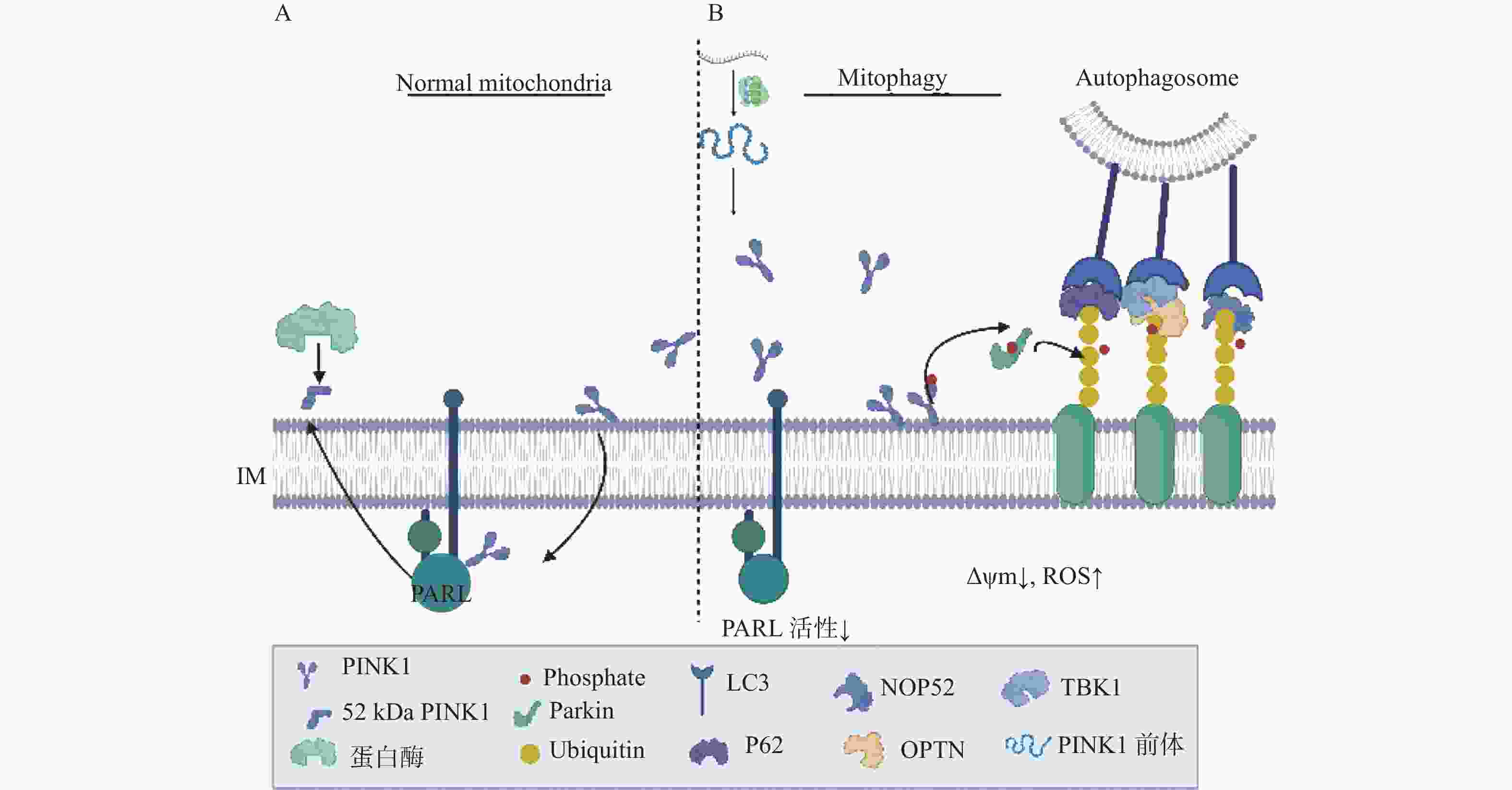
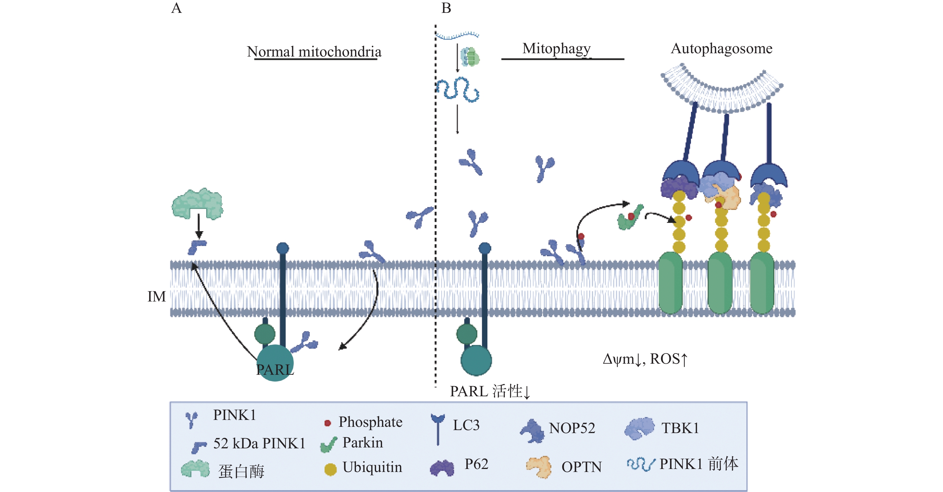
图 1 PINK1/Parkin通路介导的线粒体自噬
A:正常线粒体跨膜蛋白PARL蛋白酶切割处理PINK1;B:PINK1蛋白与Parkin介导的线粒体自噬模式图。
Figure 1. PINK1/Parkin pathway-mediated mitophagy
-
[1] Yoshii S R,Mizushima N. Autophagy machinery in the context of mammalian mitophagy[J]. Biochimica et Biophysica Acta (BBA) - Molecular Cell Research,2015,1853(10):2797-2801. doi: 10.1016/j.bbamcr.2015.01.013 [2] Gan Z Y,Komander D,Callegari S. Reassessing kinetin’ s effect on PINK1 and mitophagy[J]. Autophagy,2024,20(11):2596-2597. doi: 10.1080/15548627.2024.2395144 [3] Lu X Y,Zhu L Y,Zhu H,et al. Cyclometalated iridium(III)-lonidamine conjugates: Mitochondrial targeting and pyroptosis induction[J]. Journal of Inorganic Biochemistry,2025,266:112852. doi: 10.1016/j.jinorgbio.2025.112852 [4] Zarkovic N. Roles and functions of ROS and RNS in cellular physiology and pathology[J]. Cells,2020,9(3):767. doi: 10.3390/cells9030767 [5] Li Y,Zhang W,Zhang Q,et al. Oxidative stress of mitophagy in neurodegenerative diseases: Mechanism and potential therapeutic targets[J]. Archives of Biochemistry and Biophysics,2025,764:110283. doi: 10.1016/j.abb.2024.110283 [6] Burda R,Burda J,Morochovič R. Ischemic tolerance-a way to reduce the extent of ischemia-reperfusion damage[J]. Cells,2023,12(6):884. doi: 10.3390/cells12060884 [7] Zhu J,Xu N,Lin H,et al. Remote ischemic preconditioning plays a neuroprotective role in cerebral ischemia-reperfusion mice by inhibiting mitophagy[J]. Heliyon,2024,10(20):e39076. doi: 10.1016/j.heliyon.2024.e39076 [8] Mao Z,Tian L,Liu J,et al. Ligustilide ameliorates hippocampal neuronal injury after cerebral ischemia reperfusion through activating PINK1/Parkin-dependent mitophagy[J]. Phytomedicine,2022,101:154111. doi: 10.1016/j.phymed.2022.154111 [9] Li X,Guan L,Liu Z,et al. Ubiquitination of ATAD3A by TRIM25 exacerbates cerebral ischemia-reperfusion injury via regulating PINK1/Parkin signaling pathway-mediated mitophagy[J]. Free Radical Biology and Medicine,2024,224:757-769. doi: 10.1016/j.freeradbiomed.2024.09.029 [10] Xiao B,Cui Y,Li B,et al. ROS antagonizes the protection of Parkin-mediated mitophagy against aluminum-induced liver inflammatory injury in mice[J]. Food and Chemical Toxicology,2022,165:113126. doi: 10.1016/j.fct.2022.113126 [11] Fan P,Xie X H,Chen C H,et al. Molecular regulation mechanisms and interactions between reactive oxygen species and mitophagy[J]. DNA and Cell Biology,2019,38(1):10-22. doi: 10.1089/dna.2018.4348 [12] Tang C,He L,Liu J,et al. Mitophagy: Basic mechanism and potential role in kidney diseases[J]. Kidney Diseases,2015,1(1):71-79. doi: 10.1159/000381510 [13] Wen J,Pan T,Li H,et al. Role of mitophagy in the hallmarks of aging[J]. Journal of Biomedical Research,2022,37(1):1-14. [14] Tintos-Hernández J A,Santana A,Keller K N,et al. Lysosomal dysfunction impairs mitochondrial quality control and is associated with neurodegeneration in TBCK encephaloneuronopathy[J]. Brain Communications,2021,3(4):fcab215. doi: 10.1093/braincomms/fcab215 [15] Tang M,Rong D,Gao X,et al. A positive feedback loop between SMAD3 and PINK1 in regulation of mitophagy[J]. Cell Discovery,2025,11(1):22. doi: 10.1038/s41421-025-00774-4 [16] Jin S M,Youle R J. PINK1- and Parkin-mediated mitophagy at a glance[J]. Journal of Cell Science,2012,125(4):795-799. doi: 10.1242/jcs.093849 [17] Cheng J,Wei L,Li M. Progress in regulation of mitochondrial dynamics and mitochondrial autophagy[J]. Sheng Li Xue Bao: (Acta Physiologica Sinica),2020,72(4):475-487. [18] Hu T,Wu C,Jian W,et al. Effect of PINK1 and Parkin gene silencing on sodium arsenite-induced mitophagy in normal rat liver cells (BRL-3A)[J]. Toxicology Research,2022,11(1):52-59. doi: 10.1093/toxres/tfab110 [19] Kanki T,Furukawa K,Yamashita S. Mitophagy in yeast: Molecular mechanisms and physiological role[J]. Biochimica et Biophysica Acta (BBA) - Molecular Cell Research,2015,1853(10):2756-2765. doi: 10.1016/j.bbamcr.2015.01.005 [20] Wang Y,Shen J,Chen Y,et al. PINK1 protects against oxidative stress induced senescence of human nucleus pulposus cells via regulating mitophagy[J]. Biochemical and Biophysical Research Communications,2018,504(2):406-414. doi: 10.1016/j.bbrc.2018.06.031 [21] Bowling J L,Skolfield M C,Riley W A,et al. Temporal integration of mitochondrial stress signals by the PINK1: Parkin pathway[J]. BMC Molecular and Cell Biology,2019,20(1):33. doi: 10.1186/s12860-019-0220-5 [22] Gao J,Qin S,Jiang C. Parkin-induced ubiquitination of Mff promotes its association with p62/SQSTM1 during mitochondrial depolarization[J]. Acta Biochimica et Biophysica Sinica,2015,47(7):522-529. doi: 10.1093/abbs/gmv044 [23] Jayatunga D P W,Hone E,Bharadwaj P,et al. Targeting mitophagy in Alzheimer’ s disease[J]. Journal of Alzheimer’ s Disease,2020,78(4):1273-1297. doi: 10.3233/JAD-191258 [24] Ashok B S,Ajith T A,Sivanesan S. Hypoxia-inducible factors as neuroprotective agent in Alzheimer’ s disease[J]. Clinical and Experimental Pharmacology and Physiology,2017,44(3):327-334. doi: 10.1111/1440-1681.12717 [25] Hassan W,Noreen H,Rehman S,et al. Association of oxidative stress with neurological disorders[J]. Current Neuropharmacology,2022,20(6):1046-1072. doi: 10.2174/1570159X19666211111141246 [26] George A J,Gordon L,Beissbarth T,et al. A serial analysis of gene expression profile of the Alzheimer’ s disease Tg2576 mouse model[J]. Neurotoxicity Research,2010,17(4):360-379. doi: 10.1007/s12640-009-9112-3 [27] Mart í n-Maestro P,Gargini R,Garc í a E,et al. Mitophagy failure in APP and tau overexpression model of Alzheimer’ s disease[J]. Journal of Alzheimer’ s Disease,2019,70(2):525-540. doi: 10.3233/JAD-190086 [28] Zeng K,Yu X,Mahaman Y A R,et al. Defective mitophagy and the etiopathogenesis of Alzheimer’ s disease[J]. Translational Neurodegeneration,2022,11(1):32. doi: 10.1186/s40035-022-00305-1 [29] Quinn P M J,Moreira P I,Ambrósio A F,et al. PINK1/PARKIN signalling in neurodegeneration and neuroinflammation[J]. Acta Neuropathologica Communications,2020,8(1):189. doi: 10.1186/s40478-020-01062-w [30] Du F,Yu Q,Yan S S. PINK1 activation attenuates impaired neuronal-like differentiation and synaptogenesis and mitochondrial dysfunction in Alzheimer’ s disease trans-mitochondrial cybrid cells[J]. Journal of Alzheimer’ s Disease,2021,81(4):1749-1761. doi: 10.3233/JAD-210095 [31] Wang X juan,Qi L,Cheng Y fang,et al. PINK1 overexpression prevents forskolin-induced tau hyperphosphorylation and oxidative stress in a rat model of Alzheimer’ s disease[J]. Acta Pharmacologica Sinica,2022,43(8):1916-1927. doi: 10.1038/s41401-021-00810-5 [32] Dhapola R,Kumari S,Sharma P,et al. Advancements in autophagy perturbations in Alzheimer’ s disease: Molecular aspects and therapeutics[J]. Brain Research,2025,1851:149494. doi: 10.1016/j.brainres.2025.149494 [33] Tarakad A,Jankovic J. Recent advances in understanding and treatment of Parkinson’ s disease[J]. Faculty Reviews,2020,9:6. [34] Rango M,Dossi G,Squarcina L,et al. Brain mitochondrial impairment in early-onset Parkinson’ s disease with or without PINK1 mutation[J]. Movement Disorders,2020,35(3):504-507. doi: 10.1002/mds.27946 [35] Kazlauskaite A,Muqit M M K. PINK1 and Parkin – mitochondrial interplay between phosphorylation and ubiquitylation in Parkinson’ s disease[J]. The FEBS Journal,2015,282(2):215-223. doi: 10.1111/febs.13127 [36] Li M X,Mu D Z. Mitophagy and nervous system disease[J]. Zhongguo Dang Dai Er Ke Za Zhi = Chinese Journal of Contemporary Pediatrics,2017,19(6):724-729. [37] 沈金峰,胡芳,王福珍,等. 大蒜素调控PINK1/Parkin介导的线粒体自噬改善大鼠尿毒症心肌损伤[J]. 广州中医药大学学报,2025,2(42):448-454. [38] Rogers R S,Tungtur S,Tanaka T,et al. Impaired mitophagy plays a role in denervation of neuromuscular junctions in ALS mice[J]. Frontiers in Neuroscience,2017,11:473. doi: 10.3389/fnins.2017.00473 [39] Granatiero V,Manfredi G. Mitochondrial transport and turnover in the pathogenesis of amyotrophic lateral sclerosis[J]. Biology,2019,8(2):36. doi: 10.3390/biology8020036 [40] Zhang H,Gao C,Yang D,et al. Urolithin a improves motor dysfunction induced by copper exposure in SOD1G93A transgenic mice via activation of mitophagy[J]. Molecular Neurobiology,2024,62(6):6922-6937 [41] Gatto E M,Rojas N G,Persi G,et al. Huntington disease: Advances in the understanding of its mechanisms[J]. Clinical Parkinsonism & Related Disorders,2020,3:100056. [42] Liu T,Wetzel L,Zhu Z,et al. Disruption of mitophagy flux through the PARL-PINK1 pathway by CHCHD10 mutations or CHCHD10 depletion[J]. Cells,2023,12(24):2781. doi: 10.3390/cells12242781 [43] Joshi D C,Chavan M B,Gurow K,et al. The role of mitochondrial dysfunction in Huntington’ s disease: Implications for therapeutic targeting[J]. Biomedicine & Pharmacotherapy,2025,183:117827. [44] Khalil B,El Fissi N,Aouane A,et al. PINK1-induced mitophagy promotes neuroprotection in Huntington’ s disease[J]. Cell Death & Disease,2015,6(1):e1617. [45] Liang Z,Zhao S,Liu Y,et al. The promise of mitochondria in the treatment of glioblastoma: A brief review[J]. Discover Oncology,2025,16(1):142. doi: 10.1007/s12672-025-01891-y [46] Yao N,Wang C,Hu N,et al. Inhibition of PINK1/Parkin-dependent mitophagy sensitizes multidrug-resistant cancer cells to B5G1,a new betulinic acid analog[J]. Cell Death & Disease,2019,10(3):232. -

 下载:
下载:
 点击查看大图
图(2)
计量
- 文章访问数: 350
- HTML全文浏览量: 60
- PDF下载量: 38
- 被引次数: 0
