

Advances and Challenges of Near-infrared Fluorescence Imaging in the Diagnosis and Treatment of Bladder Cancer
-
摘要: 膀胱癌(bladder cancer,BCa)是常见的泌尿系统恶性肿瘤,早期精准诊疗对改善患者预后至关重要。近红外荧光成像(near-infrared fluorescence imaging,NIRFI)因其低背景干扰、高信噪比及实时成像能力,在膀胱癌的诊断与治疗中展现出重要应用潜力。本文综述近红外荧光成像技术在膀胱癌中的研究进展,重点总结靶向荧光探针的设计策略及其在肿瘤识别、边界判定和淋巴结定位中的应用,并归纳其在经尿道手术及相关操作中的辅助价值。在此基础上,进一步分析该技术在组织穿透深度、信号定量一致性及临床标准化方面的主要挑战,并探讨其与光热/光动力治疗、多模态成像及人工智能融合的发展趋势,旨在为近红外荧光成像在膀胱癌中的进一步研究与临床转化提供参考。Abstract: Bladder cancer (BCa) is a common malignancy of the urinary system, and early precise diagnosis and treatment are essential for improving prognosis. Near-infrared fluorescence imaging (NIR), with low background interference, high signal-to-noise ratio, and real-time imaging capability, has shown promising potential in bladder cancer management. This review summarizes recent advances in NIR imaging for bladder cancer, focusing on the design of targeted fluorescent probes and their applications in tumor detection, margin delineation, and lymph node localization, as well as their value in transurethral surgery and related procedures. Current challenges, including limited tissue penetration, inconsistency in signal quantification, and insufficient clinical standardization, are also discussed. In addition, future directions involving photothermal/photodynamic therapy, multimodal imaging, and artificial intelligence are highlighted. This review may provide a reference for further research and clinical translation of NIR imaging in bladder cancer.
-
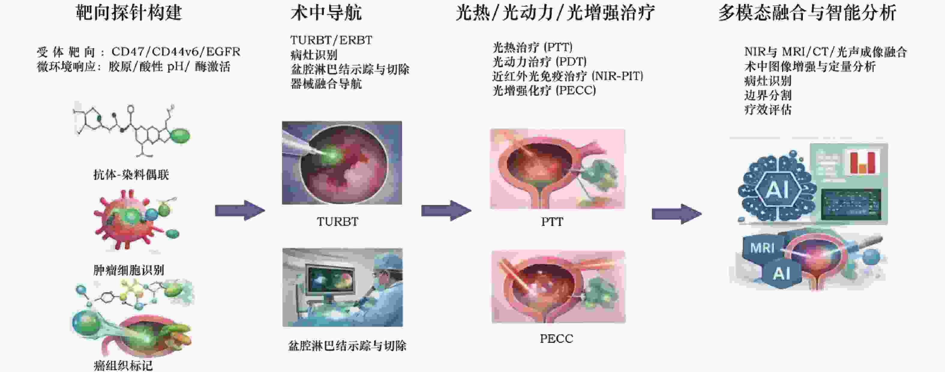
图 1 近红外荧光成像在膀胱癌诊疗中的应用流程图
Figure 1. Workflow of near-infrared fluorescence imaging in bladder cancer diagnosis and treatment
表 1 近红外荧光成像与其他常用成像技术在膀胱癌诊疗中的比较
Table 1. Comparison of near-infrared fluorescence imaging with other commonly used imaging techniques in the diagnosis and treatment of bladder cancer
技术 主要优势 主要局限 成本/便捷性 主要适用场景 白光膀胱镜 临床普及度高,操作成熟 对平坦病灶、CIS和微小病灶识别不足 成本较低,便捷性高 常规筛查、随访 超声成像 无创、动态、床旁可用 对微小/平坦病灶敏感性有限,分子特异性差 成本较低,便捷性高 初步评估、形态学观察 多参数MRI 软组织分辨率高,适合
肌层浸润与局部分期评估费用较高,术中实时性
不足成本较高,流程较复杂 术前分期、治疗反应评估 近红外荧光成像 实时成像、分子靶向、
适于术中边界识别与导航穿透深度有限,定量标准化不足 设备和探针依赖较大,但术中操作性强 术中导航、淋巴结示踪、成像引导局部治疗  下载: 导出CSV
表 2 膀胱癌不同近红外靶向探针对比
Table 2. Comparison of different near-infrared targeted probes for bladder cancer
探针类型 代表靶点/机制 主要优势 主要局限 主要适用场景 代表文献 受体靶向型 CD47、CD44v6、EGFR 特异性较强,适于高表达病灶识别 受体表达异质性
影响较大局灶病灶识别、
术后标本验证[6][9][17] 核/细胞内靶向型 c(RGDfK)-AO 细胞内定位更精细 临床转化证据仍有限 实验研究、机制验证 [18] 胶原靶向微环境型 胶原黏附/溶胶-
凝胶转变边界显示较稳定,受异质性影响较小 可能受间质改变影响 NMIBC、边界勾勒 [10] pH响应型 pHLIP-ICG、酸触发“switch-on” 背景低,适合癌前/微小病灶提示 受局部酸度波动影响 表浅病灶、
隐匿病灶识别[19][20][21]
下载: 导出CSV
-
[1] Bray F, Laversanne M, Sung H, et al. Global cancer statistics 2022: GLOBOCAN estimates of incidence and mortality worldwide for 36 cancers in 185 countries[J]. CA Cancer J Clin, 2024, 74(3): 229-263. doi: 10.3410/f.739487650.793592245 [2] Karimi A, Shobeiri P, Azadnajafabad S, et al. A global, regional, and national survey on burden and Quality of Care Index (QCI) of bladder cancer: The global burden of disease study 1990-2019[J]. PLoS One, 2022, 17(10): e0275574. doi: 10.1371/journal.pone.0275574 [3] Mulawkar PM, Sharma G, Tamhankar A, et al. Role of macroscopic image enhancement in diagnosis of non-muscle-invasive bladder cancer: an analytical review[J]. Front Surg, 2022, 9: 762027. doi: 10.3389/fsurg.2022.762027 [4] Williams S B, Gavaghan M B, Fernandez A, et al. Macro and microeconomics of blue light cystoscopy with CYSVIEW® in non-muscle-invasive bladder cancer[J]. Urol Oncol, 2022 , 40(1): 10. e7-10. e12. [5] Ge C, Zhang W, Huang J, et al. Research progress of near-infrared fluorescence imaging in accurate theranostics in bladder cancer[J]. Photodiagnosis Photodyn Ther, 2025, 52: 104480. doi: 10.1016/j.pdpdt.2025.104480 [6] Shang W, Peng L, He K, et al. A clinical study of a CD44v6-targeted fluorescent agent for the detection of non-muscle-invasive bladder cancer[J]. Eur J Nucl Med Mol Imaging, 2022, 49(9): 3033-3045. doi: 10.1007/s00259-022-05701-3 [7] Chen LL, Zhao L, Wang ZG, et al. Near-infrared-II quantum dots for in vivo imaging and cancer therapy[J]. Small, 2022, 18(8): e2104567. doi: 10.1002/smll.202104567 [8] Huang J, Pu K. Near-infrared fluorescent molecular probes for imaging and diagnosis of nephro-urological diseases[J]. Chem Sci, 2021, 12(10): 3379-3392. doi: 10.1039/D0SC02925D [9] Hao H, Wang X, Qin Y, et al. Ex vivo near-infrared targeted imaging of human bladder carcinoma by ICG-anti-CD47[J]. Front Oncol, 2023, 13: 1083553. doi: 10.3389/fonc.2023.1083553 [10] Guo P, Qi A, Shang W, et al. Targeting tumour surface collage with hydrogel probe: a new strategy to enhance intraoperative imaging sensitivity and stability of bladder cancer[J]. Eur J Nucl Med Mol Imaging, 2024, 51(13): 4165-4176. doi: 10.1007/s00259-024-06848-x [11] Zhu J, Zhu Y, Huang J, et al. Icing on the cake: integrating optical fiber with second near-infrared aggregation-induced emission luminogen for exceptional phototheranostics of bladder cancer[J]. Adv Mater, 2025, 37(29): e2502452. doi: 10.1002/adma.202502452 [12] You C, Zhu Y, Zhu J, et al. Strength in numbers: a giant NIR-II AIEgen with one-for-all phototheranostic features for exceptional orthotopic bladder cancer treatment. [J] Angew Chem Int Ed Engl, 2025 , 64(6): e202417865. [13] Fukushima H, Takao S, Furusawa A, et al. Near-infrared photoimmunotherapy targeting Nectin-4 in a preclinical model of bladder cancer[J]. Cancer Lett, 2024, 585: 216606. doi: 10.1016/j.canlet.2023.216606 [14] van Oosterom M N, van Leeuwen S I, Mazzone E, et al. Click-on fluorescence detectors: using robotic surgical instruments to characterize molecular tissue aspects[J]. J Robot Surg, 2023, 17(1): 131-140. doi: 10.1007/s11701-022-01382-0 [15] Baio R, Intilla O, Di Mauro U, et al. Near-infrared fluorescence imaging with intraoperative administration of indocyanine green for laparoscopic radical prostatectomy: Is it a useful weapon for pelvic lymph node dissection[J]. J Surg Case Rep, 2022, 2022(3): rjab614. doi: 10.1093/jscr/rjab614 [16] Railkar R, Krane L S, Li Q Q, et al. Epidermal Growth Factor Receptor (EGFR)-targeted photoimmunotherapy for the treatment of EGFR-expressing bladder cancer[J]. Mol Cancer Ther, 2017, 16(10): 2201-2214. doi: 10.1158/1535-7163.MCT-16-0924 [17] Aayush Aayush, Darji S, Dhawan D, et al. Targeted elastin-like polypeptide fusion protein for near-infrared imaging of human and canine urothelial carcinoma[J]. Oncotarget, 2022, 13: 1004-1016. doi: 10.18632/oncotarget.28271 [18] Qin J, Liang Q, Wang G, et al. Targeted delivery of nuclear targeting probe for bladder cancer using cyclic pentapeptide c(RGDfK) and acridine orange[J]. Clin Transl Oncol, 2023, 25(2): 375-383. doi: 10.1007/s12094-022-02938-0 [19] Golijanin J, Amin A, Moshnikova A, et al. Targeted imaging of urothelium carcinoma in human bladders by an ICG pHLIP peptide ex vivo[J]. Proc Natl Acad Sci U S A, 2016, 113(42): 11829-11834. doi: 10.1073/pnas.1610472113 [20] Moshnikova A, Golijanin B, Amin A, et al. Targeting Bladder Urothelial Carcinoma with pHLIP-ICG and Inhibition of Urothelial Cancer Cell Proliferation by pHLIP-amanitin[J]. Front Urol, 2022, 2: 868919. doi: 10.3389/fruro.2022.868919 [21] Luo R, Ou C, Li X, et al. An acidity-initiated self-assembly/disassembly nanoprobe to switch on fluorescence for tumor-targeted near-infrared imaging[J]. Nano Lett, 2022, 22(1): 151-156. doi: 10.1021/acs.nanolett.1c03534 [22] Yang Y, Yang X, Liu C, et al. En bloc tumor resection, optical molecular imaging, and the potential synergy of the combination of the two techniques in bladder cancer[J]. Front Oncol, 2021, 11: 638083. doi: 10.3389/fonc.2021.638083 [23] Yang Y, Yang X, Liu C, et al. Preliminary study on the application of en bloc resection combined with near-infrared molecular imaging technique in the diagnosis and treatment of bladder cancer[J]. World J Urol, 2020, 38(12): 3169-3176. doi: 10.1007/s00345-020-03143-w [24] Baart V M, van der Horst G, Deken M M, et al. A multimodal molecular imaging approach targeting urokinase plasminogen activator receptor for the diagnosis, resection and surveillance of urothelial cell carcinoma[J]. Eur J Cancer, 2021, 146: 11-20. doi: 10.1016/j.ejca.2021.01.001 [25] Sacconi A, Managò S, Calderan L, et al. Indocyanine green nanoparticles: are they compelling for diagnostic and theranostic applications[J]. Front Chem, 2020, 8: 535. [26] Zhang Y, Guo X, Zhang Y, Wei J, et al. A preliminary investigation of precise visualization, localization, and resection of pelvic lymph nodes in bladder cancer by using indocyanine green fluorescence-guided approach through intracutaneous dye injection into the lower limbs and perineum[J]. Front Oncol, 2024, 14: 1384268. doi: 10.3389/fonc.2024.1384268 [27] Püllen L, Costa P F, Darr C, et al. Near-infrared fluorescence lymph node template region dissection plus backup lymphadenectomy in open radical cystectomy for bladder cancer using an innovative handheld device: A single center experience[J]. J Surg Oncol, 2024, 129(7): 1325-1331. doi: 10.1002/jso.27618 [28] Puccetti M, Paganelli G, Bravaccini S, et al. Spotlight on PSMA as a new theranostic biomarker for bladder cancer[J]. Sci Rep, 2021, 11: 9777. doi: 10.1038/s41598-021-89160-0 [29] Li Y, Zhang X, Yang Z, et al. Prognostic value of vascular-expressed PSMA and CD248 in urothelial carcinoma of the bladder and its correlation analysis with TCGA-BLCA data[J]. Front Oncol, 2021, 11: 788822. [30] Li P, Xu Y, Yu S, et al. First-in-human study of DGPR1008 for intraoperative fluorescence imaging of prostate-specific membrane antigen-positive prostate cancer in patients undergoing radical prostatectomy[J]. Transl Androl Urol, 2025, 14(9): 2697-2709. doi: 10.21037/tau-2025-537 [31] Blair S, Garcia M, Davis T, et al. Hexachromatic bioinspired camera for image-guided cancer surgery. Sci Transl Med, 2021, 13(592): eaaw7067. [32] George M B, Lew B, Blair S, et al. Bioinspired color-near-infrared endoscopic imaging system for molecular guided cancer surgery[J]. J Biomed Opt, 2023, 28(5): 056002. [33] Zhu C, Gao Q, Wang C, et al. Quinoxalineimide-based semiconducting polymer nanoparticles as an effective phototheranostic for the second near-infrared fluorescence imaging and photothermal therapy[J]. ACS Appl Mater Interfaces, 2023, 15(24): 29396-29405. doi: 10.1021/acsami.3c06853 [34] Wang T, Li H, Chen J, et al. Recent progress in second near-infrared (NIR-II) fluorescence imaging for cancer diagnosis and therapy[J]. Front Bioeng Biotechnol, 2022, 10: 1020427. [35] Zhang Y, Zhang F, Wu X, et al. Engineering mitochondria targeting near-infrared AIE fluorescent probes and image-guided photodynamic therapy[J]. Luminescence, 2025, 40(10): e70330. [36] Wu J, Zhang Y, Wu X, et al. Near infrared aggregation-induced emission fluorescent materials for lipid droplets testing and photodynamic therapy[J]. Luminescence, 2024, 39(9): e4885. doi: 10.1002/bio.4885 [37] Xu Y, Chen B, Su D, et al. Near-infrared conjugated polymers containing thermally activated delayed fluorescence units enable enhanced photothermal therapy[J]. ACS Appl Mater Interfaces, 2023, 15(48): 56314-56327. doi: 10.1021/acsami.3c13821 [38] Jo G, Park Y, Park M H, et al. Near-infrared fluorescent hydroxyapatite nanoparticles for targeted photothermal cancer therapy[J]. Pharmaceutics, 2023, 15(5): 1374. doi: 10.3390/pharmaceutics15051374 [39] Xing X, Yang K, Li B, et al. Boron dipyrromethene-based phototheranostics for near-infrared fluorescent and photoacoustic imaging-guided synchronous photodynamic and photothermal therapy of cancer[J]. J Phys Chem Lett, 2022, 13(34): 7939-7946. doi: 10.1021/acs.jpclett.2c02122 [40] Luo H, Gao S. Recent advances in fluorescence imaging-guided photothermal therapy and photodynamic therapy for cancer: From near-infrared-I to near-infrared-II[J]. J Control Release, 2023, 362: 425-445. doi: 10.1016/j.jconrel.2023.08.056 [41] Potara M, Nagy-Simon T, Focsan M, et al. Folate-targeted Pluronic-chitosan nanocapsules loaded with IR780 for near-infrared fluorescence imaging and photothermal-photodynamic therapy of ovarian cancer[J]. Colloids Surf B Biointerfaces, 2021, 203: 111755. doi: 10.1016/j.colsurfb.2021.111755 [42] Guo P, Wang L, Shang W, et al. Intravesical in situ immunostimulatory gel for triple therapy of bladder cancer[J]. ACS Appl Mater Interfaces, 2020, 12(49): 54367-54377. doi: 10.1021/acsami.0c15176 [43] Liu Q, Pu T, Zhou X, et al. A follicle-stimulating hormone receptor-targeted near-infrared fluorescent probe for tumor-selective imaging and photothermal therapy[J]. Mater Today Bio, 2024, 24: 100904. doi: 10.1016/j.mtbio.2023.100904 [44] Zhao H, Li C, Shi X, et al. Near-infrared II fluorescence-guided glioblastoma surgery targeting monocarboxylate transporter 4 combined with photothermal therapy[J], EBioMedicine. 2024, 106: 105243. [45] Zhou F, Yang S, Zhao C, et al. γ-Glutamyl transpeptidase-activatable near-infrared nanoassembly for tumor fluorescence imaging-guided photothermal therapy[J]. Theranostics, 2021, 11(14): 7045-7056. doi: 10.7150/thno.60586 [46] Jiang M, Wu J, Liu W, et al. Novel selenium-containing photosensitizers for near-infrared fluorescence imaging-guided photodynamic therapy[J]. J Photochem Photobiol B, 2022, 233: 112488. doi: 10.1016/j.jphotobiol.2022.112488 [47] Li L, Hu R, Zhang X, et al. Carboxylesterase-activatable multi-in-one nanoplatform for near-infrared fluorescence imaging guided chemo/photodynamic/sonodynamic therapy toward cervical cancer[J]. Int J Biol Macromol, 2024, 283(Pt 4): 137899. [48] Ogawa M, Tomita Y, Nakamura Y, et al. Immunogenic cancer cell death selectively induced by near infrared photoimmunotherapy initiates host tumor immunity[J]. Oncotarget, 2017, 8(6): 10425-10436. doi: 10.18632/oncotarget.14425 [49] Powles T, Rosenberg J E, Sonpavde G P, et al. Enfortumab vedotin in previously treated advanced urothelial carcinoma[J]. N Engl J Med, 2021, 384(12): 1125-1135. doi: 10.1056/NEJMoa2035807 [50] Nagaya T, Friedman J, Maruoka Y, et al. Host immunity following near-infrared photoimmunotherapy is enhanced with PD-1 checkpoint blockade to eradicate established antigenic tumors[J]. Cancer Immunol Res, 2019, 7(3): 401-413. doi: 10.1158/2326-6066.CIR-18-0546 [51] Xiao F, Chen G, Lu H, et al. RC48-targeted two-dimensional black phosphorus nanoplatform for precise photothermal-immunotherapy of HER2-positive breast cancer[J]. J Colloid Interface Sci, 2025, 700(Pt 2): 138422. [52] Ding K, Wang L, Zhu J, et al. Photo-enhanced chemotherapy performance in bladder cancer treatment via albumin-coated AIE aggregates[J]. ACS Nano, 2022, 16(5): 7535-7546. doi: 10.1021/acsnano.1c10770 [53] Cheng Y, Meyers J D, Broome A M, et al. Deep penetration of a PDT drug into tumors by noncovalent drug loading and its light-triggered release[J]. ACS Nano, 2011, 5(6): 4653-4661. [54] Fan W, Yung B, Huang P, et al. Nanotechnology for multimodal synergistic cancer therapy[J]. Chem Rev, 2017, 117(22): 13566-13638. doi: 10.1021/acs.chemrev.7b00258 [55] Burke B P, Cawthorne C, Archibald S J. Multimodal nanoparticle imaging agents: design and applications[J]. Philos Trans A Math Phys Eng Sci, 2017, 375(2107): 20170261. doi: 10.1098/rsta.2017.0261 [56] Zheng S, Cui X, Ye Z. Integrating artificial intelligence into radiological cancer imaging: from diagnosis and treatment response to prognosis[J]. Cancer Biol Med, 2025, 22(1): 6-13. [57] Alfano M, Alchera E, Monieri M, et al. A simple and robust nanosystem for photoacoustic imaging of bladder cancer based on α5β1-targeted gold nanorods[J]. J Nanobiotechnology, 2023, 21(1): 301. doi: 10.1186/s12951-023-02028-5 [58] Alchera E, Monieri M, Maturi M, et al. Early diagnosis of bladder cancer by photoacoustic imaging of tumor-targeted gold nanorods[J]. Photoacoustics, 2022, 28: 100400. doi: 10.1016/j.pacs.2022.100400 [59] Cheng H, Xu H, Peng B, et al. Illuminating the future of precision cancer surgery with fluorescence imaging and artificial intelligence convergence[J]. NPJ Precision Oncol, 2024, 8: 196. doi: 10.1038/s41698-024-00699-3 [60] Dijkhuis T H, Bijlstra O D, Warmerdam M I, et al. Semi-automatic standardized analysis method to objectively evaluate near-infrared fluorescent dyes in image-guided surgery[J]. J Biomed Opt, 2024, 29(2): 026001. doi: 10.1117/1.jbo.29.2.026001 [61] Arita Y, Kwee T C, Akin O, et al. Multiparametric MRI and artificial intelligence in predicting and monitoring treatment response in bladder cancer[J]. Insights Imaging, 2025, 16(1): 7. doi: 10.1186/s13244-024-01884-5 [62] Yang G, Bai J, Hao M, et al. Enhancing recurrence risk prediction for bladder cancer using multi-sequence MRI radiomics[J]. Insights Imaging, 2024, 15(1): 88. doi: 10.1186/s13244-024-01662-3 -

 点击查看大图

计量
- 文章访问数: 7
- HTML全文浏览量: 4
- PDF下载量: 2
- 被引次数: 0
