




Research Progress of Ultrasound and Related Technologies in the Diagnosis and Treatment of Pancreatic Cancer
-
摘要: 胰腺癌尤其是胰腺导管腺癌(pancreatic ductal adenocarcinoma,PDAC)起病隐匿、进展迅速,早期诊断和精准治疗仍面临挑战。增强CT和MRI/MRCP仍是分期及可切除性评估的基础影像;超声相关技术的核心价值主要体现在局部高分辨率评估、组织获取和介入支持。现有证据表明,内镜超声(endoscopic ultrasound,EUS)及EUS引导细针活检(EUS-guided fine-needle biopsy,EUS-FNB)已成为小病灶识别、局部分期以及病理和分子取材的成熟关键环节;EUS引导胆道引流和腹腔神经丛毁损在特定胆道梗阻和癌痛场景中具有明确的姑息价值。造影增强EUS、弹性成像和人工智能主要承担辅助鉴别、靶区选择和质量提升作用;EUS引导射频消融、高强度聚焦超声及局部递送等治疗技术仍处探索阶段,其主要证据缺口在于患者选择、技术参数、联合治疗时机以及多中心随机研究对生存获益的验证。围绕上述证据层级,总结超声及相关技术在胰腺癌诊疗中的临床定位、关键数据和未来研究方向。Abstract: Pancreatic cancer, particularly pancreatic ductal adenocarcinoma (PDAC), has an insidious onset and rapid progression, and early diagnosis and precision treatment remain challenging. Contrast-enhanced CT and MRI/MRCP remain the basis for staging and resectability assessment, whereas ultrasound-related technologies is primarily reflected in high-resolution local evaluation, tissue acquisition, and interventional support. Current evidence indicates that endoscopic ultrasound (EUS) and EUS-guided fine-needle biopsy (EUS-FNB) have become mature key techniques for detecting small lesions, local staging, and obtaining tissue for histopathologic and molecular analyses. EUS-guided biliary drainage and celiac plexus neurolysis have established palliative roles in selected patients with biliary obstruction or cancer-related pain. By contrast, Contrast-enhanced EUS, elastography, and artificial intelligence are primarily adjunctive tools for lesion characterization, target selection, and quality improvement, while EUS-guided radiofrequency ablation, high-intensity focused ultrasound, and local delivery remain in the exploratory stage. The main evidence gaps include patient selection, technical parameters, optimal integration with systemic therapy, and confirmation of survival benefit through multicenter randomized studies. This review summarizes the clinical positioning, key quantitative evidence, and future research priorities of ultrasound and related technologies in the diagnosis and treatment of pancreatic cancer according to the above evidence levels.
-
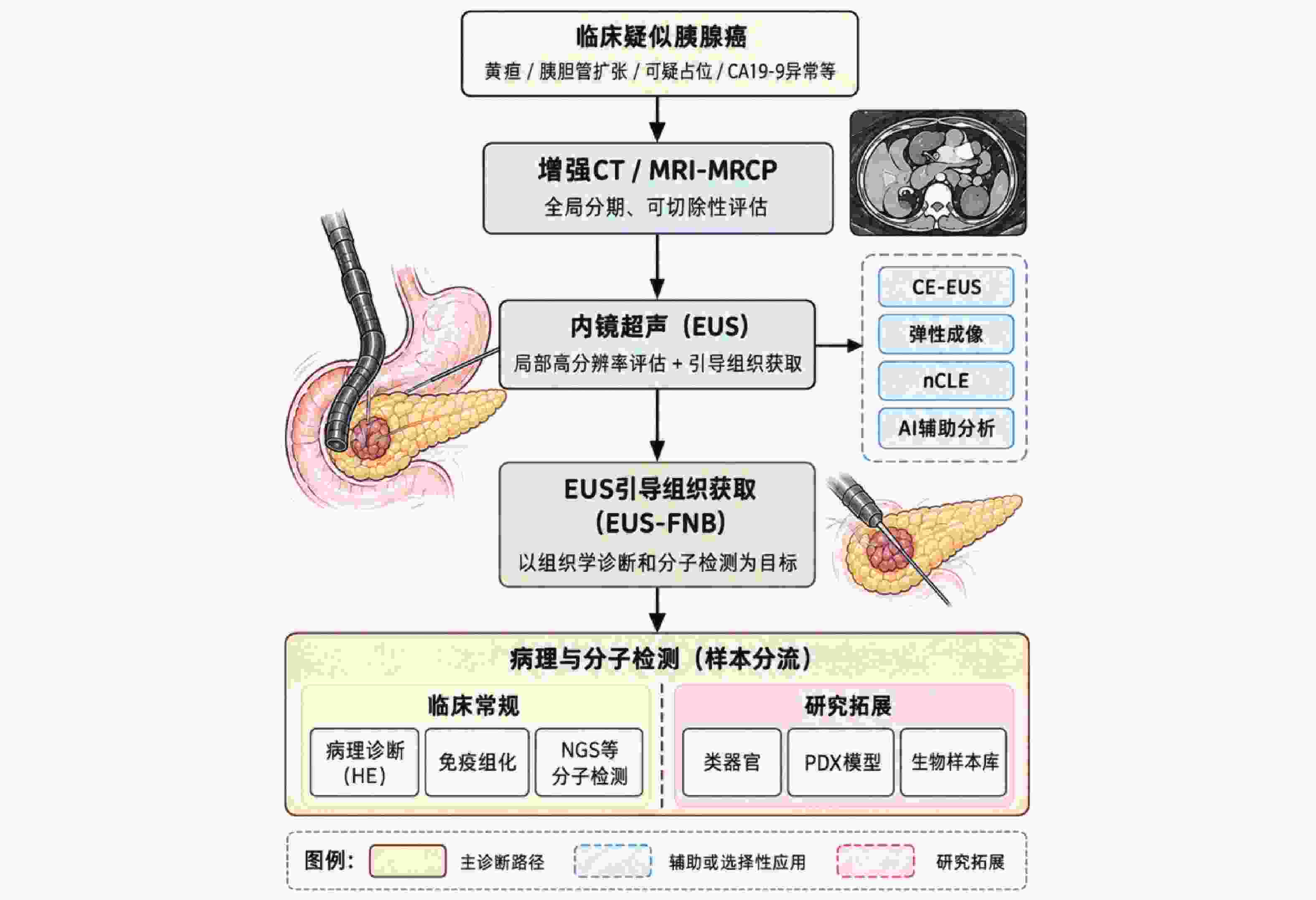
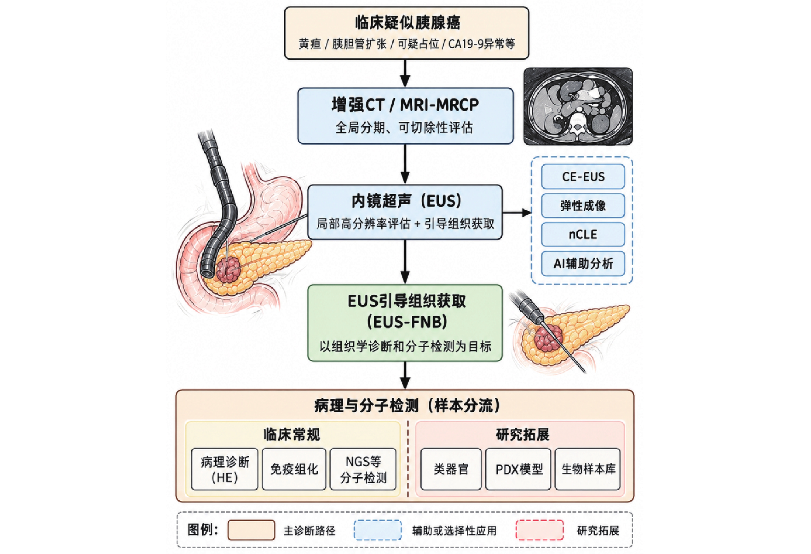
图 1 胰腺癌超声相关技术的诊断路径与组织获取策略
Figure 1. Diagnostic pathway and tissue acquisition strategy of ultrasound-related techniques in pancreatic cancer
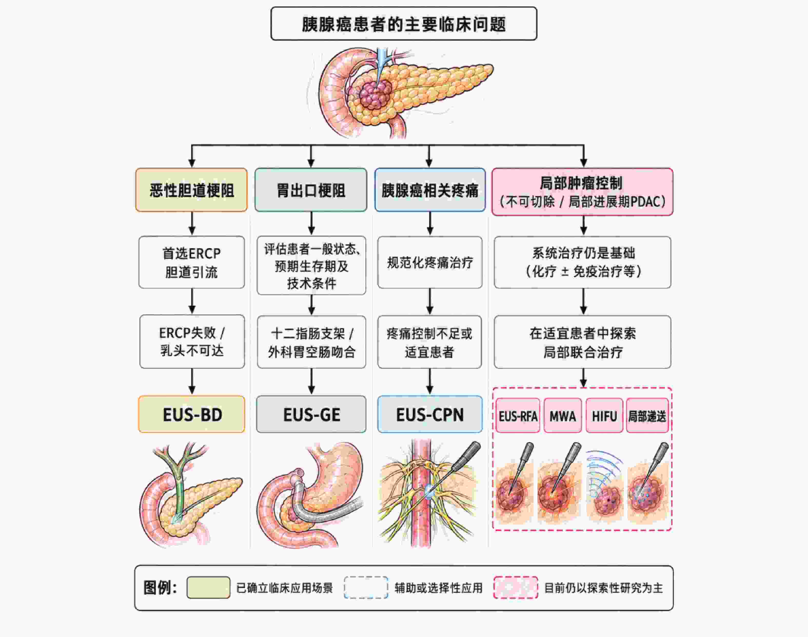
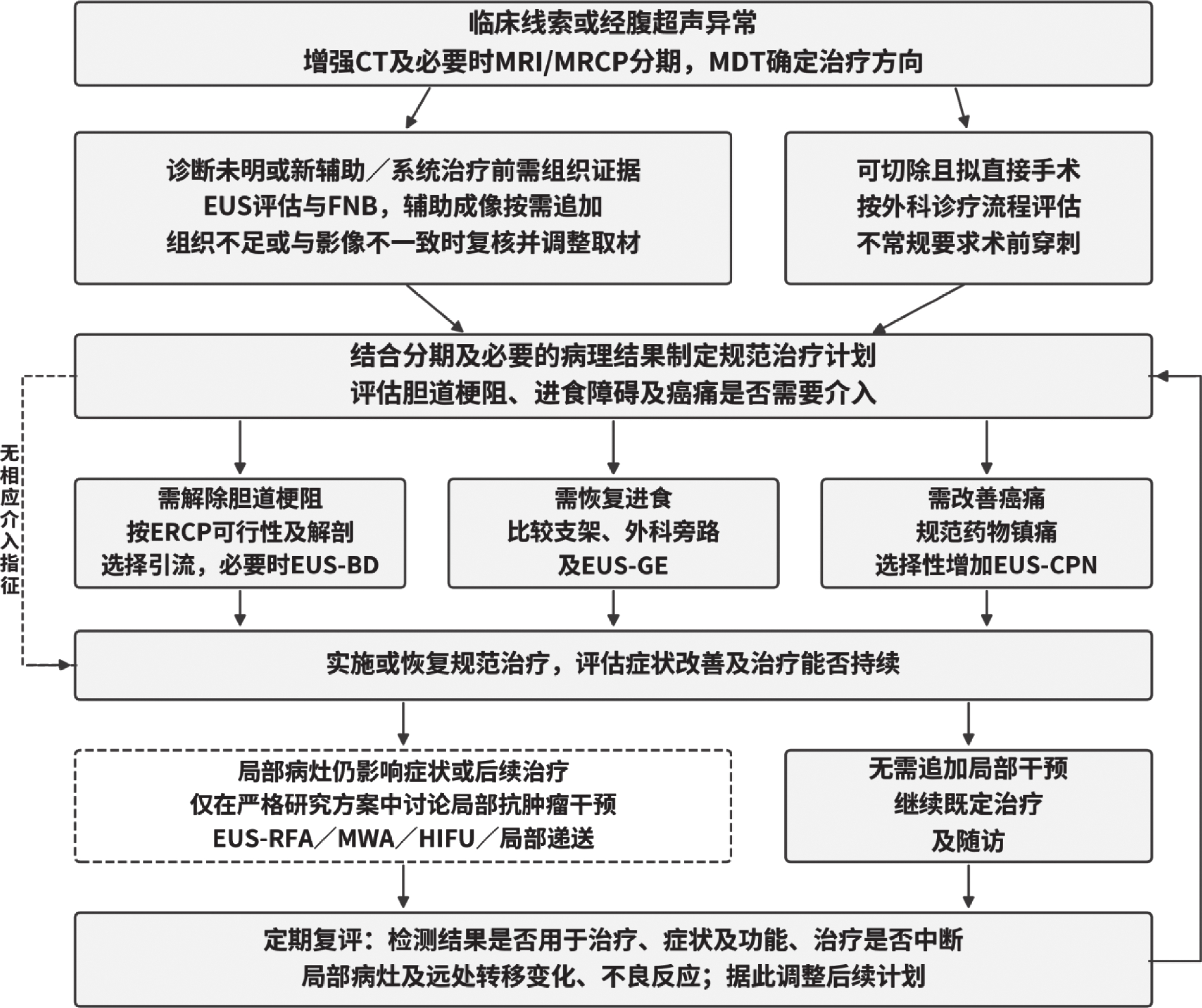
图 2 超声介导技术在胰腺癌治疗中的临床定位与决策框架
Figure 2. Clinical positioning and decision-making framework of ultrasound-mediated techniques in the treatment of pancreatic cancer
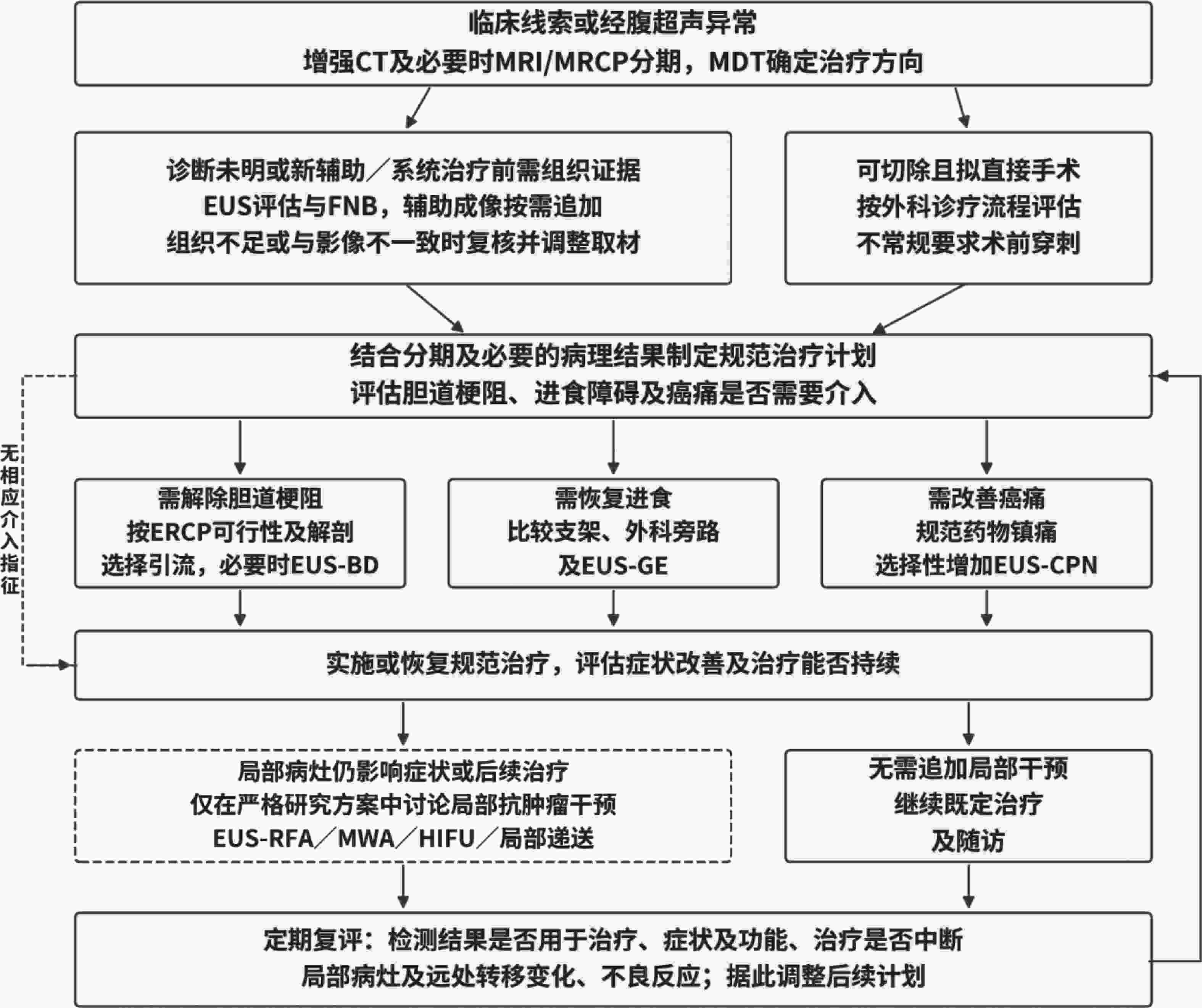
图 3 胰腺癌超声及相关技术的分阶段应用与诊疗衔接流程
Figure 3. Staged application and diagnostic-therapeutic integration workflow of ultrasound-related technologies for pancreatic cancer
表 1 胰腺癌不同超声引导/介导局部治疗技术的特点与临床定位比较
Table 1. Comparison of characteristics and clinical positioning of different ultrasound-guided or ultrasound-mediated local treatment modalities for pancreatic cancer
治疗方式 主要原理/操作 主要优势 主要局限 临床定位与证据判断 EUS-RFA EUS引导电极进入病灶,以射频热效应造成局部凝固性坏死 实时定位;可重复实施 有创;存在胰腺炎、出血及邻近胰胆管/血管热损伤风险 不可切除或局部进展期PDAC的探索性局部治疗;已有小样本研究与Meta分析,尚无随机证据证实生存获益 MWA 微波能量快速升温形成热凝固 升温快;消融范围相对较大 胰腺邻近重要结构,热损伤控制要求高;PDAC资料很少 技术探索阶段,临床证据少于EUS-RFA HIFU 体外聚焦超声产生热效应、空化效应及机械效应 无创;可重复;具有镇痛潜力 受胃肠气体、呼吸运动、病灶深度和声窗影响 可作为不可切除/局部进展期患者局部控制和镇痛的补充;已有系统评价及随机试验方案,尚缺高质量随机结果 EUS局部递送 EUS引导将药物或生物治疗制剂直接递送至病灶 提高局部药物浓度;减少全身暴露 制剂、剂量、注射方式及重复间隔尚未标准化 研究阶段,主要用于精准递送和转化研究探索  下载: 导出CSV
-
[1] Dahiya D S, Shah Y R, Ali H, et al. Basic principles and role of endoscopic ultrasound in diagnosis and differentiation of pancreatic cancer from other pancreatic lesions: A comprehensive review of endoscopic ultrasound for pancreatic cancer[J]. J Clin Med, 2024, 13(9): 2599. doi: 10.3390/jcm13092599 [2] Tempero M A, Malafa M P, Al-Hawary M, et al. Pancreatic adenocarcinoma, version 2.2021, NCCN clinical practice guidelines in oncology[J]. J Natl Compr Canc Netw, 2021, 19(4): 439-457. [3] Ciaravino V, D’Onofrio M. Pancreatic ultrasound: State of the art[J]. J Ultrasound Med, 2019, 38(5): 1125-1137. doi: 10.1002/jum.14978 [4] Zakaria A, Al-Share B, Klapman J B, et al. The role of endoscopic ultrasonography in the diagnosis and staging of pancreatic cancer[J]. Cancers, 2022, 14(6): 1373. doi: 10.3390/cancers14061373 [5] Machicado J D, Sheth S G, Chalhoub J M, et al. American Society for Gastrointestinal Endoscopy guideline on the role of endoscopy in the diagnosis and management of solid pancreatic masses: Summary and recommendations[J]. Gastrointest Endosc, 2024, 100(5): 786-796. doi: 10.1016/j.gie.2024.06.002 [6] Facciorusso A, Arvanitakis M, Crinò S F, et al. Endoscopic ultrasound-guided tissue sampling: European Society of Gastrointestinal Endoscopy (ESGE) Technical and Technology Review[J]. Endoscopy, 2025, 57(4): 390-418. doi: 10.1055/a-2524-2596 [7] D’Onofrio M, Beleù A, De Robertis R. Ultrasound-guided percutaneous procedures in pancreatic diseases: New techniques and applications[J]. Eur Radiol Exp, 2019, 3(1): 2. doi: 10.1186/s41747-018-0081-2 [8] Harmsen F R, Domagk D, Dietrich C F, et al. Discriminating chronic pancreatitis from pancreatic cancer: Contrast-enhanced EUS and multidetector computed tomography in direct comparison[J]. Endosc Ultrasound, 2018, 7(6): 395-403. doi: 10.4103/eus.eus_24_18 [9] Conti C B, Mulinacci G, Salerno R, et al. Applications of endoscopic ultrasound elastography in pancreatic diseases: From literature to real life[J]. World J Gastroenterol, 2022, 28(9): 909-917. doi: 10.3748/wjg.v28.i9.909 [10] Pawelec N, Durko Ł, Małecka-Wojciesko E. Changes connected to early chronic pancreatitis and early pancreatic cancer in endoscopic ultrasonography (EUS): Clinical implications[J]. Cancers, 2025, 17(11): 1891. doi: 10.3390/cancers17111891 [11] Yao D W, Qin M Z, Jiang H X, et al. Comparison of EUS-FNA and EUS-FNB for diagnosis of solid pancreatic mass lesions: A meta-analysis of prospective studies[J]. Scand J Gastroenterol, 2024, 59(8): 972-979. doi: 10.1080/00365521.2024.2354908 [12] Chong C C, Pittayanon R, Pausawasdi N, et al. Consensus statements on endoscopic ultrasound-guided tissue acquisition. Guidelines from the Asian Endoscopic Ultrasound Group[J]. Dig Endosc, 2024, 36(8): 871-883. doi: 10.1111/den.14768 [13] Itonaga M, Yasukawa S, Fukutake N, et al. Comparison of 22-gauge standard and Franseen needles in EUS-guided tissue acquisition for diagnosing solid pancreatic lesions: A multicenter randomized controlled trial[J]. Gastrointest Endosc, 2022, 96(1): 57-66. e2. [14] Chen Y I, Chatterjee A, Berger R, et al. Endoscopic ultrasound (EUS)-guided fine needle biopsy alone vs. EUS-guided fine needle aspiration with rapid onsite evaluation in pancreatic lesions: A multicenter randomized trial[J]. Endoscopy, 2022, 54(1): 4-12. doi: 10.1055/a-1375-9775 [15] Chalhoub J M, Hawa F, Grantham T, et al. Effect of the number of passes on diagnostic performance of EUS fine-needle biopsy of solid pancreatic masses: A systematic review and meta-analysis[J]. Gastrointest Endosc, 2024, 100(4): 595-604. e8. [16] Engh M A, Teutsch B, Schulze Wenning A, et al. Contrast-enhanced endoscopic ultrasound likely does not improve diagnostic adequacy during endoscopic ultrasound guided tissue acquisition: A systematic review and meta-analysis[J]. Pancreatology, 2024, 24(4): 649-660. doi: 10.1016/j.pan.2024.04.007 [17] Pan Y, Ran T, Zhang X, et al. Adequacy of EUS-guided fine-needle aspiration and fine-needle biopsy for next-generation sequencing in pancreatic malignancies: A systematic review and meta-analysis[J]. Endosc Ultrasound, 2024, 13(6): 366-375. doi: 10.1097/eus.0000000000000097 [18] Bang J Y, Jhala N, Seth A, et al. Standardisation of EUS-guided FNB technique for molecular profiling in pancreatic cancer: Results of a randomised trial[J]. Gut, 2023, 72(7): 1255-1257. doi: 10.1136/gutjnl-2023-329495 [19] Yan X, Lv K, Xiao M, et al. The diagnostic performance of contrast-enhanced ultrasound versus contrast-enhanced computed tomography for pancreatic carcinoma: A systematic review and meta-analysis[J]. Transl Cancer Res TCR, 2022, 11(10): 3645-3656. doi: 10.21037/tcr-22-601 [20] Shin C M, Villa E. The efficiency of contrast-enhanced endoscopic ultrasound (EUS) combined with EUS elastography for pancreatic cancer diagnosis: A systematic review and meta-analysis[J]. Ultrasonography, 2023, 42(1): 20-30. doi: 10.14366/usg.22103 [21] Wu C C H, Lim S J M, Tan D M Y. Endoscopic ultrasound-guided needle-based confocal laser endomicroscopy for pancreatic cystic lesions: Current status and future prospects[J]. Clin Endosc, 2024, 57(4): 434-445. doi: 10.5946/ce.2023.157 [22] Singh B S, Cazacu I M, Deza C A, et al. Image fusion involving real-time transabdominal or endoscopic ultrasound for gastrointestinal malignancies: Review of current and future applications[J]. Diagnostics (Basel), 2022, 12(12): 3218. doi: 10.3390/diagnostics12123218 [23] Dumitrescu E A, Ungureanu B S, Cazacu I M, et al. Diagnostic value of artificial intelligence-assisted endoscopic ultrasound for pancreatic cancer: A systematic review and meta-analysis[J]. Diagnostics (Basel), 2022, 12(2): 309. doi: 10.3390/diagnostics12020309 [24] Podină N, Gheorghe E C, Constantin A, et al. Artificial intelligence in pancreatic imaging: A systematic review[J]. United European Gastroenterol J, 2025, 13(1): 55-77. doi: 10.1002/ueg2.12723 [25] Michiels N, Doppenberg D, Groen J V, et al. Intraoperative ultrasound during surgical exploration in patients with pancreatic cancer and vascular involvement (ULTRAPANC): A prospective multicenter study[J]. Ann Surg Oncol, 2023, 30(6): 3455-3463. doi: 10.1245/s10434-023-13112-3 [26] Rimbaș M, Dumitru A C, Tripodi G, et al. EUS-guided radiofrequency ablation therapy for pancreatic neoplasia[J]. Diagnostics (Basel), 2024, 14(19): 2111. doi: 10.3390/diagnostics14192111 [27] Patel V, Abdelbaki A, Thosani N C, et al. Endoscopic ultrasound-guided radiofrequency ablation of pancreatic tumors[J]. Curr Opin Gastroenterol, 2024, 40(5): 369-378. doi: 10.1097/MOG.0000000000001026 [28] Tieranu C G, Balaban D V, Tabacelia D, et al. Endoscopic ultrasound-guided radiofrequency ablation for pancreatic adenocarcinoma: A scoping review with meta-analysis[J]. Diagnostics (Basel), 2025, 15(4): 437. doi: 10.3390/diagnostics15040437 [29] Oh D, Seo D W, Song T J, et al. Clinical outcomes of EUS-guided radiofrequency ablation for unresectable pancreatic cancer: A prospective observational study[J]. Endosc Ultrasound, 2022, 11(1): 68-74. doi: 10.4103/EUS-D-21-00049 [30] Robles-Medranda C, Del Valle R, Puga-Tejada M, et al. Assessing EUS-guided radiofrequency ablation in unresectable pancreatic ductal adenocarcinoma: A single-center historic cohort study[J]. Gastrointest Endosc, 2024, 100(2): 250-258. doi: 10.1016/j.gie.2024.03.023 [31] Costanzo A, Fulco E, Marini M, et al. Endoscopic ultrasound-guided radiofrequency ablation (EUS-RFA) for pancreatic adenocarcinoma: A review[J]. Cureus, 2024, 16(10): e70691. [32] Giovannini M, Marx M. Radiofrequency ablation of pancreatic solid tumors[J]. Gastrointest Endosc Clin N Am, 2023, 33(4): 855-865. [32] Giovannini M, Marx M. Radiofrequency ablation of pancreatic solid tumors[J]. Gastrointest Endosc Clin N Am, 2023, 33(4): 855-865. [33] Fergadi M P, Magouliotis D E, Rountas C, et al. A meta-analysis evaluating the role of high-intensity focused ultrasound (HIFU) as a fourth treatment modality for patients with locally advanced pancreatic cancer[J]. Abdom Radiol, 2022, 47(1): 254-264. [33] Fergadi M P, Magouliotis D E, Rountas C, et al. A meta-analysis evaluating the role of high-intensity focused ultrasound (HIFU) as a fourth treatment modality for patients with locally advanced pancreatic cancer[J]. Abdom Radiol, 2022, 47(1): 254-264. [34] Marinova M, Khouri D A, Küppers J, et al. Study protocol of a randomized, two-arm, phase I/II trial investigating the feasibility, safety, and efficacy of local treatment with US-guided high-intensity focused ultrasound in combination with palliative chemotherapy in inoperable pancreatic cancer[J]. J Clin Med, 2024, 13(13): 3717. [34] Marinova M, Khouri D A, Küppers J, et al. Study protocol of a randomized, two-arm, phase I/II trial investigating the feasibility, safety, and efficacy of local treatment with US-guided high-intensity focused ultrasound in combination with palliative chemotherapy in inoperable pancreatic cancer[J]. J Clin Med, 2024, 13(13): 3717. [35] Yu L, Liu Y, Li Z, et al. A retrospective comparative study on the treatment of non-metastatic pancreatic cancer using high-intensity focused ultrasound versus radical surgery[J]. Int J Hyperth, 2024, 41: 2398557. doi: 10.1080/02656736.2024.2398557 [36] Sears O, Zhang H, Blatz N, et al. Focused ultrasound in pancreatic ductal adenocarcinoma: Mechanisms, preclinical evidence, and emerging clinical applications[J]. Cancers, 2026, 18(4): 574. doi: 10.3390/cancers18040574 [37] Zhou W, Lei C, Xue X, et al. Revelations of pancreatic cancer treated by high-intensity focused ultrasound[J]. Discov Oncol, 2026, 17(1): 1211. doi: 10.1007/s12672-026-05049-2 [38] Matsumoto K, Kato H, Tsutsumi K, et al. Current status of endoscopic ultrasound-guided antitumor treatment for pancreatic cancer[J]. Dig Endosc, 2025, 37(1): 18-28. doi: 10.1111/den.14815 [39] Kerdsirichairat T, Shin E J. Endoscopic ultrasound guided interventions in the management of pancreatic cancer[J]. WJGE, 2022, 14(4): 191-204. doi: 10.4253/wjge.v14.i4.191 [40] Moond V, Maniyar B, Harne P S, et al. Harnessing endoscopic ultrasound-guided radiofrequency ablation to reshape the pancreatic ductal adenocarcinoma microenvironment and elicit systemic immunomodulation[J]. Explor Target Antitumor Ther, 2024, 5(5): 1056-1073. doi: 10.37349/etat.2024.00263 [41] Isayama H, Nakai Y, Itoi T, et al. Clinical practice guidelines for safe performance of endoscopic ultrasound/ultrasonography-guided biliary drainage: 2018[J]. J Hepatobiliary Pancreat Sci, 2019, 26(7): 249-269. doi: 10.1002/jhbp.631 [42] van der Merwe S W, van Wanrooij R L J, Bronswijk M, et al. Therapeutic endoscopic ultrasound: European society of gastrointestinal endoscopy (ESGE) guideline[J]. Endoscopy, 2022, 54(2): 185-205. doi: 10.1055/a-1717-1391 [43] Pawa S, Marya N B, Thiruvengadam N R, et al. American Society for Gastrointestinal Endoscopy guideline on the role of therapeutic EUS in the management of biliary tract disorders: Summary and recommendations[J]. Gastrointest Endosc, 2024, 100(6): 967-979. doi: 10.1016/j.gie.2024.03.027 [44] Khashab M A, Bukhari M, Baron T H, et al. EUS-guided gastroenterostomy for malignant gastric outlet obstruction: Current status and future directions[J]. Gastrointest Endosc Clin N Am, 2020, 30(3): 479-494. doi: 10.1016/j.giec.2020.02.005 [45] Asif A A, Walayat S K, Bechtold M L, et al. EUS-guided celiac plexus neurolysis for pain in pancreatic cancer patients–a meta-analysis and systematic review[J]. J Community Hosp Intern Med Perspect, 2021, 11(4): 536-542. doi: 10.1080/20009666.2021.1929049 [46] Kamata K, Kinoshita M, Kinoshita I, et al. Efficacy of EUS-guided celiac plexus neurolysis in combination with EUS-guided celiac ganglia neurolysis for pancreatic cancer-associated pain: A multicenter prospective trial[J]. Int J Clin Oncol, 2022, 27(7): 1196-1201. doi: 10.1007/s10147-022-02160-6 [47] Bharwad A V, Ahuja R, Jain P, et al. Artificial intelligence in pancreatobiliary endoscopy: Current advances, opportunities, and challenges[J]. J Clin Med, 2025, 14(21): 7519. doi: 10.3390/jcm14217519 -

 点击查看大图
计量
- 文章访问数: 3
- HTML全文浏览量: 2
- PDF下载量: 0
- 被引次数: 0
