




Spatio-Temporal Characteristics of Early Gait Abnormalities in AD Model Mice Induced by Acute Intracerebroventricular Injection of Aβ1-42
-
摘要:
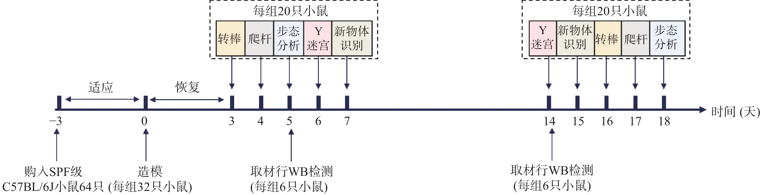
目的 探索侧脑室注射Aβ1-42对小鼠步态的急性影响,并表征其步态改变。 方法 将64只SPF级C57/BL6J小鼠随机分为Control组(n = 32)和AD组(n = 32),运用侧脑室注射Aβ1-42的方法造模,实验初期(3~7 d)及末期(14~18 d)对小鼠进行转棒实验、爬杆实验、步态分析、Y迷宫和新物体识别实验测试(n = 20),并于造模后第5天(n = 6)和第14 天(n = 6)对小鼠双侧纹状体、海马进行Aβ含量的WB检测。 结果 两组小鼠在转棒和爬杆实验中的行为表现差异无统计学意义(P > 0.05)。相较于Control组,5 d时AD组小鼠四肢步幅均明显缩短(P < 0.001)、前肢步宽(P < 0.001)、后肢步宽(P < 0.001)明显降低,四肢步幅变异系数明显增加(P < 0.05),步长周期在中、高总速度条件下明显缩短(P < 0.01),而支撑时相、摆动时相与Control组相比差异无统计学意义(P > 0.05)。造模后6~7 d AD组小鼠进入Y迷宫新异臂的次数和在新异臂的探索距离与对照组无显著差异(P > 0.05),但其在新物体识别实验中对新物体的探索次数和时间均少于Control组(P < 0.05),14~15 d AD组小鼠进入Y迷宫新异臂的次数、在新异臂的探索距离、对新物体的探索次数和时间均显著少于Control组(P < 0.05)。18 d时AD小鼠相较于Control组小鼠四肢步幅明显减小(P < 0.001)、步宽明显降低(P < 0.001)、步长周期显著缩短(P < 0.001)、四肢步幅变异系数较Control组小鼠均明显增加(P < 0.01),支撑时相、摆动时相则与Control组相比差异无统计学意义(P > 0.05)。5 d和14 d的WB结果显示AD组小鼠双侧纹状体Aβ含量无显著变化(P > 0.05),海马区Aβ含量升高(P < 0.05)。 结论 急性侧脑室注射Aβ1-42可导致小鼠步态异常,呈现出一种小步幅、窄步宽的步行模式,且步幅变异增加。 Abstract:Objective To explore the acute effect of intracerebroventricular injection of Aβ1-42 on mouseb gait and characterize the resulting gait alterations. Methods Sixty-four SPF-grade C57/BL6J mice were randomly assigned to a Control group(n = 32) and an AD group (n = 32). The AD mouse model was established by intracerebroventricular injection of Aβ1-42. Rotarod test, pole test, gait analysis, Y-maze test and novel object recognition test were performed in the early (3~7 d) and late (14~18 d) experimental stages (n = 20). Western-blotting (WB) was used to detect Aβ levels in the bilateral striatum and hippocampus at day 5 (n = 6) and day 14 (n = 6) after modeling. Results There were no statistically significant differences in the performance of rotarod and pole tests between the two groups (P > 0.05). Compared with the Control group, the AD mice exhibited significantly shortened stride length of all four limbs (P<0.001), decreased forelimb and hindlimb stride width (P < 0.001), increased coefficient of variation of stride length (P < 0.05), and shortened step cycle under medium and high total speeds(P < 0.01) at day 5. No significant differences in stance phase and swing phase were observed between groups (P > 0.05). At 6~7 d after modeling, the number of entries into and exploration distance within the novel arm of the Y-maze test showed no significant differences between the AD group and Control group (P > 0.05). However, AD mice made fewer explorations of the novel object and spent less time exploring it than Control mice (P < 0.05). At 14~15 d, AD mice showed significantly fewer entries into the novel Y-maze arm, a shorter exploration distance in that arm, and fewer and shorter explorations of the novel object than Control mice (P < 0.05). At day 18, AD group showed markedly reduced limb stride length (P < 0.001), decreased stride width (P < 0.001), shortened step cycle (P < 0.001), and elevated coefficient of variation of stride length (P < 0.01), whereas no inter-group differences were found in stance phase and swing phase (P > 0.05). Western blotting at days 5 and 14 showed no significant change in Aβ levels in the bilateral striatum of AD mice (P > 0.05), whereas hippocampal Aβ levels were increased (P < 0.05). Conclusion Acute intracerebroventricular injection of Aβ1-42 induces gait abnormalities in mice, characterized by a short-stride, narrow-based walking pattern and increased stride variability. -
Key words:
- Alzheimer's disease /
- Dementia /
- Gait analysis /
- Motor function /
- Cognitive function
-
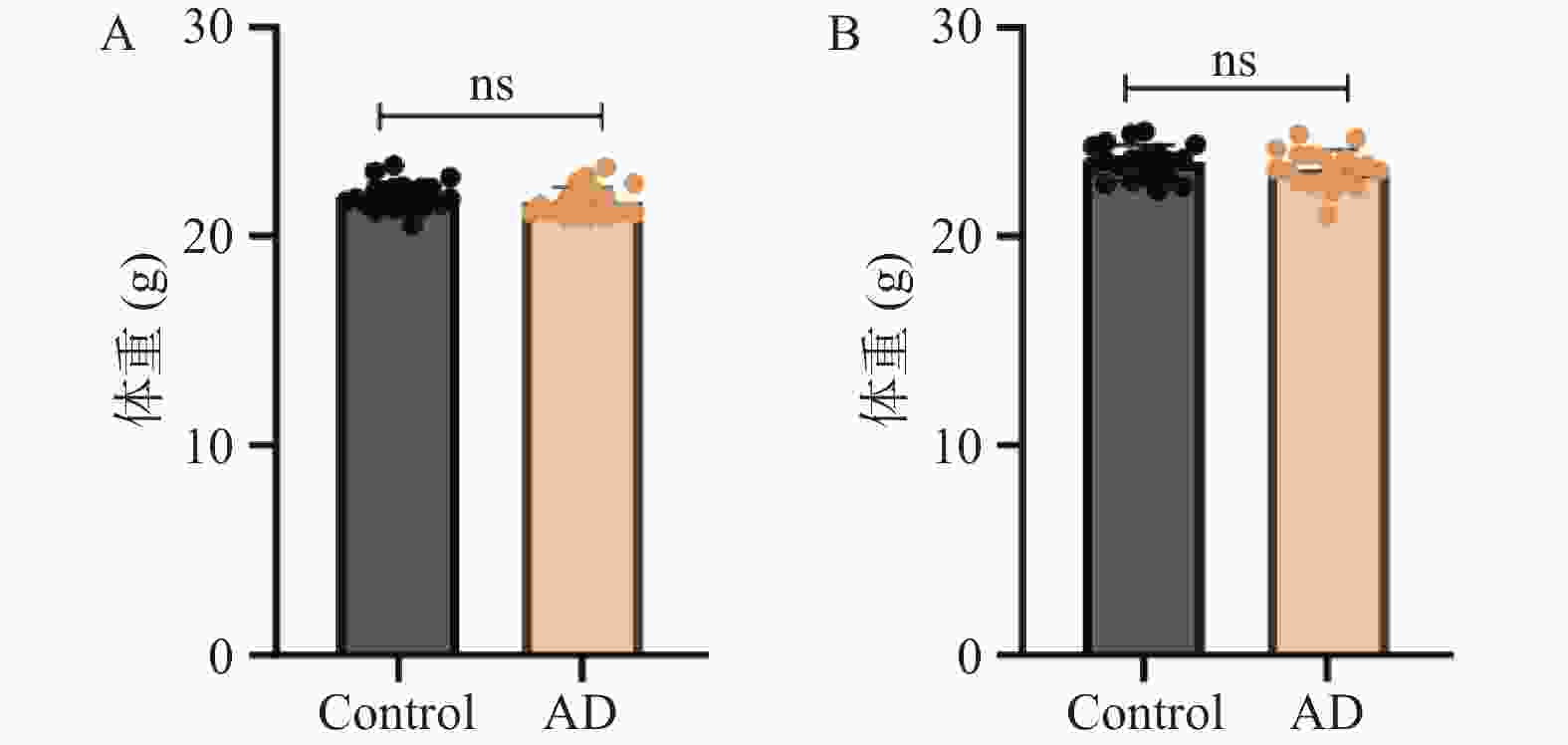
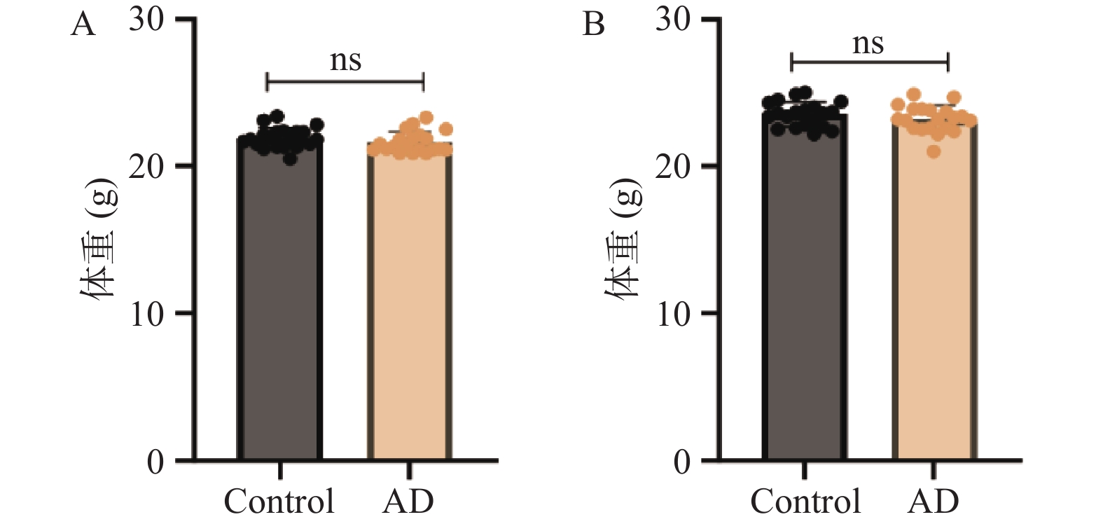
图 2 小鼠体重情况($ \bar x \pm s $,n = 20)
A:0 d两组小鼠体重对比情况;B:18 d两组小鼠体重对比情况;*P < 0.05;**P < 0.01;***P < 0.001;ns表示差异无统计学意义。
Figure 2. Body weight of mice ($ \bar x \pm s $,n = 20)
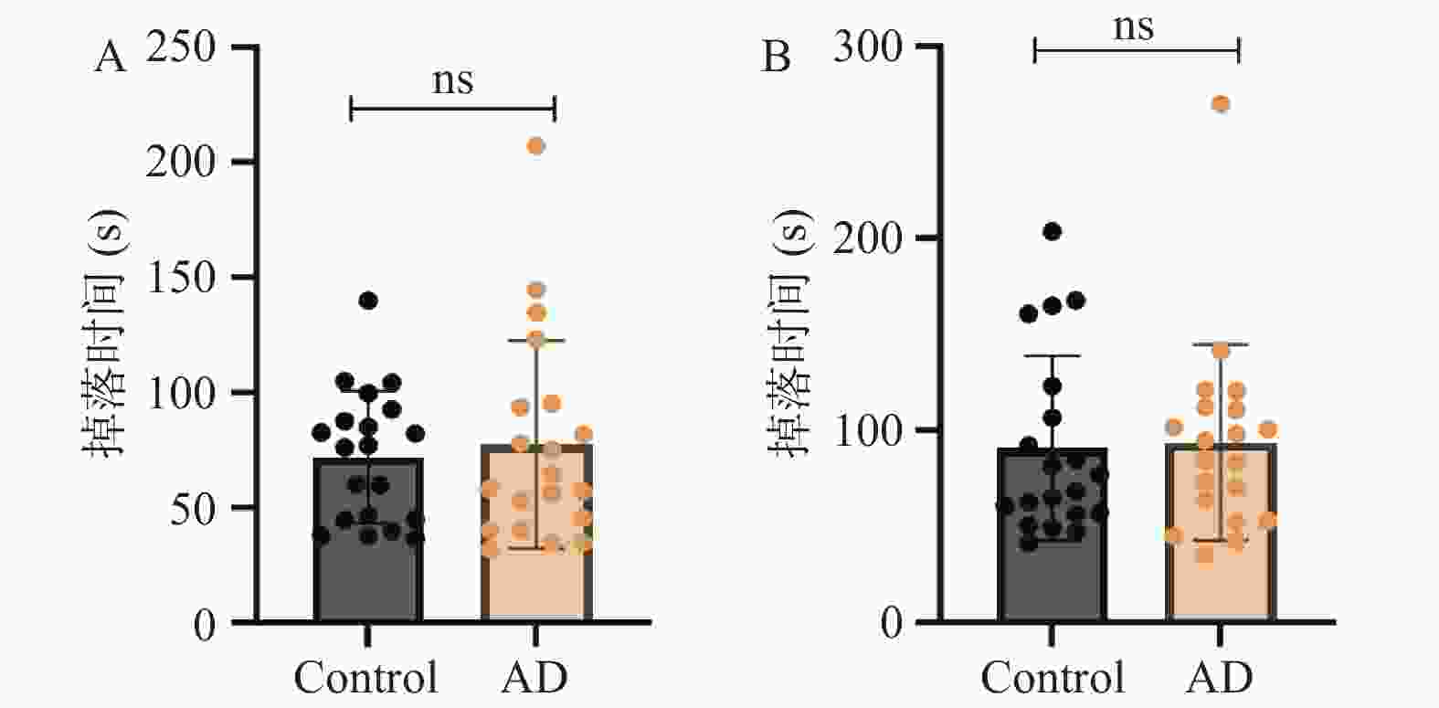
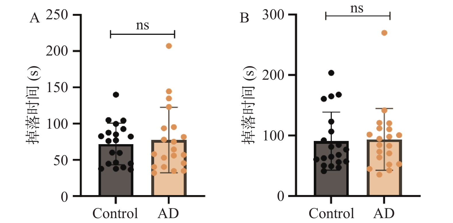
图 3 两组小鼠转棒实验结果($ \bar x \pm s $,n = 20)
A:3 d小鼠转棒实验结果;B:16 d小鼠转棒实验结果;*P < 0.05;**P < 0.01;***P < 0.001;ns表示差异无统计学意义。
Figure 3. Rotarod test results of mice in the two groups ($ \bar x \pm s $,n = 20)
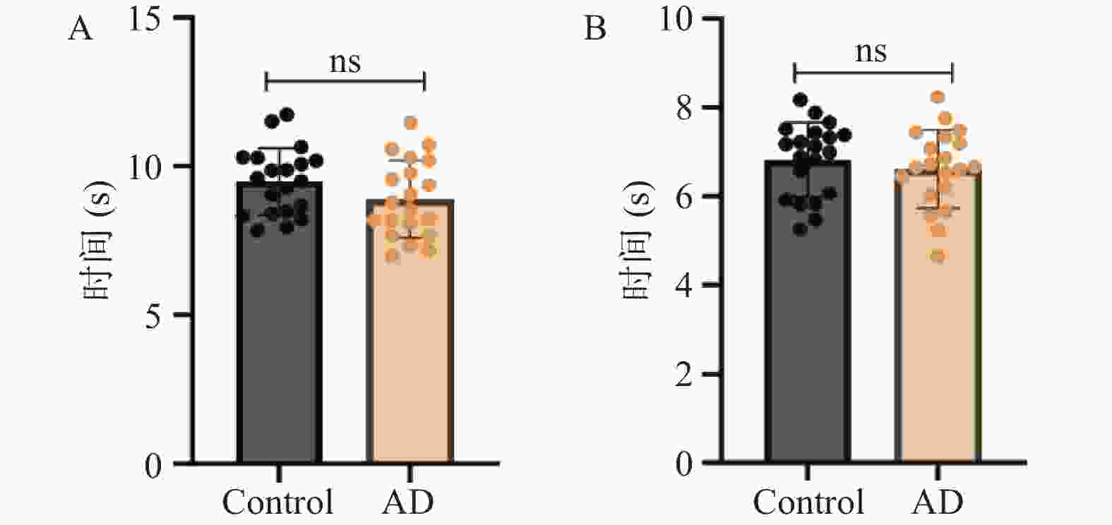
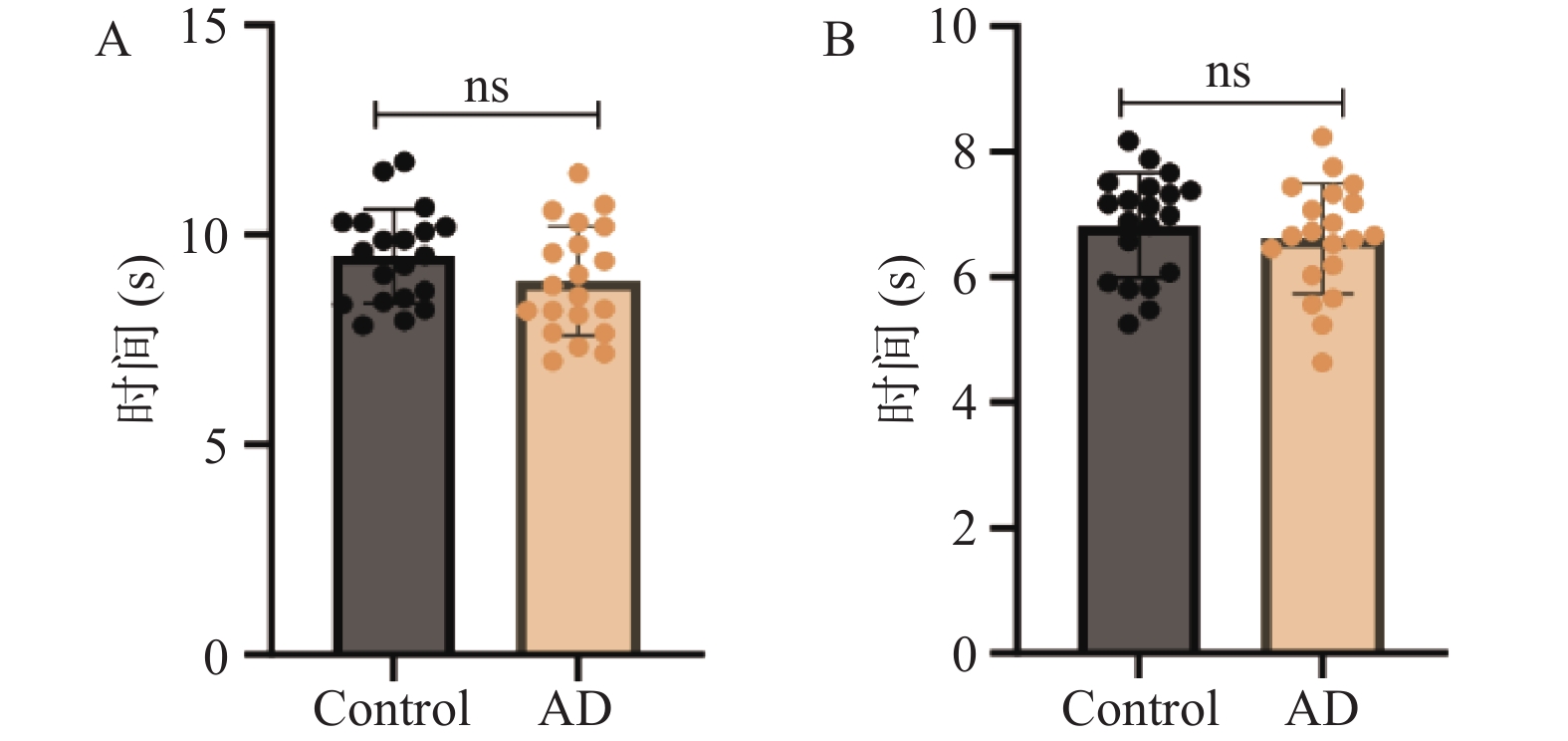
图 4 两组小鼠爬杆实验结果($ \bar x \pm s $,n = 20)
A:3 d小鼠爬杆实验结果;B:16 d小鼠爬杆实验结果;*P < 0.05;**P < 0.01;***P < 0.001;ns表示差异无统计学意义。
Figure 4. Pole test results of mice in the two groups ($ \bar x \pm s $,n = 20)
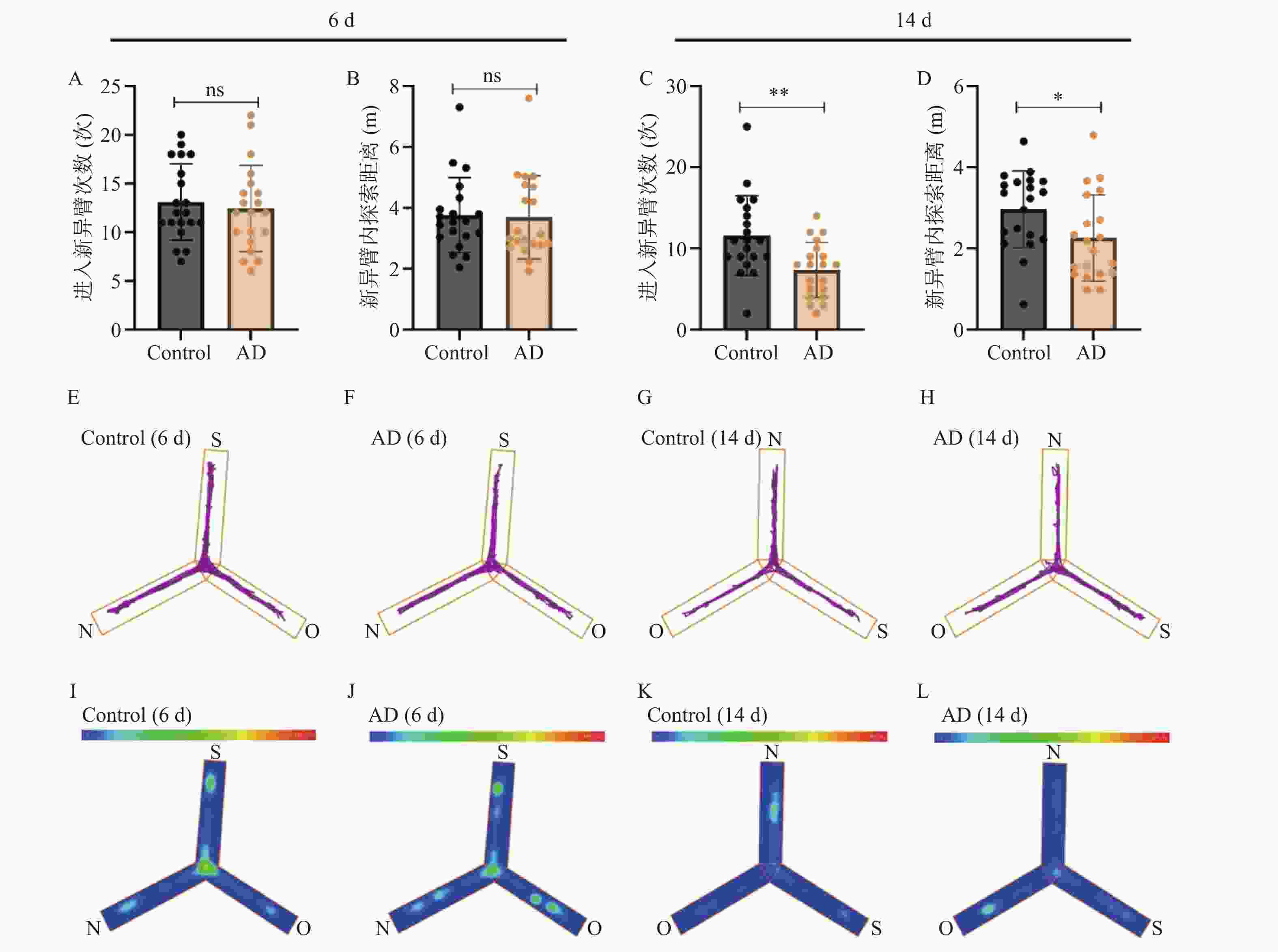
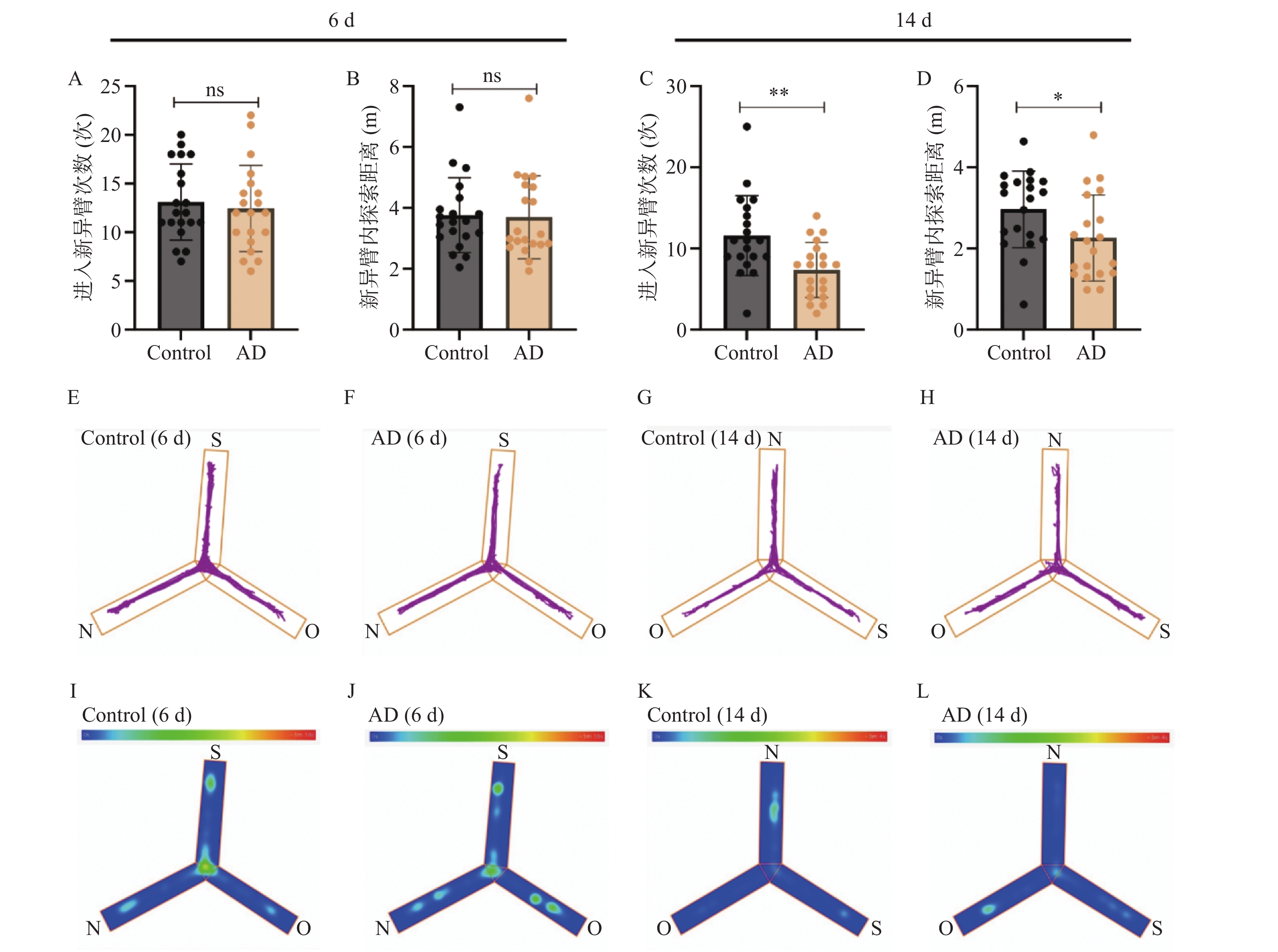
图 5 两组小鼠Y迷宫实验结果($ \bar x \pm s $,n = 20)
A~B:6 d Y迷宫实验结果统计图;C~D:14 d Y迷宫实验结果统计图;E~F:6 d Control 和AD组小鼠在Y迷宫实验中的代表性轨迹图;G~H:14 d Control 和AD组小鼠在Y迷宫实验中的代表性轨迹图;I~J:6 d Control 和AD组小鼠在Y迷宫实验中的代表性热图;K~L:14 d Control 和AD组小鼠在Y迷宫实验中的代表性热图。轨迹图显示动物中心点在测试过程中的运动轨迹,热图显示动物中心点出现在迷宫中的时间占比,色标从左到右为权重从低到高。N:新异臂;O:其他臂;S:起始臂;*P < 0.05;**P < 0.01;***P < 0.001;ns表示差异无统计学意义。
Figure 5. Y-maze test results of mice in the two groups ($ \bar x \pm s $,n = 20)
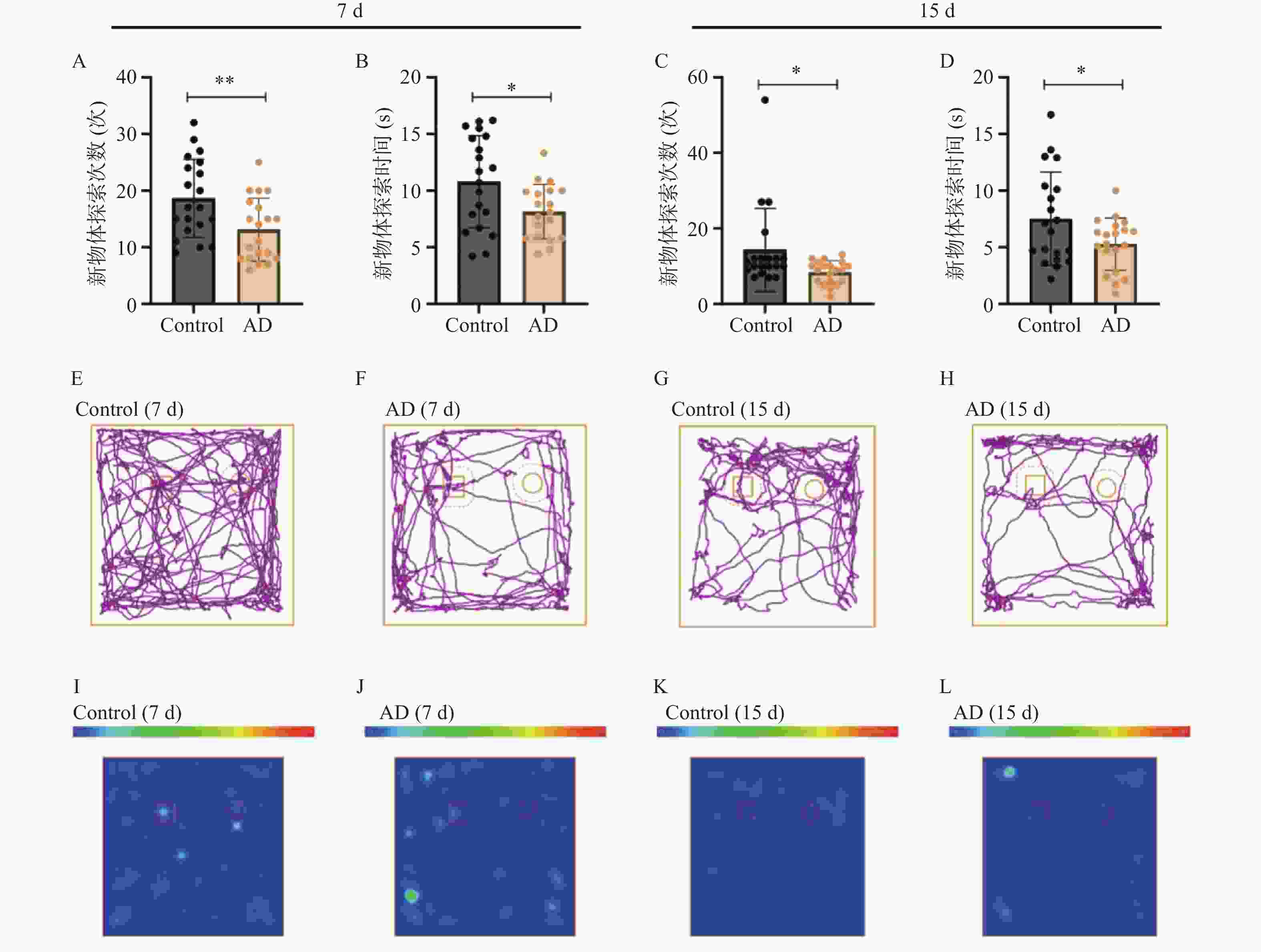
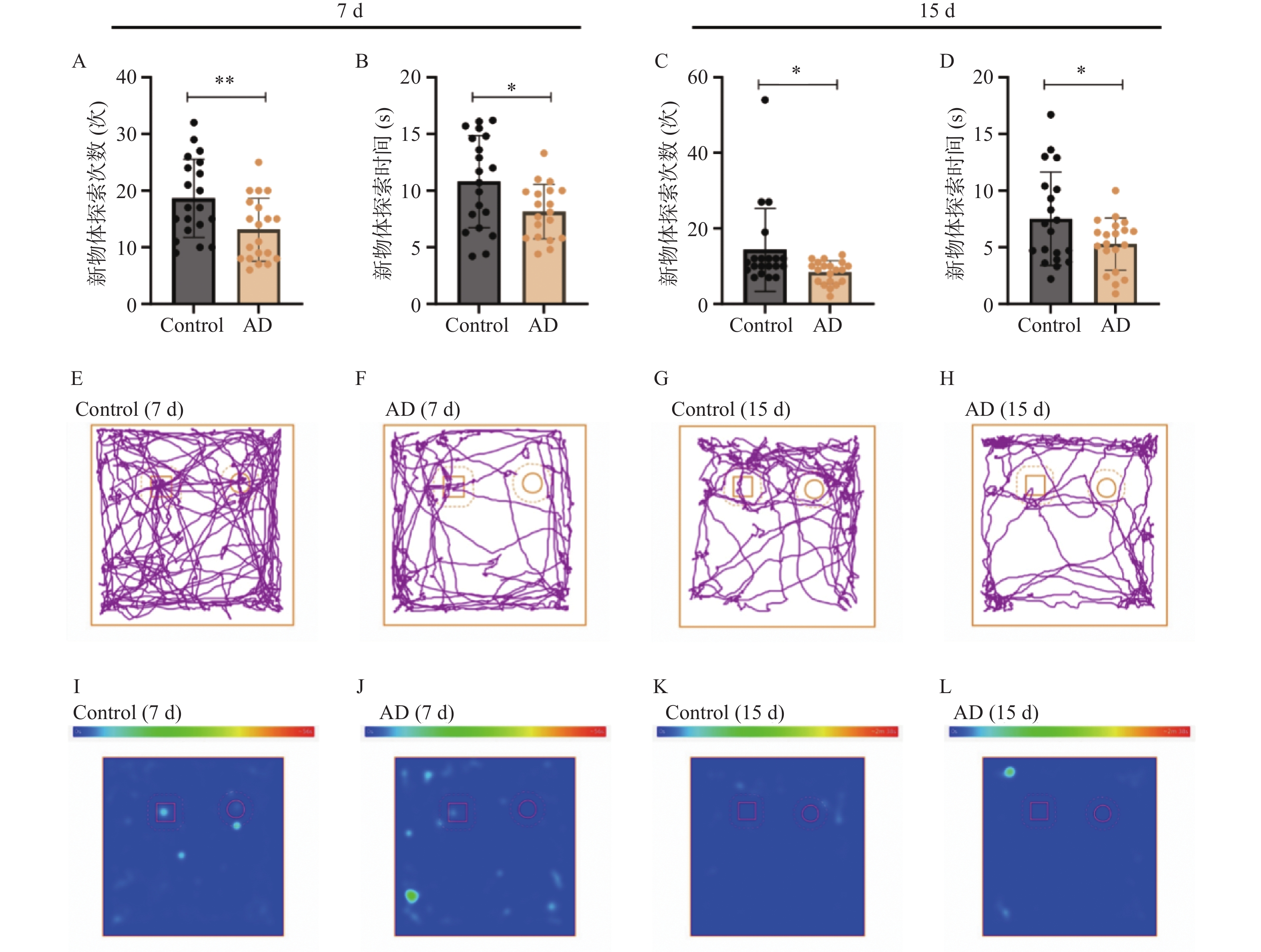
图 6 新物体识别实验结果($ \bar x \pm s $,n = 20)
A~B:7 d 新物体识别实验结果统计图;C~D:15 d 新物体识别实验结果统计图;E~F:7 d Control 和AD组小鼠在新物体识别实验中的代表性轨迹图;G~H:15 d Control 和AD组小鼠在新物体识别实验中的代表性轨迹图;I~J:7 d Control 和AD组小鼠在新物体识别实验中的代表性热图;K~L:15 d Control 和AD组小鼠在新物体识别实验中的代表性热图。轨迹图显示动物中心点在测试过程中的运动轨迹,热图显示动物中心点出现在场内的时间占比,色标从左到右为权重从低到高。方形为旧物体,圆形为新物体。*P < 0.05;**P < 0.01;***P < 0.001;ns表示差异无统计学意义。
Figure 6. Results of novel object recognition test ($ \bar x \pm s $,n = 20)
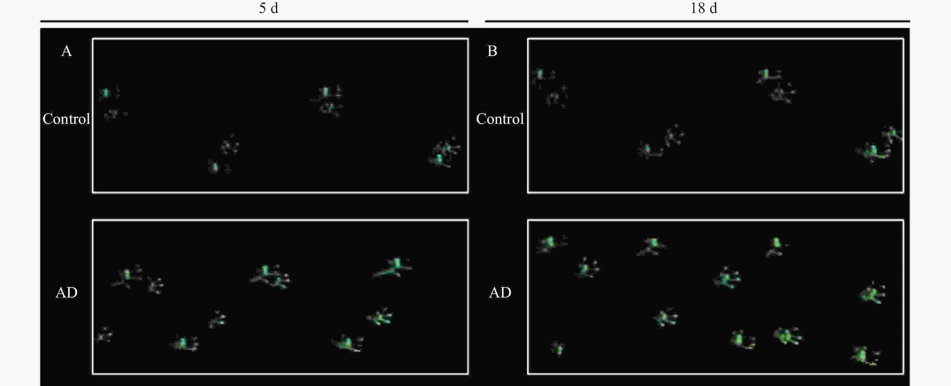
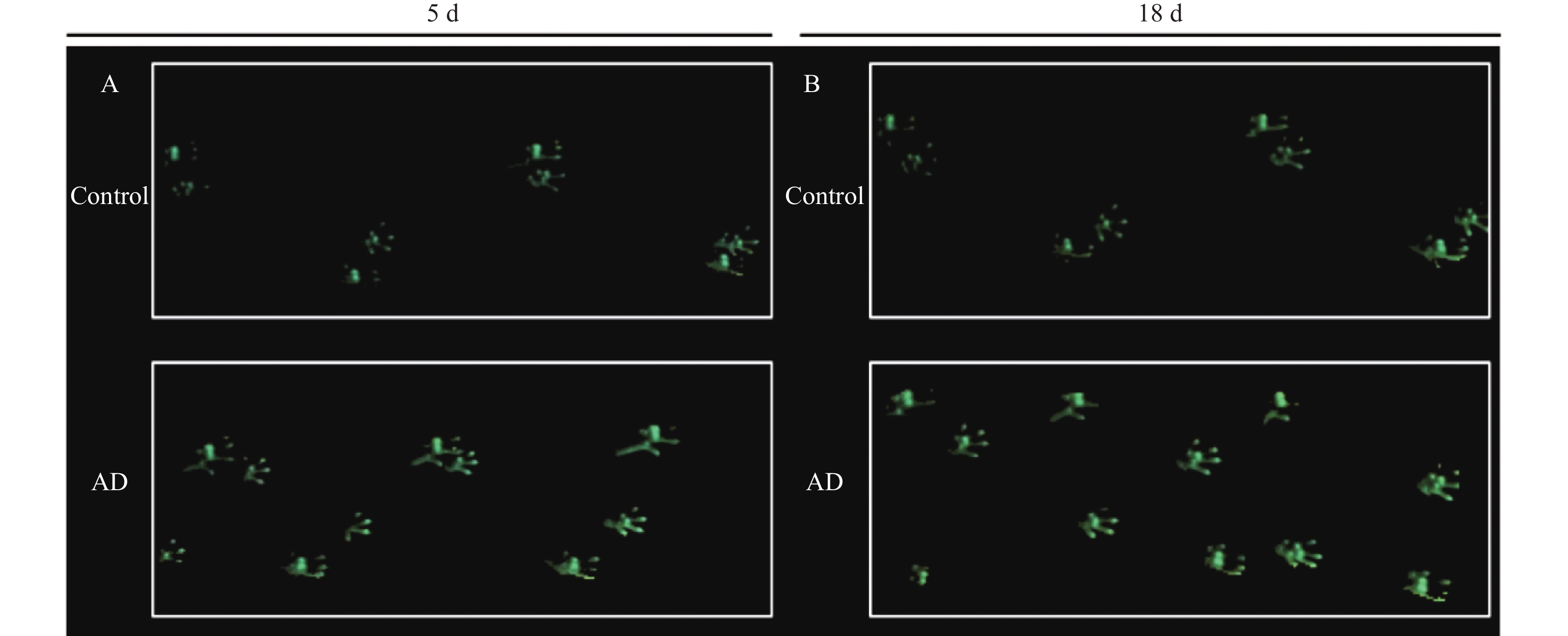
图 7 两组小鼠代表性足印图
A: 5 d Control组和AD组小鼠的代表性足印;B: 18 d Control组和AD组小鼠的代表性足印。
Figure 7. Representative foot-prints of mice in the two groups
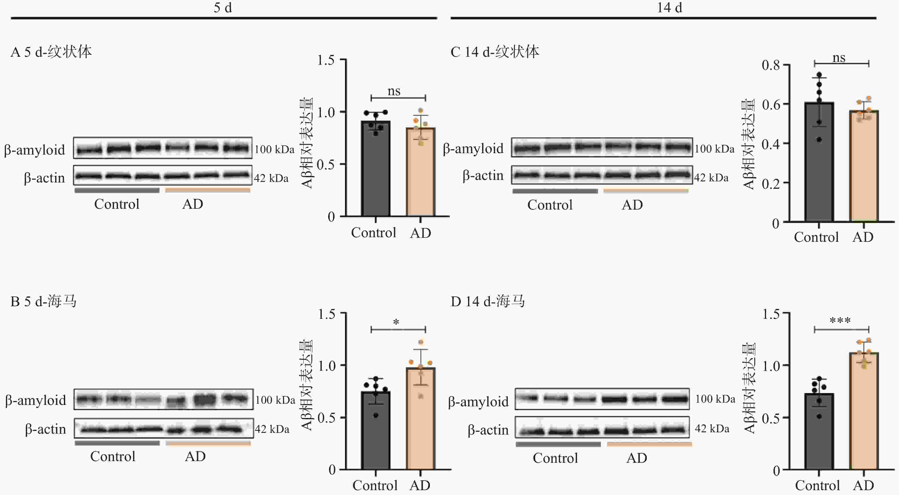
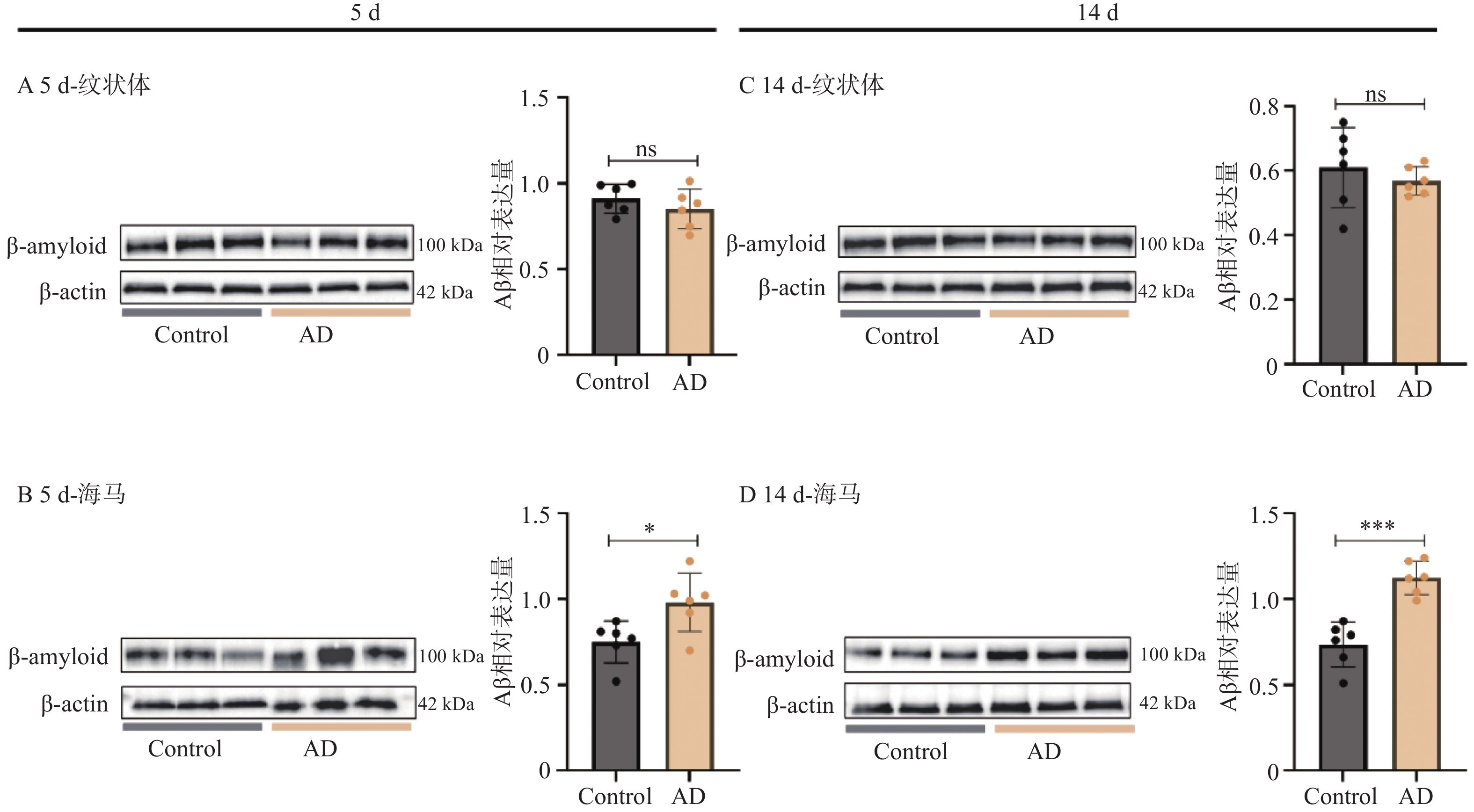
图 8 两组小鼠纹状体、海马组织中Aβ含量的WB结果($ \bar x \pm s $,n = 6)
A: 5 d 时Control和AD组小鼠纹状体中Aβ含量的WB代表性条带和统计结果;B: 5 d 时Control和AD组小鼠海马中Aβ含量的WB代表性条带和统计结果;C:14 d 时Control和AD组小鼠纹状体中Aβ含量的WB代表性条带和统计结果;D:14 d 时Control和AD组小鼠海马中Aβ含量的WB代表性条带和统计结果;*P < 0.05;**P < 0.01;***P < 0.001;ns表示差异无统计学意义。
Figure 8. WB results of Aβ contents in striatum and hippocampus tissues of mice from the two groups ($ \bar x \pm s $, n = 6)
表 1 本研究评估的步态参数及意义
Table 1. Gait parameters evaluated in this study and their implications
步态参数 意义 步幅(cm) 同一肢连续两个最大脚印横坐标中点之间的距离 步长周期(s) 单肢步幅的平均时长 支撑时相(%) 支撑时长占步幅时长的百分比 摆动时相(%) 摆动时长占步幅时长的百分比 总速度(cm/s) 步长的总和/步幅时长总和 步幅变异系数 步幅标准差/步幅平均值 步宽(cm) 在行走中左、右两足间的距离 脚步频率(步/min) 单位时间内的脚步数  下载: 导出CSV
表 2 5 d步态分析结果(协方差分析部分)
Table 2. Gait parameters of mice at 5 d after modeling (ANCOVA)
步态参数组别*总速度 Control-5d AD-5d
F(df1,df2)
PF P 原始值/校正值 原始值/校正值 左前爪步态数据 步长周期(s) 4.537 0.040* 0.095±0.016/ — 0.103±0.014/ — — — 步幅(cm) 0.008 0.931 6.424±0.550/6.233±0.100 5.366±0.386/5.557±0.100 18.778(1,37) <0.001*** 支撑时相(%) 0.695 0.410 54.995±4.648/56.882±1.123 59.500±5.568/57.613±1.123 0.175(1,37) 0.678 摆动时相(%) 0.695 0.410 45.005±4.648/43.118±1.123 40.500±5.568/42.387±1.123 0.175(1,37) 0.678 步幅变异系数 0.043 0.836 0.029±0.012/0.027±0.016 0.125±0.090/0.127±0.016 15.297(1,37) <0.001*** 总速度(cm/s) — — 69.756±14.582/ — 52.945±7.989/ — — — 右前爪步态数据 步长周期(s) 9.284 0.004* 0.097±0.016/ — 0.105±0.020/ — — — 步幅(cm) 0.280 0.600 6.426±0.549/6.258±0.098 5.328±0.395/5.495±0.098 26.142(1,37) <0.001*** 支撑时相(%) 0.019 0.891 56.972±4.046/57.822±1.508 60.686±7.828/59.836±1.508 0.764(1,37) 0.388 摆动时相(%) 0.019 0.891 43.028±4.046/42.178±1.508 39.314±7.828/40.164±1.508 0.764(1,37) 0.388 步幅变异系数 0.191 0.665 0.035±0.027/0.039±0.016 0.110±0.086/0.106±0.016 7.619(1,37) 0.009** 总速度(cm/s) — — 68.853±14.759/ — 52.576±11.428/ — — — 左后爪步态数据 步长周期(s) 0.243 0.625 0.095±0.017/0.104±0.002 0.098±0.016/0.089±0.002 14.518(1,37) 0.001** 步幅(cm) 3.062 0.089 6.462±0.510/6.229±0.134 5.069±0.724/5.303±0.134 19.747(1,37) <0.001*** 支撑时相(%) 2.446 0.127 59.273±4.922/60.888±1.269 63.676±6.039/62.061±1.269 0.356(1,37) 0.555 摆动时相(%) 2.446 0.127 40.727±4.922/39.112±1.269 36.324±6.039/37.939±1.269 0.356(1,37) 0.555 步幅变异系数 0.255 0.616 0.035±0.020/0.040±0.021 0.180±0.113/0.174±0.021 6.255(1,37) 0.017* 总速度(cm/s) — — 70.704±14.688/ — 52.733±10.829/ — — — 右后爪步态数据 步长周期(s) 11.278 0.002* 0.094±0.016/ — 0.104±0.019/ — — — 步幅(cm) 0.319 0.576 6.436±0.556/6.230±0.102 5.265±0.394/5.471±0.102 22.477(1,37) <0.001*** 支撑时相(%) 1.142 0.241 57.309±6.316/58.481±1.896 63.014±8.544/61.842±1.896 1.278(1,37) 0.266 摆动时相(%) 1.142 0.241 42.691±6.316/41.519±1.896 36.987±8.544/38.158±1.896 1.278(1,37) 0.266 步幅变异系数 1.353 0.252 0.035±0.034/0.037±0.015 0.124±0.083/0.115±0.015 10.450(1,37) 0.003** 总速度(cm/s) — — 70.307±14.170/ — 52.231±9.442/ — — — 多爪步态数据 前肢步宽(cm) 0.326 0.572 3.570±0.289/3.539±0.076 2.991±0.243/3.022±0.076 20.982(1,37) <0.001*** 后肢步宽(cm) 1.157 0.289 4.177±0.265/4.149±0.065 3.574±0.294/3.602±0.065 31.572(1,37) <0.001*** 脚步频率 — — 32.600±6.038/ — 27.800±4.021/ — — — 注:采用协方差分析,原始值以$ \bar x \pm s $表示;校正后值以均数±标准误(SE)表示*P < 0.05;**P < 0.01;***P < 0.001;n=20。
下载: 导出CSV
表 3 5 d步态分析结果(简单斜率分析部分)
Table 3. Gait parameters of mice at 5 d after modeling (simple slopes analysis)
步态参数 总速度水平 对比组别 回归系数 标准误(SE) t P 95% CI 左前爪步态数据 步长周期(s) 低速($ \overline{x}\text{-s} $) AD vs. Control −0.005 0.004 −1.419 0.165 (−0.013,0.002) 中等速度($ \overline{x} $) AD vs. Control −0.012 0.003 −4.114 <0.001*** (−0.019,−0.006) 高速($ \overline{x}\text{+s} $) AD vs. Control −0.020 0.005 −3.722 <0.001*** (−0.030,−0.009) 右前爪步态数据 步长周期(s) 低速($ \overline{x}\text{-s} $) AD vs. Control −0.003 0.004 −0.838 0.408 (−0.011,0.005) 中等速度($ \overline{x} $) AD vs. Control −0.013 0.003 −4.278 <0.001*** (−0.018,−0.007) 高速($ \overline{x}\text{+s} $) AD vs. Control −0.022 0.005 −4.84 <0.001*** (−0.031,−0.013) 右后爪步态数据 步长周期(s) 低速($ \overline{x}\text{-s} $) AD vs. Control −0.004 0.004 −0.839 0.407 (−0.012,0.005) 中等速度($ \overline{x} $) AD vs. Control −0.016 0.003 −4.634 <0.001*** (−0.023,−0.009) 高速($ \overline{x}\text{+s} $) AD vs. Control −0.028 0.006 −5.035 <0.001*** (−0.039,−0.017) 注:采用简单斜率分析,*P < 0.05;**P < 0.01;***P < 0.001;n=20。
下载: 导出CSV
表 4 18 d步态分析结果
Table 4. Gait analysis results at 18 d
步态参数组别*总速度 Control-18d AD-18d
F(df1,df2)
PF P 原始值/校正值 原始值/校正值) 左前爪步态数据 步长周期(s) 0.071 0.791 0.089±0.022/0.105±0.002 0.104±0.020/0.088±0.002 30.778(1,37) <0.001*** 步幅(cm) 3.184 0.083 6.750±0.310/6.639±0.126 5.317±0.629/5.428±0.126 36.571(1,37) <0.001*** 支撑时相(%) 0.301 0.587 54.448±3.987/55.736±1.406 57.937±6.759/56.649±1.406 0.167(1,37) 0.685 摆动时相(%) 0.301 0.587 45.552±3.987/44.264±1.406 42.063±6.759/43.351±1.406 0.167(1,37) 0.685 步幅变异系数 2.529 0.121 0.039±0.018/0.047±0.010 0.113±0.055/0.104±0.010 11.471(1,37) 0.002** 总速度(cm/s) — — 80.162±17.751/ — 53.601±14.297/ — — — 右前爪步态数据 步长周期(s) 0.177 0.677 0.089±0.022/0.105±0.002 0.105±0.019/0.089±0.002 27.608(1,37) <0.001*** 步幅(cm) 0.092 0.764 6.782±0.339/6.713±0.132 5.354±0.627/5.423±0.132 37.556(1,37) <0.001*** 支撑时相(%) 0.866 0.358 56.004±4.539/57.561±1.477 61.107±6.940/59.549±1.477 0.712(1,37) 0.404 摆动时相(%) 0.866 0.358 43.996±4.539/42.439±1.477 38.893±6.940/40.451±1.477 0.712(1,37) 0.404 步幅变异系数 1.201 0.280 0.038±0.024/0.046±0.012 0.122±0.058/0.114±0.012 13.626(1,37) 0.001** 总速度(cm/s) — — 80.546±17.984/ — 53.205±14.302/ — — — 左后爪步态数据 步长周期(s) 0.692 0.411 0.089±0.021/0.103±0.002 0.103±0.020/0.089±0.002 16.587(1,37) <0.001*** 步幅(cm) 2.596 0.116 6.609±0.472/6.414±0.119 5.297±0.588/5.491±0.119 24.706(1,37) <0.001*** 支撑时相(%) 1.280 0.265 56.454±7.126/59.892±1.483 59.655±7.733/56.218±1.483 2.512(1,37) 0.122 摆动时相(%) 1.280 0.265 43.546±7.126/40.108±1.483 40.345±7.733/43.782±1.483 2.512(1,37) 0.122 步幅变异系数 3.537 0.068 0.033±0.020/0.042±0.016 0.135±0.085/0.126±0.016 11.879(1,37) 0.001** 总速度(cm/s) — — 78.048±17.994/ — 53.871±14.770/ — — — 右后爪步态数据 步长周期(s) 1.268 0.268 0.088±0.020/0.104±0.002 0.103±0.020/0.087±0.002 28.245(1,37) <0.001*** 步幅(cm) 2.903 0.097 6.710±0.442/6.529±0.123 5.203±0.568/5.384±0.123 33.631(1,37) <0.001*** 支撑时相(%) 2.223 0.145 52.818±6.457/56.157±1.930 59.269±9.674/55.930±1.930 0.005(1,37) 0.942 摆动时相(%) 2.223 0.145 47.183±6.457/43.843±1.930 40.731±9.674/44.070±1.930 0.005(1,37) 0.942 步幅变异系数 0.963 0.333 0.036±0.028/0.053±0.026 0.188±0.140/0.171±0.026 7.763(1,37) 0.008** 总速度(cm/s) — — 79.849±17.373/ — 52.912±13.954/ — — — 多爪步态数据 前肢步宽(cm) 0.110 0.742 3.683±0.190/3.672±0.076 3.067±0.407/3.077±0.076 28.115(1,37) <0.001*** 后肢步宽(cm) 0.165 0.687 4.140±0.231/4.120±0.069 3.623±0.340/3.643±0.069 21.940(1,37) <0.001*** 脚步频率 — — 31.100±8.626/ — 28.950±6.894/ — — — 注:采用协方差分析,原始值以($ \bar x \pm s $)表示;校正后值以均数±标准误(SE)表示;*P < 0.05;**P < 0.01;***P < 0.001;n=20。
下载: 导出CSV
表 5 AD小鼠步态分析前后对比(5d 与18d对比)
Table 5. Comparison of gait parameters in AD mice (comparison between 5 d and 18 d)
步态参数AD-5d
($ \bar x \pm s $)AD-18d
($ \bar x \pm s $)时间效应 总速度/脚步频率协变量 成对比较
(5 d vs. 18 d)PaF(df1,df2) P F(df1,df2) P 左前爪步态数据 步长周期(s) 0.103±0.014 0.104±0.020 0.351(1,18.804) 0.560 137.121(1,36.743) <0.001*** 0.560 步幅(cm) 5.366±0.386 5.317±0.629 0.273(1,18.561) 0.608 21.092(1,36.410) <0.001*** 0.608 支撑时相(%) 59.500±5.568 57.937±6.759 0.587(1,18.231) 0.453 4.534(1,36.997) 0.040* 0.453 摆动时相(%) 40.500±5.568 42.063±6.759 0.584(1,18.231) 0.453 4.534(1,37.000) 0.040* 0.453 步幅变异系数 0.125±0.090 0.113±0.055 0.207(1,18.312) 0.655 0.420(1,34.378) 0.521 0.655 总速度(cm/s) 52.945±7.989 53.601±14.297 — — — — — 右前爪步态数据 步长周期(s) 0.105±0.020 0.105±0.019 0.089(1,18.766) 0.768 165.145(1,36.619) <0.001*** 0.768 步幅(cm) 5.328±0.395 5.354±0.627 0.006(1,18.207) 0.940 27.693(1,36.213) <0.001*** 0.940 支撑时相(%) 60.686±7.828 61.107±6.940 0.053(1,18.731) 0.820 2.978(1,36.899) 0.093 0.820 摆动时相(%) 39.314±7.828 38.893±6.940 0.053(1,18.731) 0.820 2.978(1,36.892) 0.093 0.820 步幅变异系数 0.110±0.086 0.122±0.058 0.320(1,23.752) 0.577 2.622(1,56.260) 0.111 0.577 总速度(cm/s) 52.576±11.428 53.205±14.302 — — — — — 左后爪步态数据 步长周期(s) 0.098±0.016 0.103±0.020 5.415(1,18.782) 0.031* 76.147(1,36.970) <0.001*** 0.031* 步幅(cm) 5.069±0.724 5.297±0.588 1.873(1,17.939) 0.188 25.735(1,36.839) <0.001*** 0.188 支撑时相(%) 63.676±6.039 59.655±7.733 4.141(1,15.597) 0.059 24.647(1,30.705) <0.001*** 0.059 摆动时相(%) 36.324±6.039 40.345±7.733 4.141(1,15.597) 0.059 24.647(1,30.708) <0.001*** 0.059 步幅变异系数 0.180±0.113 0.135±0.085 1.945(1,18.742) 0.179 3.105(1,34.973) 0.087 0.179 总速度(cm/s) 52.733±10.829 53.871±14.770 — — — — — 右后爪步态数据 步长周期(s) 0.104±0.019 0.103±0.020 0.000(1,18.867) 0.992 136.621(1,34.548) <0.001*** 0.992 步幅(cm) 5.265±0.394 5.203±0.568 0.316(1,17.367) 0.581 18.658(1,34.759) <0.001*** 0.581 支撑时相(%) 63.014±8.544 59.269±9.674 2.387(1,17.345) 0.140 13.968(1,35.469) 0.001** 0.140 摆动时相(%) 36.987±8.544 40.731±9.674 2.387(1,17.345) 0.140 13.968(1,35.469) 0.001** 0.140 步幅变异系数 0.124±0.083 0.188±0.140 3.809(1,18.573) 0.066 2.924(1,36.880) 0.096 0.066 总速度(cm/s) 52.231±9.442 52.912±13.954 — — — — — 多爪步态数据 前肢步宽(cm) 2.991±0.243 3.067±0.407 0.417(1,18.881) 0.526 0.299(1,36.695) 0.588 0.526 后肢步宽(cm) 3.574±0.294 3.623±0.340 0.101(1,18.866) 0.754 1.802(1,36.163) 0.188 0.754 脚步频率 27.800±4.021 28.950±6.894 — — — — — 注:采用线性混合效应模型分析,aSidak法进行比较校正;*P < 0.05;**P < 0.01;***P < 0.001;n=20。
下载: 导出CSV
-
[1] Zhang J, Zhang Y, Wang J, et al. Recent advances in Alzheimer’ s disease: Mechanisms, clinical trials and new drug development strategies[J]. Signal Transduct Target Ther, 2024, 9(1): 211. doi: 10.1038/s41392-024-01911-3 [2] Scheltens P, De Strooper B, Kivipelto M, et al. Alzheimer’ s disease[J]. Lancet, 2021, 397(10284): 1577-1590. doi: 10.1016/S0140-6736(20)32205-4 [3] 田娜, 朱敏. 阿尔茨海默病发病机制研究进展[J]. 中国实用内科杂志, 2025, 45(10): 817-822. [4] Zhao J, Liu X, Xia W, et al. Targeting amyloidogenic processing of APP in Alzheimer’ s disease[J]. Front Mol Neurosci, 2020, 13: 137. doi: 10.3389/fnmol.2020.00137 [5] Andrade-Guerrero J, Martínez-Orozco H, Villegas-Rojas M M, et al. Alzheimer’ s disease: Understanding motor impairments[J]. Brain Sci, 2024, 14(11): 1054. doi: 10.3390/brainsci14111054 [6] Montero-Odasso M, Pieruccini-Faria F, Ismail Z, et al. CCCDTD5 recommendations on early non cognitive markers of dementia: A Canadian consensus[J]. Alzheimers Dement, 2020, 6(1): e12068. [7] Huang S, Zhou X, Liu Y, et al. High fall risk associated with memory deficit and brain lobes atrophy among elderly with amnestic mild cognitive impairment and mild Alzheimer’ s disease[J]. Front Neurosci, 2022, 16: 896437. doi: 10.3389/fnins.2022.896437 [8] Pieruccini-Faria F, Black S E, Masellis M, et al. Gait variability across neurodegenerative and cognitive disorders: Results from the Canadian Consortium of Neurodegeneration in Aging (CCNA) and the Gait and Brain Study[J]. Alzheimers Dement, 2021, 17(8): 1317-1328. doi: 10.1002/alz.12298 [9] Yamada Y, Shinkawa K, Kobayashi M, et al. Combining multimodal behavioral data of gait, speech, and drawing for classification of Alzheimer’ s disease and mild cognitive impairment[J]. J Alzheimers Dis, 2021, 84(1): 315-327. doi: 10.3233/JAD-210684 [10] Castillo-Mariqueo L, Giménez-Llort L. Kyphosis and bizarre patterns impair spontaneous gait performance in end-of-life mice with Alzheimer’ s disease pathology while gait is preserved in normal aging[J]. Neurosci Lett, 2022, 767: 136280. doi: 10.1016/j.neulet.2021.136280 [11] Seo J S, Leem Y H, Lee K W, et al. Severe motor neuron degeneration in the spinal cord of the Tg2576 mouse model of Alzheimer disease[J]. J Alzheimers Dis, 2010, 21(1): 263-276. [12] Luo K, Wang Y, Chen W S, et al. Treatment combining focused ultrasound with gastrodin alleviates memory deficit and neuropathology in an Alzheimer’ s disease-like experimental mouse model[J]. Neural Plast, 2022, 2022: 5241449. [13] Zhou H, Niu L, Meng L, et al. Noninvasive ultrasound deep brain stimulation for the treatment of Parkinson’ s disease model mouse[J]. Research, 2019, 2019: 1748489. [14] 许畅, 牛洁琪, 张淑琛, 等. 慢性肾脏病对子代鼠生长发育和认知功能的影响及艾沙利酮在其中的作用[J]. 中国病理生理杂志, 2025, 41(2): 261-267. doi: 10.3969/j.issn.1000-4718.2025.02.006 [15] 凌梦钰, 杨一卓, 刘帅, 等. 运动训练对脑缺血再灌注大鼠认知功能及前额皮层神经元核抗原和突触素1表达的效果[J]. 中国康复理论与实践, 2021, 27(11): 1272-1281. doi: 10.3969/j.issn.1006⁃9771.2021.11.006 [16] 尚培骏, 姬乃春, 杨锦雯, 等. 基于APP/PS1双转基因小鼠与野生型小鼠比较综述学习记忆相关行为学实验优缺点和注意事项[J]. 中国实验动物学报, 2023, 31(8): 969-984. doi: 10.3969/j.issn.1005-4847.2023.08.001 [17] Kuan Y C, Huang L K, Wang Y H, et al. Balance and gait performance in older adults with early-stage cognitive impairment[J]. Eur J Phys Rehabil Med, 2021, 57(4): 560-567. [18] Verghese J, Wang C, Lipton R B, et al. Quantitative gait dysfunction and risk of cognitive decline and dementia[J]. J Neurol Neurosurg Psychiatry, 2007, 78(9): 929-935. [19] Scarmeas N, Albert M, Brandt J, et al. Motor signs predict poor outcomes in Alzheimer disease[J]. Neurology, 2005, 64(10): 1696-1703. doi: 10.1212/01.WNL.0000162054.15428.E9 [20] Nyul-Toth A, DelFavero J, Mukli P, et al. Early manifestation of gait alterations in the Tg2576 mouse model of Alzheimer’ s disease[J]. Geroscience, 2021, 43(4): 1947-1957. doi: 10.1007/s11357-021-00401-6 [21] Allali G, Kressig R W, Assal F, et al. Changes in gait while backward counting in demented older adults with frontal lobe dysfunction[J]. Gait Posture, 2007, 26(4): 572-576. doi: 10.1016/j.gaitpost.2006.12.011 [22] Beauchet O, Allali G, Berrut G, et al. Gait analysis in demented subjects: Interests and perspectives[J]. Neuropsychiatr Dis Treat, 2008, 4(1): 155-160. [23] Hausdorff J M, Yogev G, Springer S, et al. Walking is more like catching than tapping: Gait in the elderly as a complex cognitive task[J]. Exp Brain Res, 2005, 164(4): 541-548. doi: 10.1007/s00221-005-2280-3 [24] Scherder E, Eggermont L, Swaab D, et al. Gait in ageing and associated dementias; its relationship with cognition[J]. Neurosci Biobehav Rev, 2007, 31(4): 485-497. doi: 10.1016/j.neubiorev.2006.11.007 [25] Sharma B, Gee M, Nelles K, et al. Gait in cerebral amyloid angiopathy[J]. J Am Heart Assoc, 2022, 11(19): e025886. doi: 10.1161/JAHA.121.025886 [26] Gomez G T, Gottesman R F, Gabriel K P, et al. The association of motoric cognitive risk with incident dementia and neuroimaging characteristics: The Atherosclerosis Risk in Communities Study[J]. Alzheimers Dement, 2022, 18(3): 434-444. doi: 10.1002/alz.12412 [27] Annweiler C, Beauchet O, Bartha R, et al. Motor cortex and gait in mild cognitive impairment: A magnetic resonance spectroscopy and volumetric imaging study[J]. Brain, 2013, 136(Pt 3): 859-871. [28] Allali G, van der Meulen M, Beauchet O, et al. The neural basis of age-related changes in motor imagery of gait: An fMRI study[J]. J Gerontol A Biol Sci Med Sci, 2014, 69(11): 1389-1398. doi: 10.1093/gerona/glt207 [29] Ali N, Liu J, Tian H, et al. A novel dual-task paradigm with story recall shows significant differences in the gait kinematics in older adults with cognitive impairment: A cross-sectional study[J]. Front Aging Neurosci, 2022, 14: 992873. doi: 10.3389/fnagi.2022.992873 [30] Song Y N, Li H Z, Zhu J N, et al. Histamine improves rat Rota-rod and balance beam performances through H2 receptors in the cerebellar interpositus nucleus[J]. Neuroscience, 2006, 140(1): 33-43. doi: 10.1016/j.neuroscience.2006.01.045 [31] Zhang H, Su Y, Wu X, et al. The regulation of rhythmic locomotion by motor cortical and dopaminergic inputs in the mouse striatum[J]. Mol Brain, 2025, 18(1): 63. doi: 10.1186/s13041-025-01232-8 [32] Lewis A M, Kopec C D. Dopamine lesions alter the striatal encoding of single-limb gait[J]. Cell Reports, 2023, 42(11): 113342. doi: 10.1016/j.celrep.2023.113342 [33] Choudhury N, Chen L, Al-Harthi L, et al. Hyperactivity of medial prefrontal cortex pyramidal neurons occurs in a mouse model of early-stage Alzheimer’ s disease without β-amyloid accumulation[J]. Front Pharmacol, 2023, 14: 1194869. doi: 10.3389/fphar.2023.1194869 [34] Kim S W, Kim D H, Hong J Y, et al. Gait impairment associated with neuroimaging biomarkers in Alzheimer’ s disease[J]. Sci Rep, 2025, 15: 5539. doi: 10.1038/s41598-025-90020-4 [35] Joshi A, Denovellis E L, Mankili A, et al. Dynamic synchronization between hippocampal representations and stepping[J]. Nature, 2023, 617(7959): 125-131. doi: 10.1038/s41586-023-05928-6 -

 点击查看大图
计量
- 文章访问数: 3
- HTML全文浏览量: 2
- PDF下载量: 0
- 被引次数: 0
