




Panax notoginseng Saponins-induced Autophagy-mediated Microglial Polarization via Activation of the AMPK Signaling Pathway and Its Effects on Neurons in a Cellular Model of Neuropathic Pain
-
摘要:
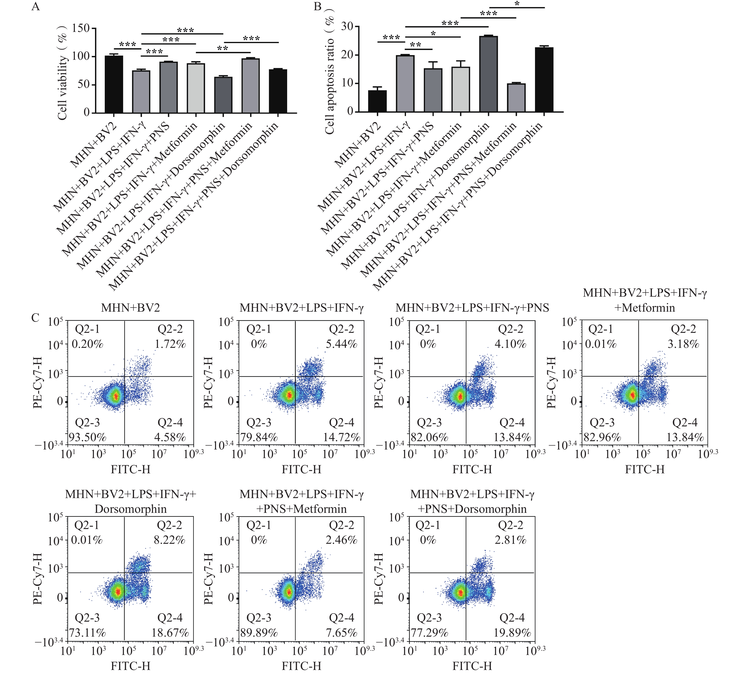
目的 探讨三七总皂苷(panax notoginseng saponins,PNS)通过AMPK信号通路调节自噬介导神经病理性疼痛(neuropathic pain,NP)细胞模型中小胶质细胞极化及其对神经元细胞的作用。 方法 采用不同浓度(0、50、100、200、400 μg/mL)PNS、AMPK激活剂Metformin或抑制剂Dorsomorphin预处理BV2细胞,再联合LPS和IFN-γ处理24 h,构建NP细胞模型。通过流式细胞术、JC-1染色、DCFH-DA荧光探针、Western blot及ELISA法,检测PNS对BV2细胞极化的影响及对AMPK/mTOR信号通路的调控作用。将小鼠海马神经元(mouse hippocampal neurons,MHN)与经上述处理的BV2细胞共培养,采用CCK-8法和Annexin V-FITC/PI凋亡检测试剂盒评估BV2细胞极化对MHN细胞活性及凋亡的影响。 结果 LPS和IFN-γ处理可增加BV2细胞中的INOS细胞比例(P < 0.001),上调TNF-α、IL-1β水平(P < 0.001),抑制CD206细胞比例(P < 0.001)、IL-10和IL-4浓度(P < 0.001),降低线粒体膜电位(P < 0.001),增加ROS水平和P62蛋白表达(P < 0.001),抑制LC3Ⅱ/Ⅰ表达(P < 0.001);同时,该处理抑制p-AMPK表达(P < 0.001),上调p-mTOR表达(P < 0.001),并降低共培养的MHN细胞活性(P < 0.001),增加细胞凋亡率(P < 0.001)。3-MA预处理可抑制细胞LC3Ⅱ/Ⅰ表达(P < 0.05),上调P62蛋白表达(P < 0.01);经PNS或Metformin预处理可促进p-AMPK表达(P < 0.01),抑制p-mTOR表达(P < 0.001),降低LPS和IFN-γ诱导的BV2细胞中INOS细胞比例和TNF-α、IL-1β水平(P < 0.001),促进CD206细胞比例、IL-10和IL-4浓度(P < 0.001);增加BV2细胞线粒体膜电位和自噬(P < 0.001),降低ROS水平(P < 0.01),增加共培养MHN细胞的活性并减少凋亡(P < 0.05)。Dorsomorphin预处理则进一步增强LPS和IFN-γ对BV2细胞中INOS比例、TNF-α、IL-1β水平的促进作用(P < 0.05),抑制CD206细胞比例、IL-10和IL-4水平(P < 0.05);降低细胞线粒体膜电位并促进ROS生成(P < 0.01),降低共培养MHN细胞的活性并加剧凋亡(P < 0.05)。PNS与Metformin联合预处理可增强Metformin对BV2及MHN细胞的保护作用(P < 0.001);而PNS与Dorsomorphin联合预处理则可部分逆转Dorsomorphin的损伤效应(P < 0.05)。 结论 PNS通过激活AMPK并抑制mTOR信号通路,抑制LPS和IFN-γ诱导的BV2细胞M1极化,缓解细胞线粒体损伤,并增强MHN细胞活性、减少其凋亡。 Abstract:Objective To investigate the effects of Panax notoginseng saponins (PNS) on microglial polarization and its effects on neuronal cells in a neuropathic pain (NP) cell model through autophagy regulation mediated by the AMPK signaling pathway. Methods BV2 cells were pretreated with PNS at different concentrations (0, 50, 100, 200, and 400 μg/mL), the AMPK activator metformin, or the AMPK inhibitor dorsomorphin, followed by co-stimulation with LPS and IFN-γ for 24 h to establish an NP cell model. Flow cytometry, JC-1 staining, DCFH-DA fluorescent probe, Western blot, and ELISA were employed to assess the effects of PNS on BV2 cell polarization and its modulation of the AMPK/mTOR signaling pathway. Mouse hippocampal neurons (MHN) were co-cultured with BV2 cells subjected to the above treatments. The effects of BV2 cell polarization on MHN cell viability and apoptosis were evaluated using the CCK-8 assay and the Annexin V-FITC/PI apoptosis detection kit. Results LPS and IFN-γ treatment increased the proportion of INOS-positive BV2 cells (P < 0.001), increased TNF-α and IL-1β levels (P < 0.001), decreased the proportion of CD206-positive cells (P < 0.001) and the concentrations of IL-10 and IL-4 (P < 0.001), reduced mitochondrial membrane potential (P < 0.001), increased ROS levels and P62 protein expression (P < 0.001), and inhibited LC3Ⅱ/Ⅰ expression (P < 0.001). Meanwhile, this treatment inhibited p-AMPK expression (P<0.001), increased p-mTOR expression (P < 0.001), decreased the viability of cocultured MHNs (P < 0.001), and increased their apoptosis rate (P < 0.001). Pretreatment with 3-MA inhibited LC3 II/I expression (P < 0.05) and increased P62 protein expression (P < 0.01). Pretreatment with PNS or metformin promoted p-AMPK expression (P < 0.01), inhibited p-mTOR expression (P < 0.001), decreased the proportion of iNOS-positive BV2 cells and the levels of TNF-α and IL-1β induced by LPS and IFN-γ (P < 0.001), and increased the proportion of CD206-positive cells and the concentrations of IL-10 and IL-4 (P < 0.001). It also increased mitochondrial membrane potential and autophagy in BV2 cells (P < 0.001), reduced ROS levels (P < 0.01), increased cocultured MHN viability, and reduced apoptosis (P < 0.05). Pretreatment with dorsomorphin further enhanced the effects of LPS and IFN-γ on increasing the proportion of iNOS-positive BV2 cells and the levels of TNF-α and IL-1β (P < 0.05), while reducing the proportion of CD206-positive cells and the levels of IL-10 and IL-4 (P < 0.05). It also decreased mitochondrial membrane potential, promoted ROS generation (P < 0.01), reduced cocultured MHN viability, and aggravated apoptosis (P < 0.05). Combined pretreatment with PNS and metformin enhanced the protective effects of metformin on BV2 and MHN cells (P < 0.001), whereas combined pretreatment with PNS and dorsomorphin partially reversed the deleterious effects of dorsomorphin (P < 0.05). Conclusion PNS inhibits LPS/IFN-γ-induced M1 polarization of BV2 cells, alleviates mitochondrial injury, enhances MHN cell viability, and reduces their apoptosis by activating AMPK and inhibiting the mTOR signaling pathway. -
Key words:
- Microglia /
- Autophagy /
- Panax notoginseng saponins /
- AMPK signaling pathway /
- Neurons
-
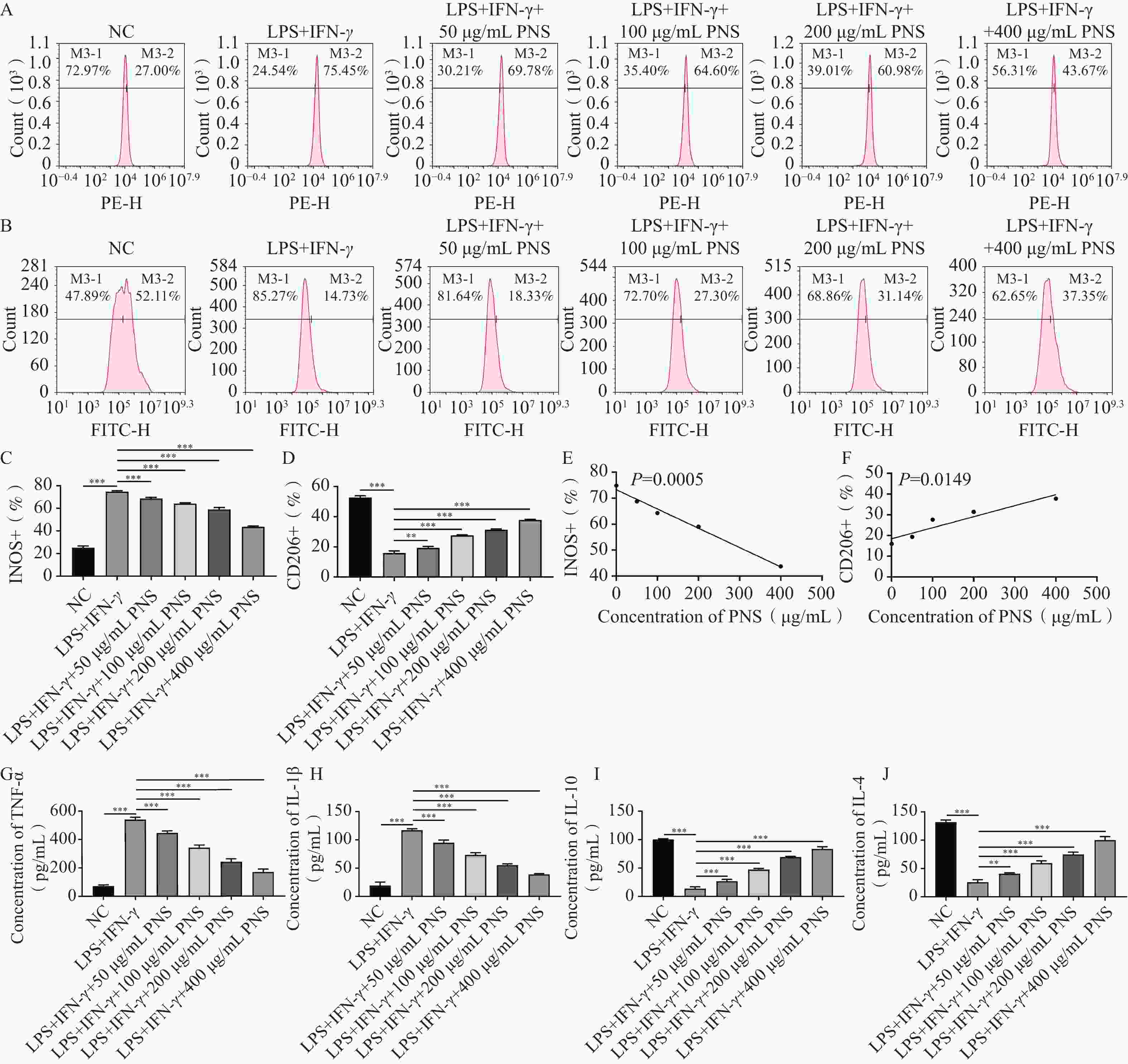
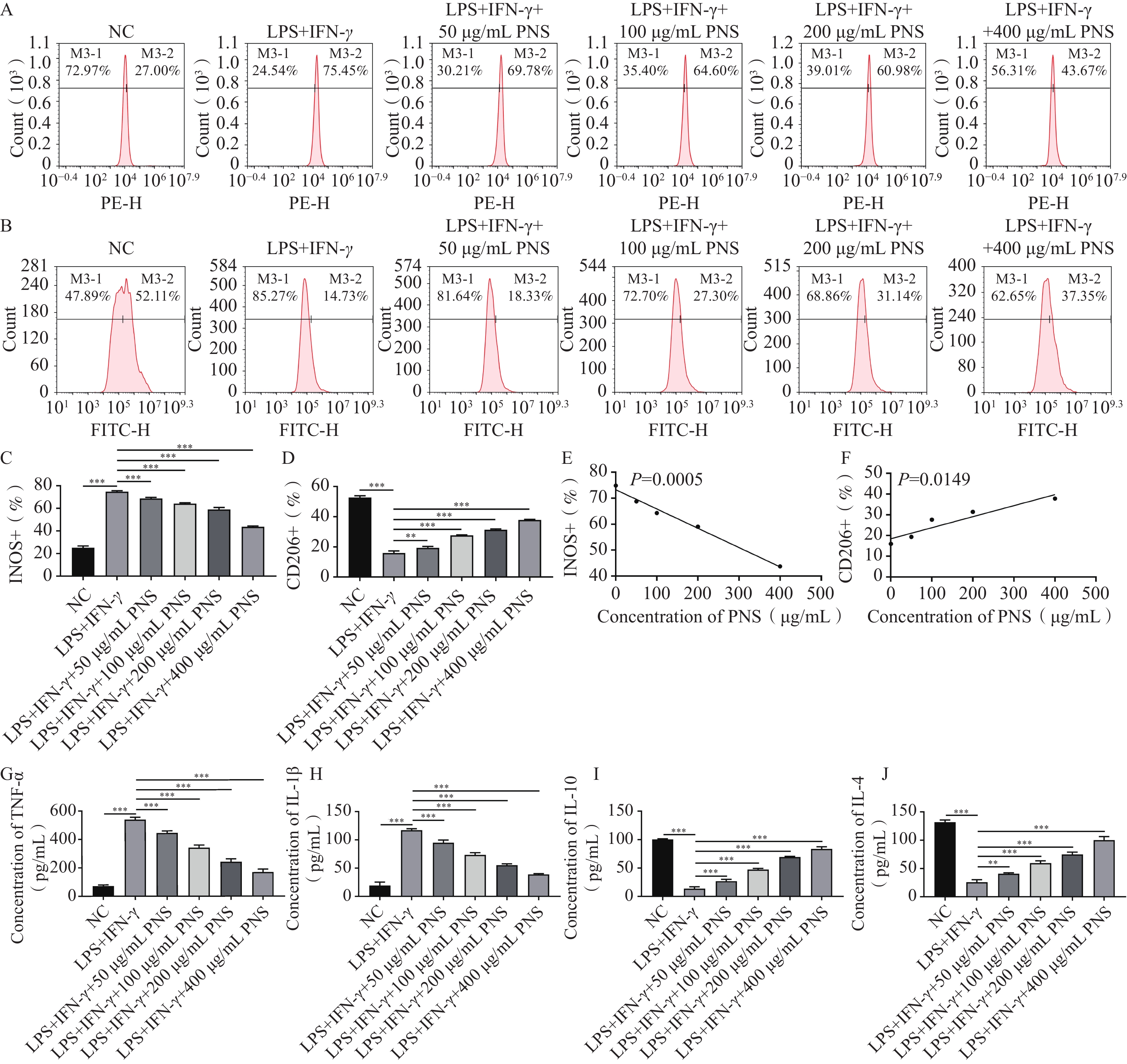
图 1 PNS抑制LPS和IFN-γ诱导BV2小胶质细胞M1极化($\bar x \pm s $,n = 3)
A~D:流式细胞术检测BV2细胞中的INOS和CD206阳性率;E~F:PNS浓度与INOS和CD206的线性相关性分析;G~J:ELISA试剂盒检测细胞培养上清液中的TNF-α、IL-1β、IL-10和IL-4浓度;**P < 0.01;***P < 0.001。
Figure 1. PNS inhibits LPS- and IFN-γ-induced M1 polarization of BV2 microglia ($\bar x \pm s $,n = 3)
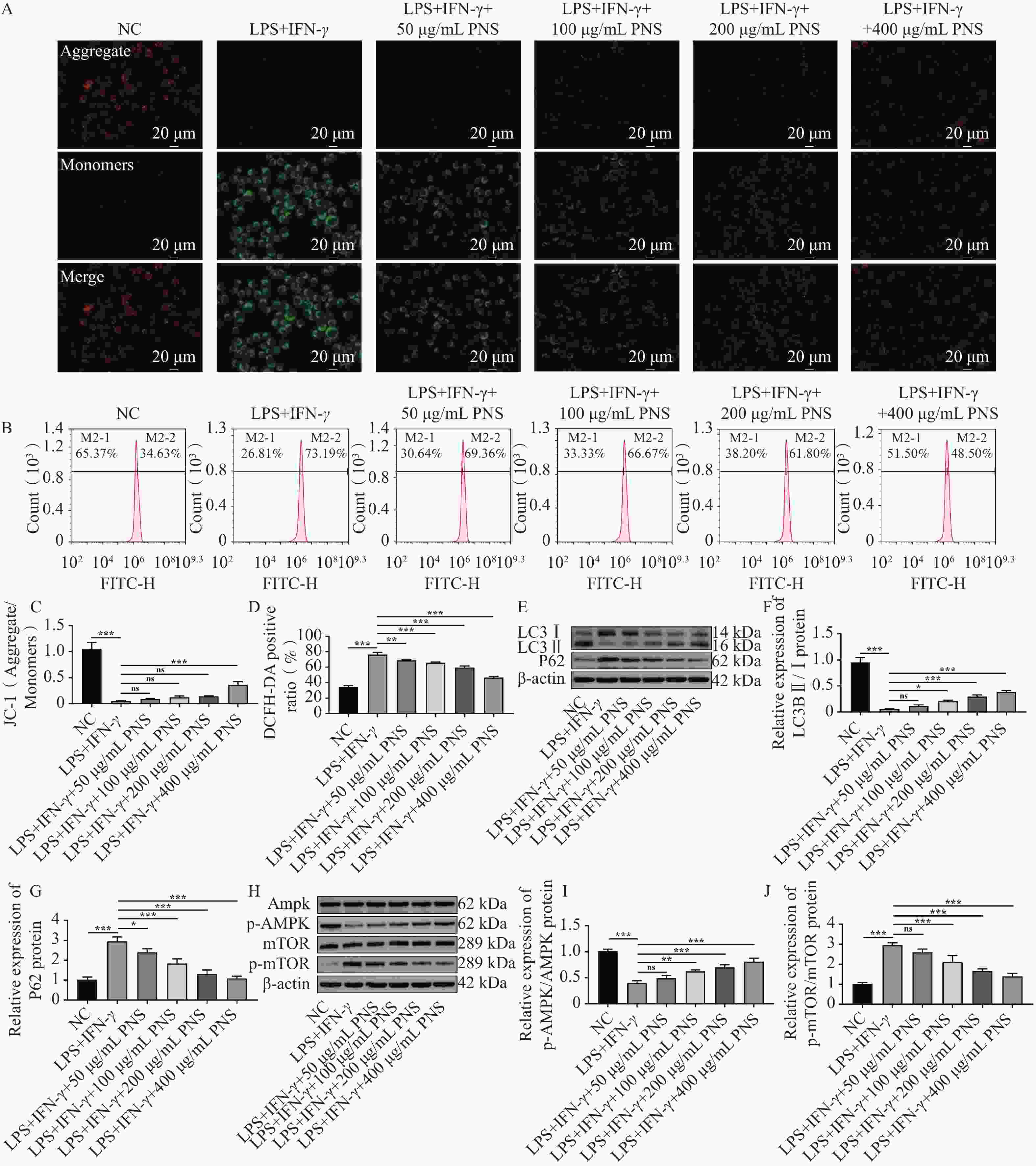
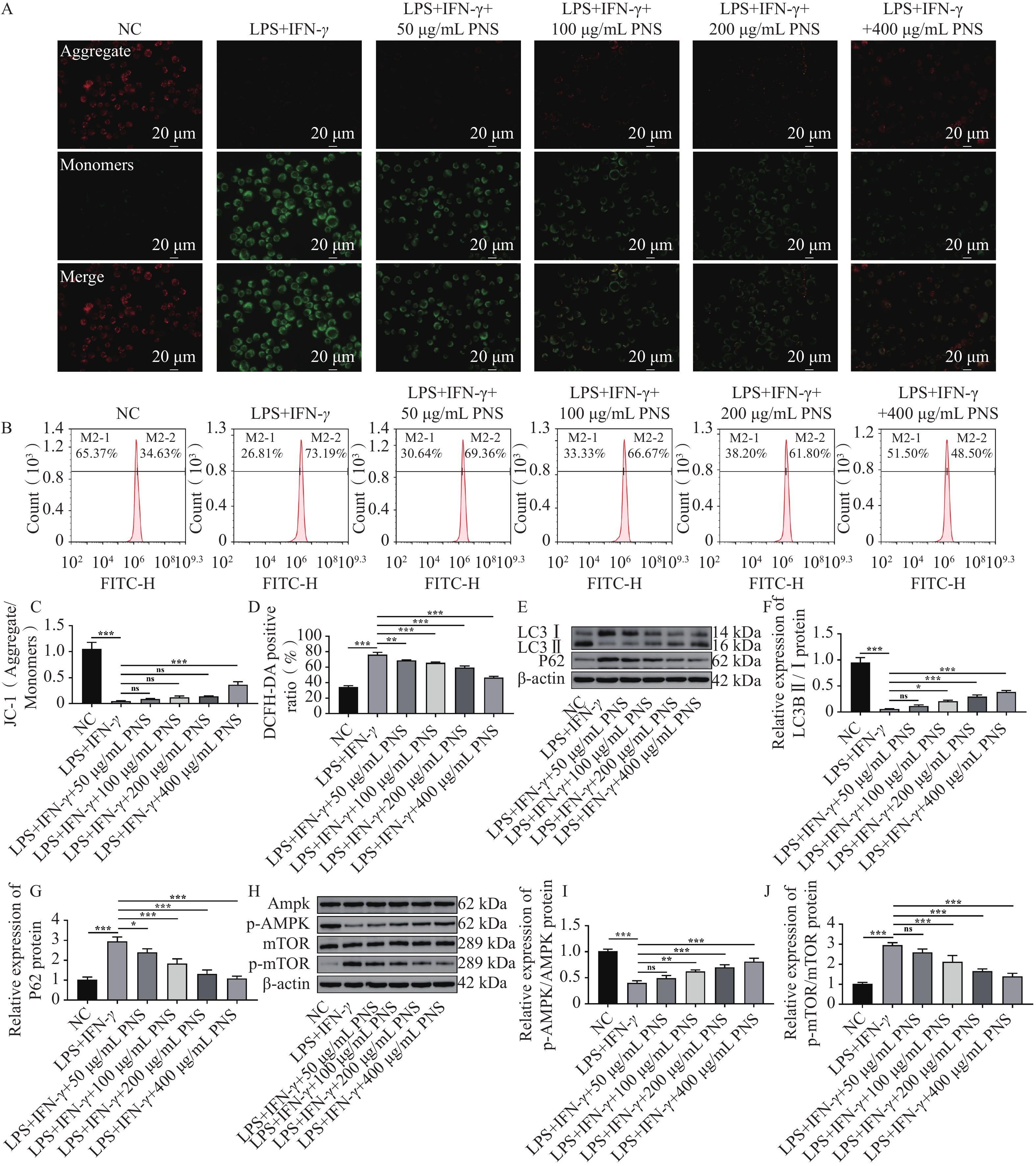
图 2 PNS抑制LPS和IFN-γ诱导的BV2小胶质细胞线粒体损伤并增强自噬($\bar x \pm s $,n = 3)
A:BV2细胞JC-1染色代表性图像;B:流式细胞术检测细胞中的ROS水平;C:JC-1染色统计结果;D:ROS水平统计结果;E~G:Western blot检测细胞中的LC3B Ⅰ/Ⅱ和P62蛋白表达;H~J:Western blot检测细胞中的AMPK、mTOR蛋白及其磷酸化水平;*P < 0.05;**P < 0.01;***P < 0.001;ns:P > 0.05。
Figure 2. PNS inhibits LPS- and IFN-γ-induced mitochondrial damage and enhances autophagy in BV2 cells ($\bar x \pm s $,n = 3)
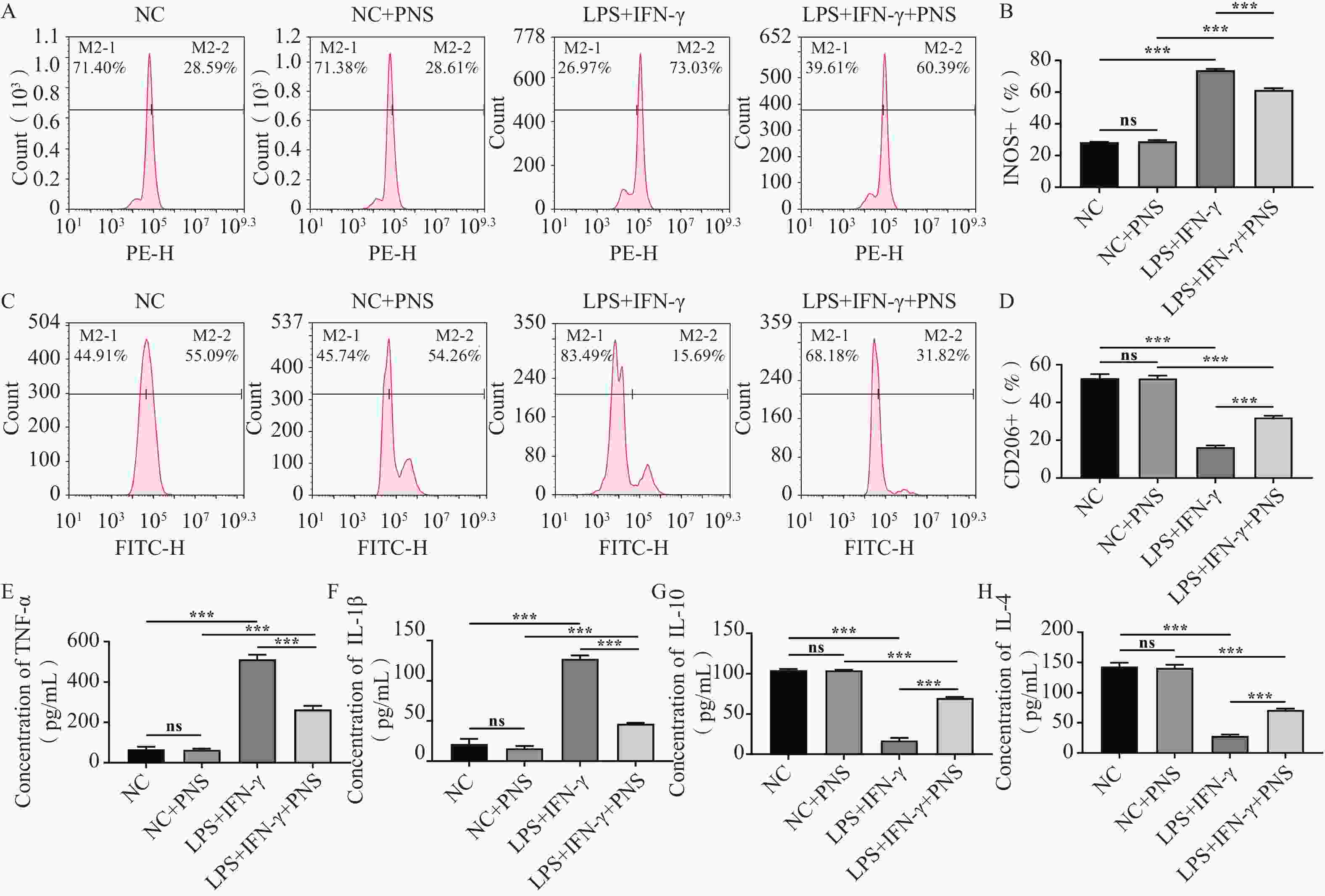
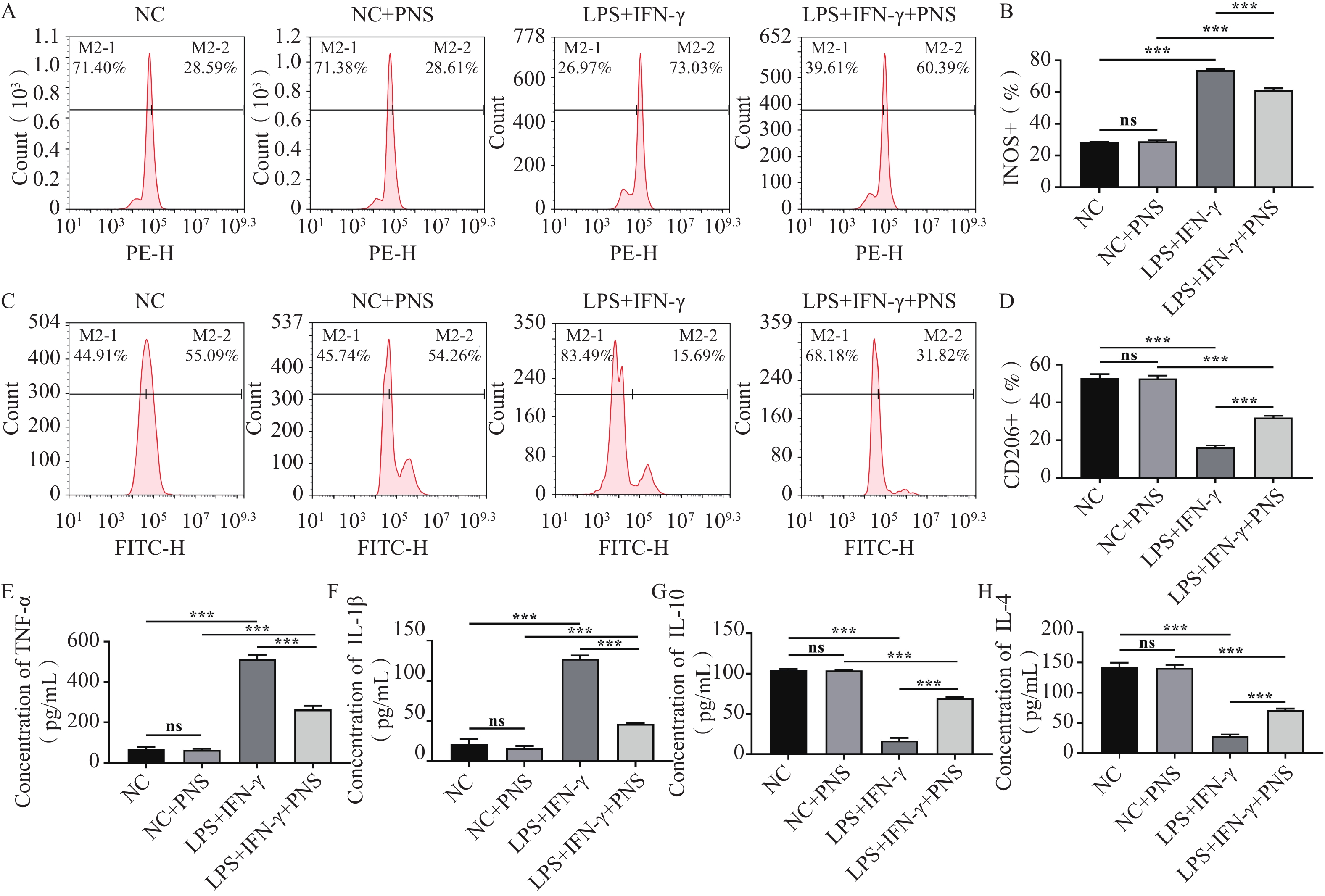
图 3 PNS对未激活的BV2细胞无明显作用($\bar x \pm s $,n = 3)
A~D:流式细胞术检测INOS和CD206阳性细胞率;E~H:ELISA试剂盒检测细胞上清液中的TNF-α、IL-1β、IL-10和IL-4水平;***P < 0.001;ns:P > 0.05。
Figure 3. PNS has no significant effect on unactivated BV2 cells ($\bar x \pm s $,n = 3)
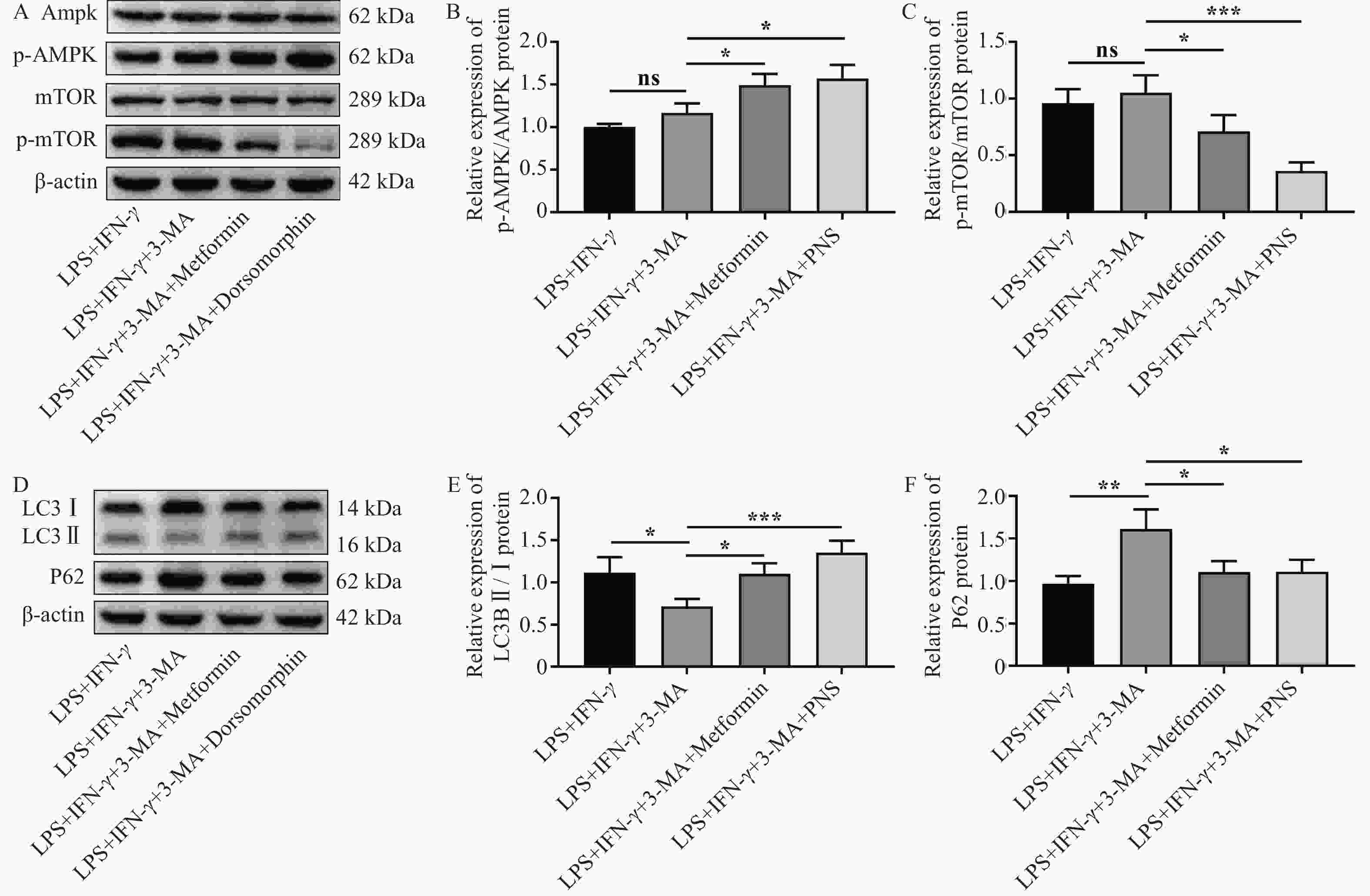
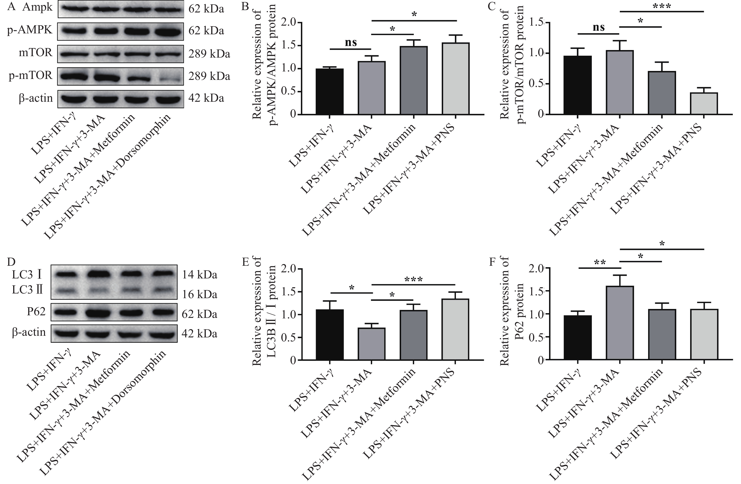
图 4 PNS通过激活AMPK/mTOR信号通路促进BV2细胞自噬($\bar x \pm s $,n = 3)
A~C:Western blot检测BV2细胞中AMPK、p-AMPK、mTOR、p-mTOR蛋白相对表达;D~F:Western blot检测BV2细胞中LC3BⅠ、LC3BⅡ和P62蛋白相对表达;*P < 0.05;**P < 0.01;***P < 0.001;ns:P > 0.05。
Figure 4. PNS promotes autophagy in BV2 cells by activating the AMPK/mTOR signaling pathway ($\bar x \pm s $,n = 3)
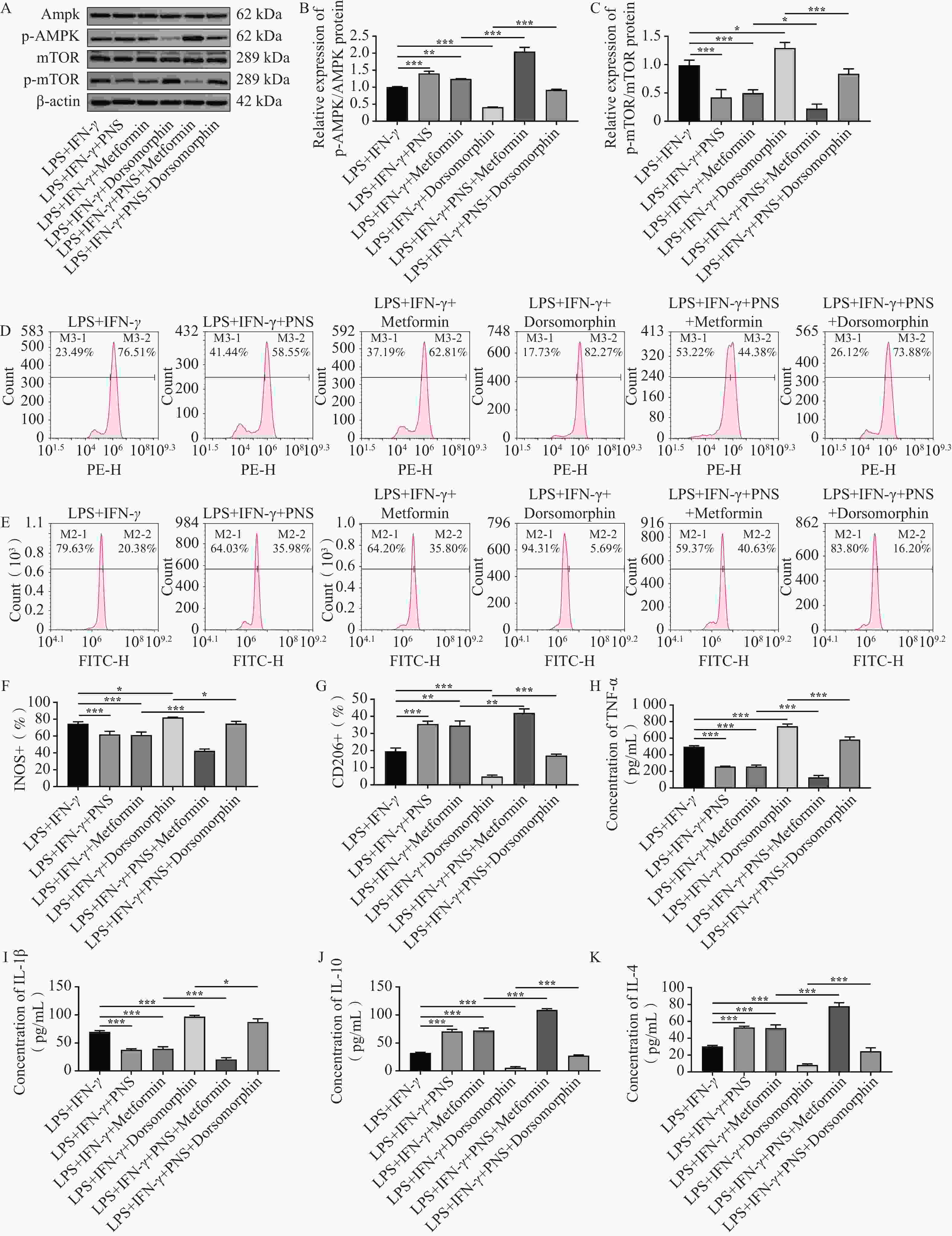
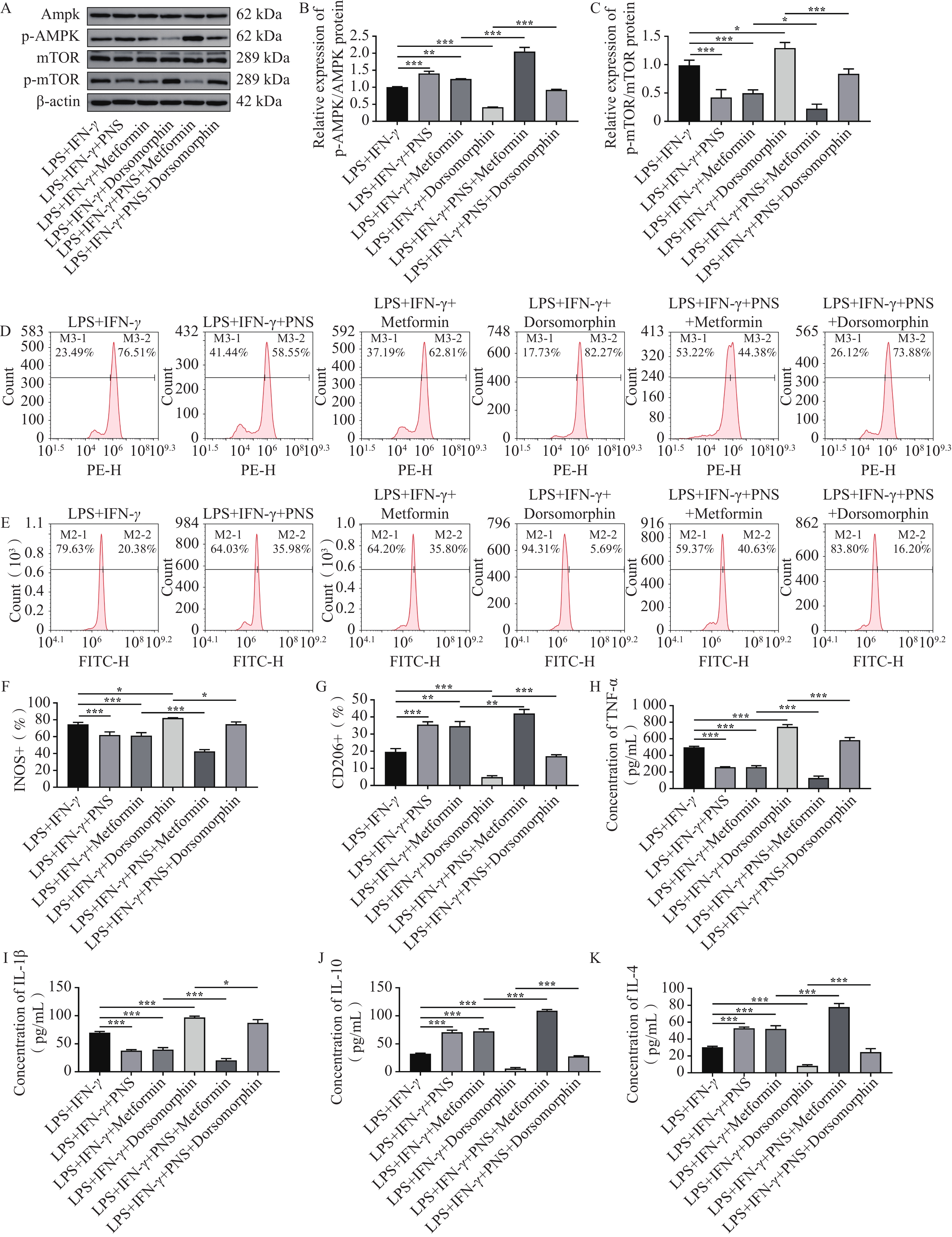
图 5 PNS激活AMPK通路促进BV2细胞M2极化($\bar x \pm s $,n = 3)
A~C:Western blot检测AMPK和mTOR蛋白及其磷酸化水平的相对表达;D~G:流式细胞术检测细胞中INOS和CD206阳性细胞比例;H~K:ELISA检测细胞培养上清中TNF-α、IL-1β、IL-10和IL-4浓度;*P < 0.05;**P < 0.01;***P < 0.001。
Figure 5. PNS activates the AMPK pathway to promote M2 polarization of BV2 cells ($\bar x \pm s $,n = 3)
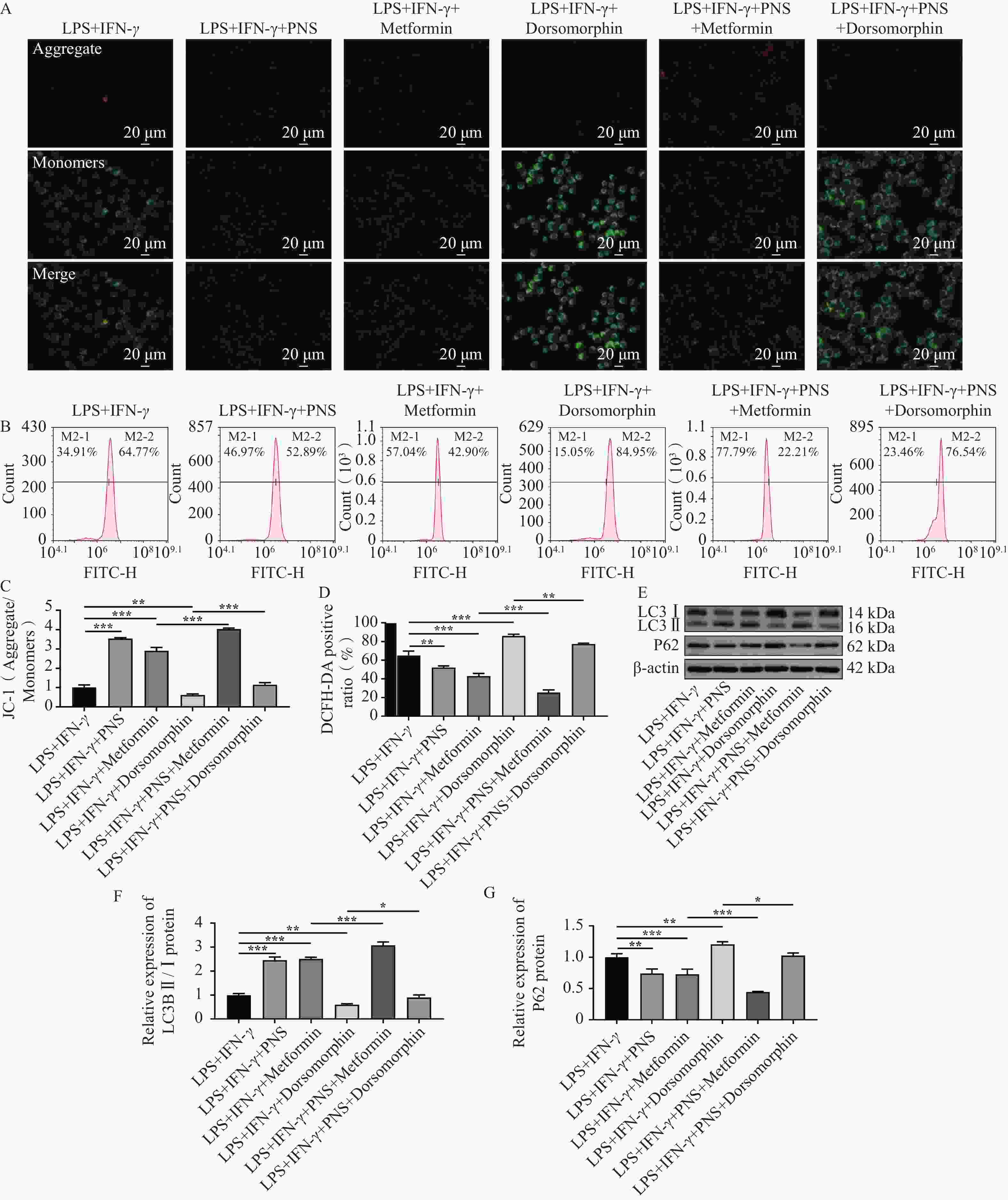
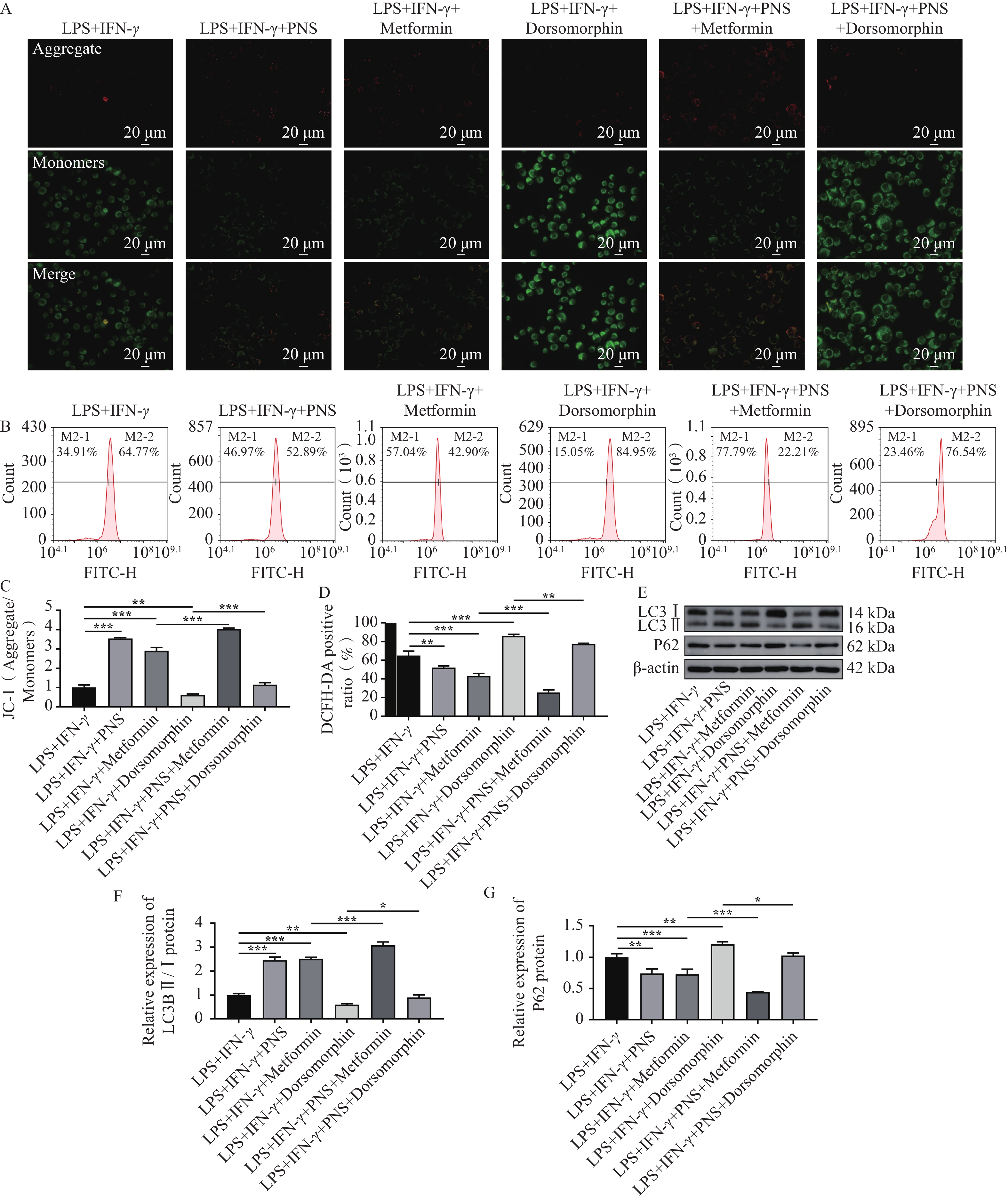
图 6 PNS通过AMPK/mTOR通路调控BV2细胞线粒体损伤和自噬($\bar x \pm s $,n = 3)
A:JC-1染色检测BV2细胞线粒体膜电位代表性图像;B:DCFH-DA荧光探针法检测细胞中的ROS水平;C:JC-1染色结果统计;D:ROS阳性细胞统计;E~G:Western blot检测细胞中自噬相关蛋白LC3B Ⅰ/Ⅱ和P62表达;*P < 0.05;**P < 0.01;***P < 0.001。
Figure 6. PNS regulates mitochondrial damage and autophagy in BV2 cells through the AMPK/mTOR pathway ($\bar x \pm s $,n = 3)
-
[1] Kaye A D, Armistead G, Amedio L S, et al. Evolving treatment strategies for neuropathic pain: A narrative review[J]. Medicina, 2025, 61(6): 1063. doi: 10.3390/medicina61061063 [2] Hua T, Kong E, Zhang H, et al. PRMT6 deficiency or inhibition alleviates neuropathic pain by decreasing glycolysis and inflammation in microglia[J]. Brain Behav Immun, 2024, 118: 101-114. doi: 10.1016/j.bbi.2024.02.027 [3] Kuffler D P. Techniques and factors for reducing chronic neuropathic pain: A review[J]. Neural Regen Res, 2026, 21(4): 1353-1358. doi: 10.4103/NRR.NRR-D-22-00015 [4] Chen L, Zeng L, Li W, et al. The activation of hippocampal microglial cells and their role in the regulation of pain[J]. J Integr Neurosci, 2025, 24(6): 27730. doi: 10.31083/JIN27730 [5] Zhang T, Liang W, Gao J, et al. Microglia polarization: A key regulatory mechanism of neuropathic pain[J]. J Pain Res, 2026, 19: 557153. [6] Wang G, Chen Z, Song Y, et al. Xueshuantong injection alleviates cerebral microcirculation disorder in middle cerebral artery occlusion/reperfusion rats by suppressing inflammation via JNK mediated JAK2/STAT3 and NF-κB signaling pathways[J]. J Ethnopharmacol, 2022, 298: 115592. doi: 10.1016/j.jep.2022.115592 [7] Wang X, Zhang R, Zeng N, et al. Panax notoginseng saponins dually modulates autophagy in gastric precancerous lesions complicated with myocardial ischemia-reperfusion injury model through the PI3K/AKT/mTOR pathway[J]. Biomed Pharmacother, 2024, 178: 117268. doi: 10.1016/j.biopha.2024.117268 [8] Li W, Shi H, Wu X. A narrative review of Panax notoginseng: Unique saponins and their pharmacological activities[J]. J Ginseng Res, 2025, 49(2): 118-133. doi: 10.1016/j.jgr.2024.12.005 [9] Al-Musailem M, Masocha W, Al-Romaiyan A. Notoginsenoside R1, a metabolite from Panax notoginseng, prevents paclitaxel-induced peripheral neuropathic pain in mice[J]. Molecules, 2025, 30(17): 3613. doi: 10.3390/molecules30173613 [10] Li Y, Zhou Y, Liu D, et al. Glutathione Peroxidase 3 induced mitochondria-mediated apoptosis via AMPK/ERK1/2 pathway and resisted autophagy-related ferroptosis via AMPK/mTOR pathway in hyperplastic prostate[J]. J Transl Med, 2023, 21(1): 575. doi: 10.1186/s12967-023-04432-9 [11] Li B, Yu K, Zhou X, et al. Increased TSPO alleviates neuropathic pain by preventing pyroptosis via the AMPK-PGC-1α pathway[J]. J Headache Pain, 2025, 26(1): 16. doi: 10.1186/s10194-025-01953-0 [12] Zhang Y, Song Y, Wang C, et al. Panax notoginseng saponin R1 attenuates allergic rhinitis through AMPK/Drp1 mediated mitochondrial fission[J]. Biochem Pharmacol, 2022, 202: 115106. doi: 10.1016/j.bcp.2022.115106 [13] Tang K, Qin W, Wei R, et al. Ginsenoside Rd ameliorates high glucose-induced retinal endothelial injury through AMPK-STRT1 interdependence[J]. Pharmacol Res, 2022, 179: 106123. doi: 10.1016/j.phrs.2022.106123 [14] Pan F, Lu Y, Yang H. Panax notoginseng saponins treat steroid-resistant lupus nephritis by inhibiting macrophage-derived exosome-induced injury in glomerular endothelial cells via the mitochondrial Autophagy-NLRP3 pathway[J]. J Ethnopharmacol, 2025, 343: 119475. doi: 10.1016/j.jep.2025.119475 [15] Li R, Ma J, Li M, et al. Panax notoginseng saponins ameliorate high-fat diet-induced liver injury via mechanisms involving TLR4-mediated signaling and lipid metabolism[J]. Food Sci Nutr, 2026, 14(2): e71544. doi: 10.1002/fsn3.71544 [16] Wu Y Q, Xiong J, He Z L, et al. Metformin promotes microglial cells to facilitate myelin debris clearance and accelerate nerve repairment after spinal cord injury[J]. Acta Pharmacol Sin, 2022, 43(6): 1360-1371. doi: 10.1038/s41401-021-00759-5 [17] Zhao P, Li X, Yang Q, et al. Malvidin alleviates mitochondrial dysfunction and ROS accumulation through activating AMPK-α/UCP2 axis, thereby resisting inflammation and apoptosis in SAE mice[J]. Front Pharmacol, 2023, 13: 1038802. doi: 10.3389/fphar.2022.1038802 [18] Li Y, Yin C, Jiang J, et al. Tumor necrosis factor α-induced protein 8-like-2 controls microglia phenotype via metabolic reprogramming in BV2 microglial cells and responses to neuropathic pain[J]. Int J Biochem Cell Biol, 2024, 169: 106541. doi: 10.1016/j.biocel.2024.106541 [19] Atta A A, Ibrahim W W, Mohamed A F, et al. Microglia polarization in nociplastic pain: Mechanisms and perspectives[J]. Inflammopharmacology, 2023, 31(3): 1053-1067. doi: 10.1007/s10787-023-01216-x [20] Sun T, Wang P, Deng T, et al. Effect of Panax notoginseng saponins on focal cerebral ischemia-reperfusion in rat models: A meta-analysis[J]. Front Pharmacol, 2021, 11: 572304. doi: 10.3389/fphar.2020.572304 [21] Zhang X, Zhang B, Zhang C, et al. Effect of Panax notoginseng saponins and major anti-obesity components on weight loss[J]. Front Pharmacol, 2021, 11: 601751. doi: 10.3389/fphar.2020.601751 [22] Liu Y, Niu P, Ji H, et al. The use of Panax notoginseng saponins injections after intravenous thrombolysis in acute ischemic stroke: A systematic review and meta-analysis[J]. Front Pharmacol, 2024, 15: 1376025. doi: 10.3389/fphar.2024.1376025 [23] Ma X, Zhang X, Kong Y, et al. Therapeutic effects of Panax notoginseng saponins in rheumatoid arthritis: Network pharmacology and experimental validation[J]. Bioengineered, 2022, 13(6): 14438-14449. doi: 10.1080/21655979.2026.2667590 [24] Gao J, Yao M, Zhang W, et al. Panax notoginseng saponins alleviates inflammation induced by microglial activation and protects against ischemic brain injury via inhibiting HIF-1α/PKM2/STAT3 signaling[J]. Biomed Pharmacother, 2022, 155: 113479. doi: 10.1016/j.biopha.2022.113479 [25] Fan L, Qu S, Miao C, et al. Panax notoginseng saponins protect the blood-brain barrier against oxidative stress by activating the Akap12-PI3K/AKT-AQP1 signaling axis[J]. Int J Biol Macromol, 2026, 353: 151045. doi: 10.1016/j.ijbiomac.2026.151045 [26] Wu H, Ni C, Zhang Y, et al. Stem-leaf saponins of Panax notoginseng attenuate experimental Parkinson’s disease progression in mice by inhibiting microglia-mediated neuroinflammation via P2Y2R/PI3K/AKT/NFκB signaling pathway[J]. Chin J Nat Med, 2025, 23(1): 43-53. [27] Zhou Z, He M, Zhao Q, et al. Panax notoginseng saponins attenuate neuroinflammation through TXNIP-mediated NLRP3 inflammasome activation in aging rats[J]. Curr Pharm Biotechnol, 2021, 22(10): 1369-1379. [28] Liu W, Zhao Y, Wang G, et al. TRIM22 inhibits osteosarcoma progression through destabilizing NRF2 and thus activation of ROS/AMPK/mTOR/autophagy signaling[J]. Redox Biol, 2022, 53: 102344. doi: 10.1016/j.redox.2022.102344 [29] Liang X, Zhang J, Yu J, et al. Quercetin ameliorates ox-LDL-induced cellular senescence of aortic endothelial cells and macrophages by p16/p21, p53/SERPINE1, and AMPK/mTOR pathways[J]. Eur J Med Res, 2025, 30(1): 359. doi: 10.1186/s40001-025-02562-y [30] Shen Q, Ma J, Zhang Y, et al. Platelet-rich plasma combined with a low-frequency pulsed electromagnetic field alleviates sciatic nerve injury by regulating the AMPK/mTOR signaling pathway[J]. Photobiomodul Photomed Laser Surg, 2025, 43(8): 354-362. [31] Zhang N, Ji Q, Chen Y, et al. TREM2 deficiency impairs the energy metabolism of Schwann cells and exacerbates peripheral neurological deficits[J]. Cell Death Dis, 2024, 15(3): 193. -

 下载:
下载:
 点击查看大图
计量
- 文章访问数: 11
- HTML全文浏览量: 4
- PDF下载量: 0
- 被引次数: 0
