CBCT Imaging Study of the Correlation between the Anatomy of Greater Palatine Foramen and the Morphology of Palatal Vault in Adults
-
摘要:
目的 研究成人腭大孔( greater palatine foramen,GPF )解剖位置及其与腭穹窿高低的相关性,为涉及上腭区域的临床操作提供理论依据。 方法 从昆明医科大学附属口腔医院影像科收集2020年1~6月符合纳入标准的180名成人患者的CBCT图像,评估双侧360例腭大孔相对于上颌磨牙的位置以及距上颌腭中缝线和釉牙骨质界的距离,并分析不同腭穹窿高低与腭大孔分布的关联。 结果 腭大孔在第2磨牙腭侧、第3磨牙近中腭侧、第3磨牙腭侧的分布率分别是21.39%、21.11%和57.50%。男性和女性腭大孔与腭中缝( midsagittal suture,MMS )的距离(GPF-MMS)分别为(16.31±1.18) mm和(15.82±1.32) mm,腭大孔至对应磨牙釉牙骨质界(enamelo-cemental junction,CEJ )的距离(GPF-M)为(17.11±2.50) mm 和(15.79±2.57) mm,均显示男性大于女性(P < 0.05)。GPF-MMS在高腭组中略低于低腭组(P < 0.05);然而,GPF-M在高腭组中明显高于低腭组(P < 0.05)。 结论 成人腭大孔位置与其性别和腭穹隆高低有关。 Abstract:Objective To explore the greater palatine foramen anatomical position and its relationship with the morphology of palatal vault in adults, so as to provide theoretical basis for the clinic operation related to palate. Methods We collected CBCT images of 180 adult patients, marked both 360 sides greater palatine foramen location, analyzed the relative position of greater palatine foramen to maxillary molar and the distance between the longitudinal suture of palate and longitudinal suture of palate and checked the relationship between different morphology of palatal vault and the distribution of greater palatine foramen. Results The distribution rates of the greater palatine foramen at the palatal side of second molar, third molar mesial side and palatal side of third molar were 21.39%, 21.11% and 57.50% respectively. The (GPF-MMS) distances of male and female were (16.31±1.18) mm and (15.82±1.32) mm, and the distance from the palatal foramen to the corresponding enamel cementum boundary (GPF-M) was (17.11±2.50) mm and (15.79±2.57) mm, All showed that male was larger than female (P < 0.05). The distance of GPF-MMS in high palate group was lower than that in low palate group; On the contrary (P < 0.05), GPF-M in high palate group was significantly higher than that in low palate group (P < 0.05). Conclusion The position of the greater palatine foramen in adults patients is related to gender and the palatal vault arch. -
Key words:
- Cone beam computed tomography /
- Palatal vault /
- Greater palatine foramen
-
表 1 纳入影像资料的年龄统计 (
$\bar x \pm s $ )Table 1. Age statistics of included image data (
$\bar x \pm s $ )性别 n 年龄范围(岁) 年龄(岁) t P 男性 85 18~50 30.20 ± 7.82 1.316 0.192 女性 95 18~56 27.92 ± 7.62 总体 180 18~56 29.00 ± 7.78 表 2 腭大孔相对于磨牙的分布 [n(%)]
Table 2. Distribution of the great palatal foramen relative to the molar [n(%)]
项目 M2腭侧 M3近中 M3腭侧 男 左(n) 13 19 53 右(n) 16 17 52 合计 29(17.06) 36(21.18) 105(61.76) 女 左(n) 26 18 51 右(n) 22 22 51 合计 48(25.26) 40(21.05) 102(53.68) 总体 左(n) 39 37 104 右(n) 38 39 103 合计 77(21.39) 76(21.11) 207(57.50) 表 3 腭穹隆形态 [(
$\bar x \pm s $ ),mm]Table 3. Morphology of palatal vault [(
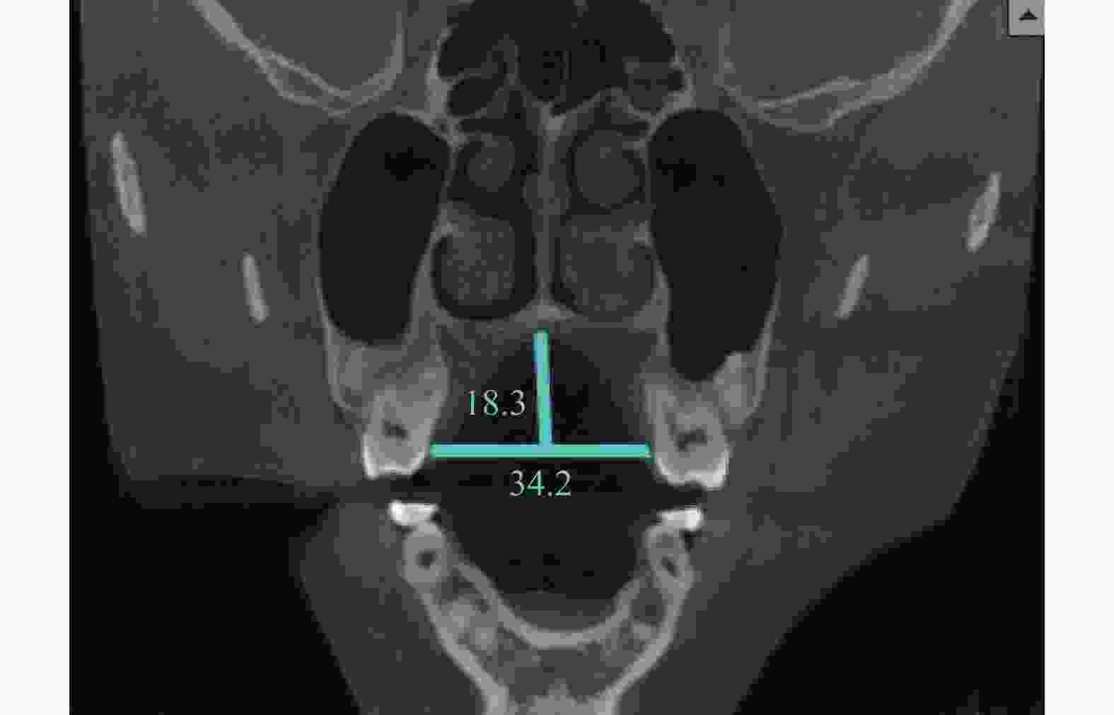
$\bar x \pm s$ ),mm]项目 男 女 t P 高度 15.42 ± 2.67 14.42 ± 2.22 2.089 0.045* 宽度 37.74 ± 2.68 36.35 ± 2.58 3.023 0.03* 高度:宽度 0.41 ± 0.07 0.40 ± 0.06 0.279 0.781 *P < 0.05。 表 4 腭大孔与腭中缝及磨牙CEJ的距离 [(
$\bar x \pm s $ ),mm]Table 4. Distance between greater palatal foramen and midpalatal suture and molar CEJ [(
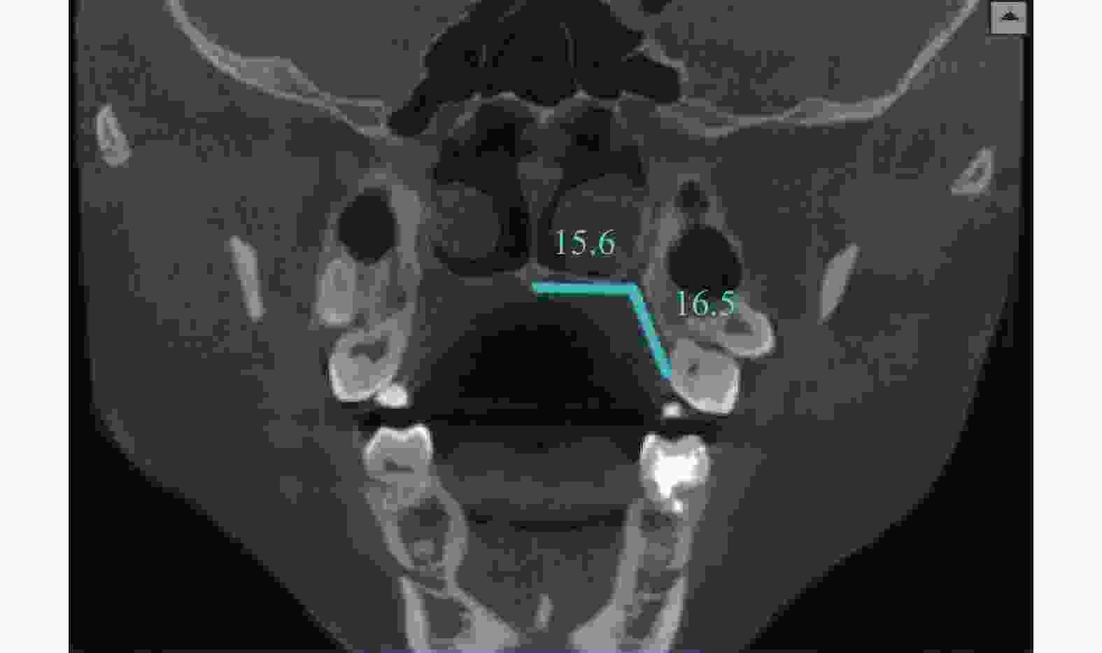
$\bar x \pm s $ ),mm]项目 位置 男性 女性 t P GPF-MSS(mm) 左 16.34 ± 1.12 15.86 ± 1.30 右 16.29 ± 1.14 15.78 ± 1.35 合计 16.31 ± 1.18 15.82 ± 1.32* 3.558 < 0.001* GPF-M(mm) 左 17.12 ± 2.55 15.81 ± 2.65 右 17.10 ± 2.46 15.76 ± 2.50 合计 17.11 ± 2.50 15.79 ± 2.57* 3.482 0.001* MSS/M 左 0.98 ± 0.17 1.03 ± 0.19 右 0.97 ± 0.17 1.03 ± 0.18 合计 0.98 ± 0.17 1.03 ± 0.19* −2.807 0.04* *P < 0.05。 表 5 高、低腭组腭大孔与腭中缝及磨牙CEJ的距离 [(
$\bar x \pm s $ ),mm]Table 5. The distance between the greater palatal foramen and the midpalatal suture and the CEJ of molars in the high and low palatal groups [(
$\bar x \pm s $ ),mm]项目 组别 男 女 GPF-MSS(mm) 高腭组 16.19 ± 1.11 15.77 ± 1.37* 低腭组 16.44 ± 1.06● 15.88 ± 1.25●* GPF-M(mm) 高腭组 17.90 ± 2.28 16.89 ± 2.20* 低腭组 16.22 ± 2.45● 14.94 ± 2.51●* MSS/M 高腭组 0.92 ± 0.15 0.94 ± 0.13* 低腭组 1.04 ± 0.17● 1.08 ± 0.20●* 与男性相比,*P < 0.05;与高腭组相比,●P < 0.05。 -
[1] Cagimni P,Govsa F,Ozer M A,et al. Computerized analysis of the greater palatine foramen to gain the palatine neurovascular bundle during palatal surgery[J]. Surgical and Radiologic Anatomy,2017,39(2):177-184. doi: 10.1007/s00276-016-1691-0 [2] 满毅,Huangphattarakul V. 腭部作为口腔软组织供区的实践要点[J]. 口腔颌面外科杂志,2020,30(5):265-271. doi: 10.3969/j.issn.1005-4979.2020.05.001 [3] Carrier S,Castagneyrol B,Beylacq L,et al. Anatomical landmarks for maxillary nerve block in the pterygopalatine fossa:A radiological study[J]. Journal of Stomatology Oral & Maxillofacial Surgery,2017,118(2):90-94. [4] 吴洁林,高莺. 硬腭获取游离软组织移植物的应用进展[J]. 国际口腔医学杂志,2020,47(6):686-692. doi: 10.7518/gjkq.2020087 [5] Costa H,Zenha H,Sequeira H,et al. Microsurgical reconstruction of the maxilla:Algorithm and concepts[J]. J Plast Reconstr Aesthet Surg,2015,68(5):e89-e104. doi: 10.1016/j.bjps.2014.12.002 [6] Hassanali J,Mwaniki D. Palatal analysis and osteology of the hard palate of the Kenyan African skulls[J]. Anat Rec,1984,209(2):273-280. doi: 10.1002/ar.1092090213 [7] Klosek S K, Rungruang T. Anatomical study of the greater palatine artery and related structures of the palatal vault: considerations for palate as the subepithelial connective tissue graft donor site. Surg Radiol Anat[J], 2009, 31(6): 245-250. [8] Arnold F,West D C. Angiogenesis in Wound Healing[J]. Pharmacol Ther,1991,52(3):407-422. doi: 10.1016/0163-7258(91)90034-J [9] Tomaszewska I M,Tomaszewski K A,Kmiotek E K,et al. Anatomical landmarks for the localization of the greater palatine foramen - a study of 1200 head CTs,150 dry skulls,systematic review of literature and meta-analysis[J]. Journal of Anatomy,2014,225(4):419-435. doi: 10.1111/joa.12221 [10] Damgaard C,Caspersen L M,Kjaer I. Maxillary sagittal growth evaluated on dry skulls from children and adolescents[J]. Acta Odontologica Scandinavica,2011,69(5):274-278. doi: 10.3109/00016357.2011.563243 [11] 肖剑峰,牛涛. 锥形束CT对鼻腭管与种植相关解剖结构的测量研究[J]. 昆明医科大学学报,2015,36(4):41-44. doi: 10.3969/j.issn.1003-4706.2015.04.011 [12] 沈晨露,高碧聪,吕柯佳. 浙江地区人群上颌腭侧咀嚼黏膜厚度与腭穹窿解剖形态的量化分析[J]. 浙江大学学报:医学版,2022,51(1):87-94. [13] 解危,唐月华. 腭大孔、翼腭管和翼上颌裂解剖学——观测与临床意义[J]. 昆明医科大学学报,1988,9(2):22-26. [14] 薛绯,段晋瑜,张瑞. 汉族人群腭大孔解剖位置及其与腭穹隆形态关系的CBCT研究[J]. 实用口腔医学杂志,2018,34(3):364-367. doi: 10.3969/j.issn.1001-3733.2018.03.016 [15] Bermúdez De Castro J M,Nicolas M E. Posterior dental size reduction in hominids:The Atapuerca evidence[J]. American Journal of Physical Anthropology,1995,96(4):335-356. doi: 10.1002/ajpa.1330960403 [16] Aoun G, Nasseh I, Saadeh M, et al. Analysis of the greater palatine foramen in a Lebanese population using cone-beam computed tomography technology[J]. Journal of International Society of Preventive & Community Dentistry, 2015, 5(Suppl 2): S82-S88. [17] 杨佳文,孙涛,和红兵. 腭大神经血管沟解剖位置的CBCT研究[J]. 昆明医科大学学报,2019,40(10):1-5. doi: 10.3969/j.issn.1003-4706.2019.10.002 -


 下载:
下载:


