Pelvic Floor Tthree-dimensional Ultrasound Evaluating Postpartum Pelvic Floor Dysfunction in Elderly Parturient Women
-
摘要:
目的 探析对比盆底二维,三维超声评估高龄产妇产后早期盆底结构及功能的变化。 方法 选择玉溪市人民医院2021年7月至2023年1月就诊的产后早期(6~8周)高龄产妇86例作为观察组,选择同期就诊的产后早期适龄产妇50例作为对照组。所有研究对象均接受盆底二维、三维超声诊断;对比两组产妇盆底二维超声评估参数、三维超声评估参数;对比盆底二维、三维超声对适龄、高龄产妇产后盆底功能障碍性疾病(pelvic fIoor dysfunction,PFD)的诊断效能。 结果 观察组静息状态膀胱颈位置(bladder neck position,BNP)、膀胱尿道后角(Posterior urethravesical angel,PUA)、Valsalva动作BNP水平、膀胱颈移动度(bladder neck descent,BND)水平明显高于对照组(P < 0.05)。观察组Valsalva动作肛提肌裂孔前后径 ( levator hiatal anteroposterior diameter,LHAP)、肛提肌裂孔左右径(levator hiatal lateral diameter,LHLP)、肛提肌裂孔面积(levator hiatal area,LHA)水平均明显高于对照组(P < 0.05)。盆底三维超声诊断灵敏度(92.59%)、准确性(91.53%)明显高于二维超声(70.37%、77.91%,P < 0.05。 结论 高龄产妇产后早期更易出现盆底功能及结构变化,相较于二维超声,盆底三维超声更有助于提高PFD诊断效能。 Abstract:Objective To explore the changes of pelvic floor structure and function in early postpartum period of elderly parturient women by two-dimensional and three-dimensional ultrasound. Method A total of 86 early postpartum (6-8 weeks) elderly parturients admitted to our hospital from July 2021 to January 2023 were selected as the observation group, and 50 early postpartum parturients admitted during the same period were selected as the control group. All study subjects underwent pelvic floor two-dimensional and three-dimensional ultrasound diagnosis. The two groups of maternal pelvic floor two-dimensional ultrasound evaluation parameters, three-dimensional ultrasound evaluation parameters were compared. The diagnostic efficacy of pelvic floor 2D and 3D ultrasound was compared for puerperal pelvic fIoor dysfunction (PFD) in women of appropriate age and advanced age. Result The observation group showed significantly higher levels of resting bladder neck position (BNP), posterior urethral angle (PUA), Valsalva maneuver BNP level, and bladder neck desent (BND) compared to the control group (P < 0.05). Levator hiatal anteroposterior diameter (LHAP), levator hiatal lateral diameter (LHLP), and levator hiatal area (LHA) when performing Valsalva maneuver were significantly higher than that of the control group (P < 0.05). The sensitivity and accuracy of pelvic floor three-dimensional ultrasound (92.59%) were significantly higher than those of two-dimensional ultrasound (70.37%, 77.91%) (P < 0.05). Conclusion Functional and structural changes of the pelvic floor are more likely to occur in older women in the early postpartum period. Compared with two-dimensional ultrasound, three-dimensional pelvic floor ultrasound is more helpful to improve the diagnostic efficiency of PFD. -
随着生育政策及社会经济形态的转变,高龄产妇(年龄≥35岁)数量日益增多。在妊娠及分娩过程中易损伤到盆底结构及功能,患盆底功能障碍性疾病(pelvic fIoor dysfunction,PFD)风险较高。诸多研究发现[1-2],高龄产妇产后患盆底疾病风险远高于适龄产妇,且影响因素较多,一旦发生势必会对产妇生活质量造成严重影响,应给予高度重视。早期评估诊断盆底功能及结构变化对于预防及降低PFD疾病发生有着重要意义。盆底超声为评估PFD疾病的主要辅助检查技术之一,具有安全无创等优势。二维超声[3]、三维超声[4]为临床常用盆底超声技术,可量化评估观察盆底解剖结构形态学变化情况。但由于盆底结构较为复杂,误诊漏诊风险较高,仍需对超声技术诊断效能作进一步明确。基于此,本研究将对比盆底二维,三维超声评估适龄、高龄产妇产后早期盆底结构及功能的变化,旨在明确盆底超声诊断效能,为临床精准诊疗提供参考依据,现报道如下。
1. 资料与方法
1.1 一般资料
选择本院2021年7月至2022年12月就诊的产后早期(6~8周)高龄产妇86例作为观察组,选择同期就诊的产后早期适龄产妇50例作为对照组。观察组年龄35~43岁(38.67±4.35)岁;BMI(24.14±2.58)kg/m2;孕次0~3次(1.38±0.14)次,人工流产史6例;对照组年龄22~34岁(27.15±4.21)岁;BMI(23.97±2.64)kg/m2;孕次0~3次(1.29±0.18)次,人工流产史5例。两组除年龄资料以外各项资料进行匹配,差异无统计学意义(P > 0.05)。
纳入标准:(1)符合盆底超声诊断适应证[5];(2)高龄产妇年龄≥35岁;(3)单胎妊娠、足月、阴道自然分娩;(4)患者家属签署知情同意书。
排除标准:(1)妊娠前存在PFD疾病;(2)存在妊娠合并症、产后大出血、持续恶露、泌尿系统炎症者;(3)既往有盆腔手术史、占位性病变者;(4)产后接受盆底功能恢复治疗者。
1.2 方法
所有研究对象均接受盆底二维、三维超声诊断,所有操作均由同一位超声科医生完成。仪器选择:GE Voluson E10彩色多普勒超声诊断仪,并配备经阴道探头RM6C-D及编码对比成像软件。具体操作如下:叮嘱产妇排空大便,并保持膀胱适度充盈,选择截石体位,保持髋部微曲及轻度外展,充分暴露会阴部。检查者采用耦合剂均匀涂抹于探头表面上,并采用安全套包裹,将探头轻柔置入阴道,对其子宫双附件进行检查后,将其放置于受检者会阴部。首先进行二维超声扫描,选择盆底正中矢状切面(显示膀胱颈、膀胱、尿道、耻骨联合前下缘、后间隙等结构)进行扫描,观察盆底器官位置及运动情况,并于静息状态、Valsalva动作(深吸气后屏气向下用力,持续6 s)下测量膀胱颈位置(bladder neck position,BNP)、膀胱尿道后角(posterior urethravesical angel,PUA),并计算膀胱颈移动度(bladder neck descent,BND)、尿道旋转角(urethral rotation angel,URA)。接着开启三维超声扫描,进行盆底正中矢状切面、肛管横切面扫查(显示耻骨、直肠、尿道、阴道、肛门括约肌、肛提肌等结构),获取肛提肌裂孔图像,测量静息状态、Valsalva动作下肛提肌裂孔前后径 ( levator hiatal anteroposterior diameter,LHAP)、肛提肌裂孔左右径(levator hiatal lateral diameter,LHLP)、肛提肌裂孔面积(levator hiatal area,LHA)。
1.3 观察指标
(1)对比2组产妇盆底二维超声评估参数;(2)对比2组产妇盆底三维超声评估参数;(3)对比盆底二维、三维超声对适龄、高龄产妇产后PFD的诊断效能:以PFD疾病相关权威指南诊断标准(盆腔脏器脱垂[6]、压力性尿失禁等)为金标准,患PFD为阳性,未患为阴性,计算两种技术诊断灵敏度(真阳性/(真阳性+假阴性))、特异度(真阴性/(真阴性+假阳性))及准确性((真阳性+真阴性)/总样本数);(4)盆底二维、三维超声诊断图像。
1.4 统计学处理
将数据纳入SPSS23.0软件中分析,计量资料(盆底二维、三维超声评估参数)比较采用t检验,并以(
$\bar x \pm s$ )表示,计数资料(诊断效能)采用χ2检验,并以率(%)表示,(P < 0.05)为差异有统计学意义。2. 结果
2.1 2组产妇盆底二维超声评估参数比较
观察组静息状态BNP、PUA、Valsalva动作BNP水平、BND水平明显高于对照组(P < 0.05);而Valsalva动作PUA及URA水平对比差异无统计学意义(P > 0.05),见表1。
表 1 盆底二维、三维超声对适龄产妇产后PFD的诊断效能比较(n)Table 1. Comparison of the diagnostic efficacy of two-dimensional and three-dimensional ultrasound (n)诊断技术 金标准 灵敏度(%) 特异度(%) 准确性(%) 阳性 阴性 合计 二维超声 阳性 7 3 10 77.78(7/9) 92.68(38/41) 90.00(45/50) 阴性 2 38 40 合计 9 41 50 三维超声 阳性 8 1 9 88.89(8/9) 97.56(40/41) 96.00(48/50) 阴性 1 40 41 合计 9 41 50 χ2 − − − − 0.400 1.051 1.383 P − − − − 0.527 0.305 0.240 2.2 2组产妇盆底三维超声评估参数比较
观察组Valsalva动作LHAP、LHLP、LHA水平均明显高于对照组(P < 0.05);而静息状态LHAP、LHLP、LHA水平对比差异无统计学意义(P > 0.05),见表2。
表 2 2组产妇盆底二维超声评估参数比较($ \bar x \pm s $ )Table 2. Comparison of the two groups of puerpera pelvic floor two-dimensional ultrasound evaluation parameter between the two groups ($ \bar x \pm s $ )组别 n 静息状态 Valsalva动作 BND(cm) URA(°) BNP(cm) PUA(°) BNP(cm) PUA(°) 观察组 86 −2.34 ± 0.47* 120.47 ± 25.34* −0.59 ± 0.23* 137.45 ± 24.32 1.75 ± 0.35* 30.78 ± 6.53 对照组 50 −3.11 ± 0.56 104.68 ± 20.15 −1.74 ± 0.39 139.54 ± 25.47 1.37 ± 0.44 29.45 ± 5.84 t − 8.577 3.766 21.654 0.475 5.545 1.190 P − <0.001 <0.001 <0.001 0.636 <0.001 0.236 与对照组比较,*P < 0.05。 2.3 盆底二维、三维超声对高龄产妇产后PFD的诊断效能比较
50例适龄产妇中PFD患者9例,膀胱脱垂5例,压力性尿失禁4例,及阴道、直肠脱垂。盆底二维、三维对比无差异(P > 0.05)。86例高龄产妇中PFD患者27例,其中盆底脱垂15例,压力性尿失禁12例。盆底三维超声诊断灵敏度(92.59%)、准确性(91.53%)明显高于二维超声(70.37%、77.91%),P < 0.05,见表3、表4。
表 3 2组产妇盆底三维超声评估参数比较($ \bar x \pm s $ )Table 3. Comparison of pelvic floor three-dimensional ultrasound evaluation parameters between the two groups ($ \bar x \pm s $ )组别 n 静息状态 Valsalva动作 LHAP(cm) LHLP(cm) LHA(cm2) LHAP(cm) LHLP(cm) LHA(cm2) 观察组 86 5.45 ± 1.26 3.01 ± 0.84 16.41 ± 2.54 6.07 ± 1.34* 4.16 ± 1.22* 25.25 ± 2.76* 对照组 50 5.12 ± 1.23 3.07 ± 0.58 15.72 ± 2.36 5.54 ± 1.25 3.73 ± 0.85 20.66 ± 2.52 t − 1.486 0.447 1.567 2.279 2.200 9.649 P − 0.140 0.656 0.119 0.024 0.030 <0.001 与对照组比较,*P < 0.05。 表 4 盆底二维、三维超声对高龄产妇产后PFD的诊断效能比较(n)Table 4. Comparison of the diagnostic effcacy of two-dimensional and three-dimensional ultrasound of pelvic floor for PFD in older women (n)诊断技术 金标准 灵敏度(%) 特异度(%) 准确性(%) 阳性 阴性 合计 二维超声 阳性 19 11 30 70.37(19/27) 70.37(48/59) 77.91(67/86) 阴性 8 48 56 合计 27 59 86 三维超声 阳性 25 5 30 92.59(25/27) 91.53(54/59) 91.86(79/86) 阴性 2 54 56 合计 27 59 86 χ2 − − − − 4.418 2.603 6.525 P − − − − 0.036 0.107 0.011 2.4 盆底二维、三维超声诊断图像
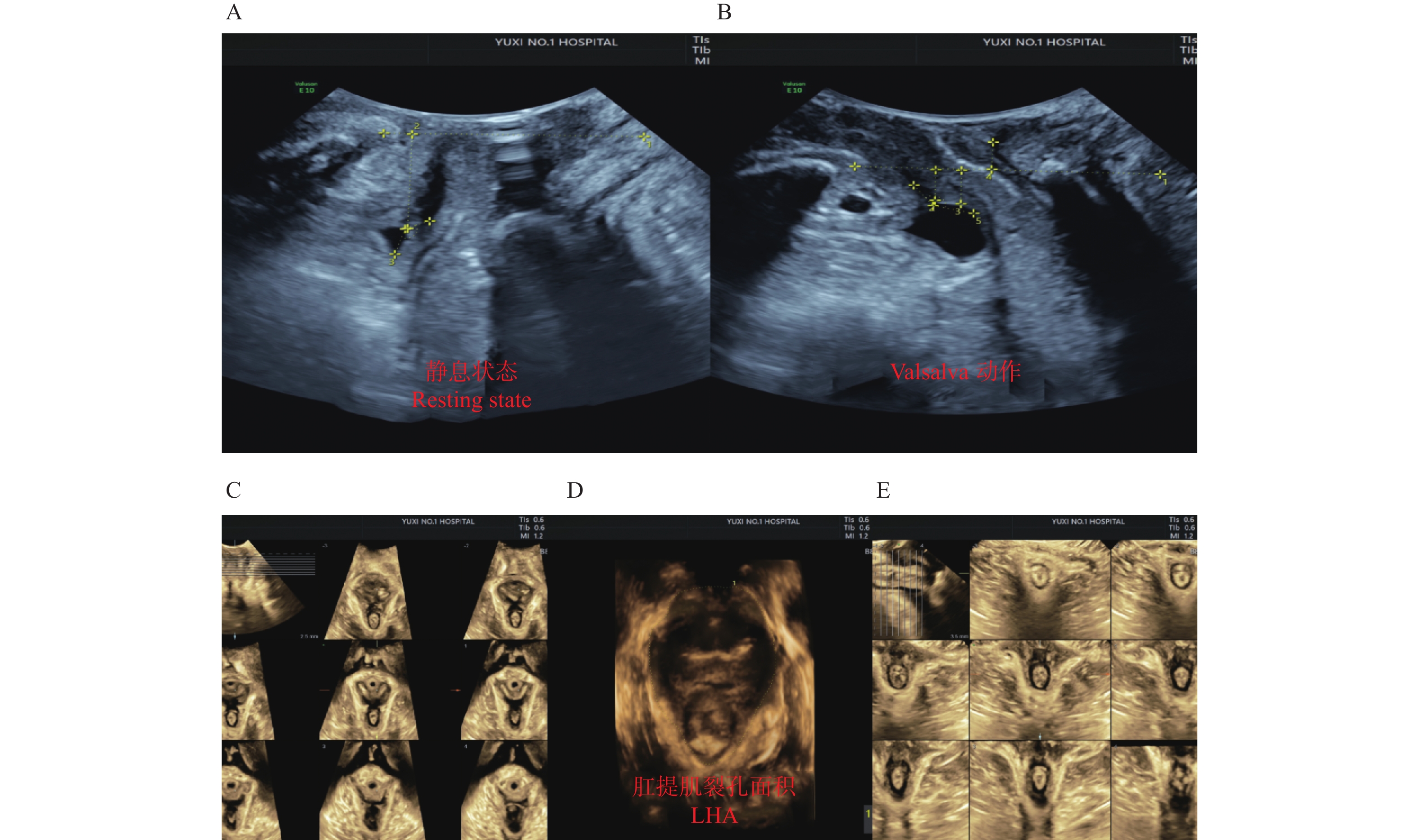
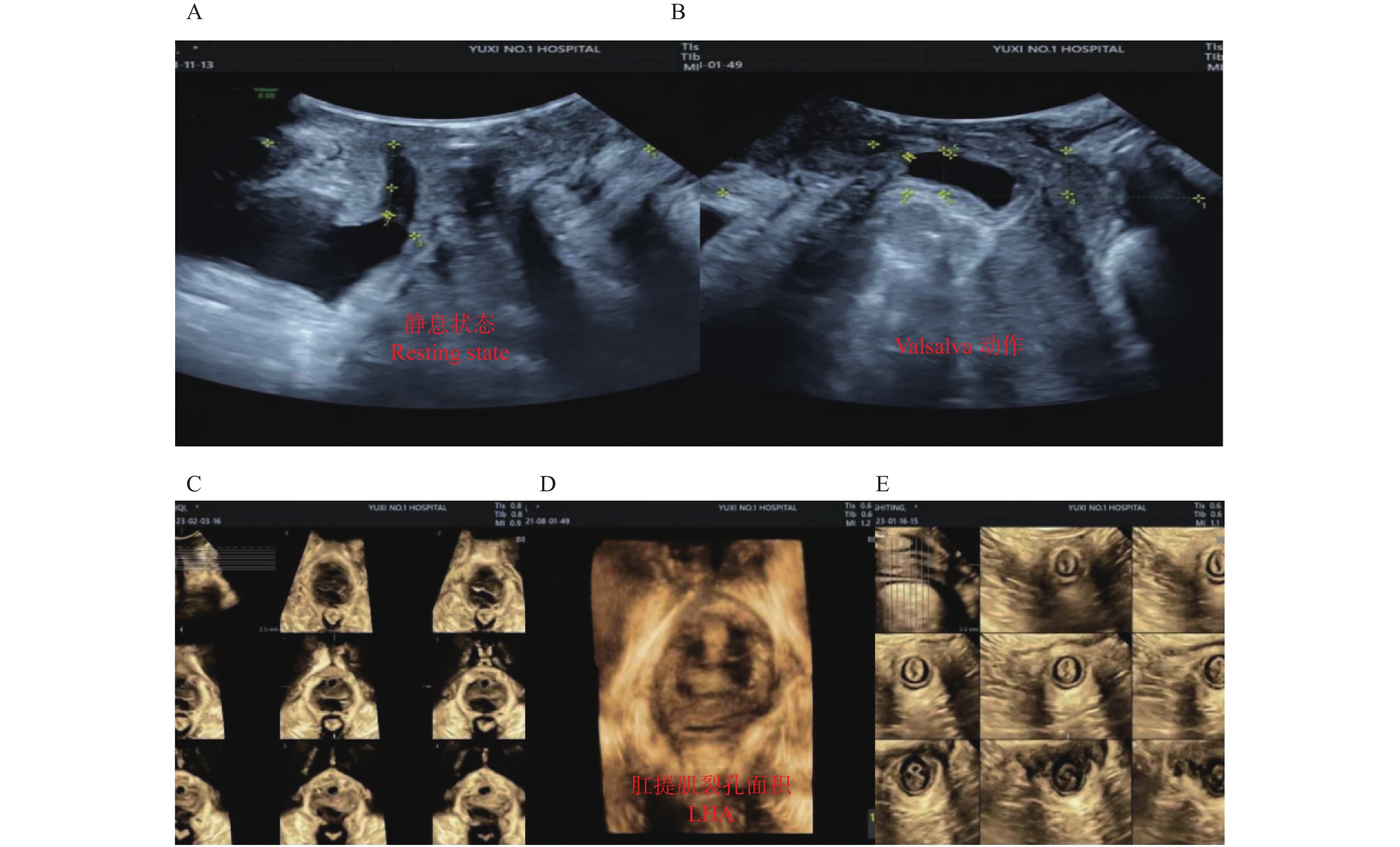
适龄、高龄产妇患者盆底二维、三维超声诊断图像分别见图1、图2,其中A为静息状态下盆底正中矢状切面;B为Valsalva动作下盆底正中矢状切面;C为断层成像模式观察肛提肌连续性,层间距2.5 mm(中间3幅图依次显示耻骨联合开放、正在关闭、已关闭状态;D为肛提肌裂孔;E为断层成像模式观察肛门括约肌连续性,其中左侧为肛门内括约肌下缘,右侧为外括约肌上缘。
3. 讨论
妊娠及分娩为引起女性盆底结构及功能损伤的主要因素,妊娠期间由于子宫体积增长,机体为适应妊娠会出现支持盆腔器官组织过度延伸,达到一定程度时将肌肉将可能丧失收缩恢复能力;分娩过程中阴道周围支持组织受到牵拉、扩张,甚或肌肉纤维断裂,继而导致盆底肌损伤。而进行产后盆底功能检测对于PFD早期诊断及预防有着重要意义。盆底超声为临床产后检查评估盆底结构主要技术,可观察盆底解剖结构形态学变化,继而评估其组织功能状态,为临床诊治及疗效评估提供客观依据。二维经阴道超声是评估的主要筛查工具,具有简单、可重复性、成像清晰等特征,可用于评估膀胱、膀胱颈、尿道等组织形态变化,辅助盆底功能及结构评估[7-9]。三维超声具有较高空间分辨率,可通过多平面成像及图像重建后处理,为临床评估盆底结构及功能提供可靠数据[10-12]。
本研究显示,观察组静息状态BNP、PUA、Valsalva动作BNP水平、BND水平明显高于对照组(P < 0.05);而Valsalva动作PUA及URA水平对比差异无统计学意义(P > 0.05)。观察组Valsalva动作LHAP、LHLP、LHA水平均明显高于对照组(P < 0.05);而静息状态LHAP、LHLP、LHA水平对比差异无统计学意义(P > 0.05)。说明相较于适龄产妇,高龄产妇产后早期更易出现肛提肌、膀胱等盆底组织功能及结构变化,适龄产妇变化不大。李宁等[13]研究报道,高龄产妇的Valsalva动作LHA水平高于适龄产妇,该结果与本研究结果一致。其原因在于相较于适龄产妇,高龄产妇的生理功能出现逐步下降,尤其是盆底肌群收缩反应时间延长,速度减慢,盆底肌肉、神经长时间处于压迫状态,出现盆底组织损伤概率较高;此外盆底肌肉组织中胶原、弹性蛋白含量降低,无法维持正常收缩功能,出现超声异常征象。盆底三维超声诊断灵敏度、准确性明显高于二维超声。此处已删减研究报道,孕妇的盆底肌肉不仅受到与分娩相关的机械损伤的影响,还受到怀孕期间生理变化的影响,继而使得提肌裂孔增大[14]。其原因在于虽然二维超声可以全面评估显示盆底结构,但该技术无法显示示肛提肌、盆膈裂孔等结构,而三维超声可通过多平面成像及图像重建后处理,更加准确、立体显示盆底结构组织及结构变化,该技术对于测量肛提肌裂孔各参数有着较高精确度,其中肛提肌裂孔可进一步反映肛提肌顺应性,继而提高临床诊断效能[15]。
综上所述,高龄产妇产后早期更易出现盆底功能及结构变化,相较于二维超声,盆底三维超声更有助于提高PFD诊断效能。
-
表 1 盆底二维、三维超声对适龄产妇产后PFD的诊断效能比较(n)
Table 1. Comparison of the diagnostic efficacy of two-dimensional and three-dimensional ultrasound (n)
诊断技术 金标准 灵敏度(%) 特异度(%) 准确性(%) 阳性 阴性 合计 二维超声 阳性 7 3 10 77.78(7/9) 92.68(38/41) 90.00(45/50) 阴性 2 38 40 合计 9 41 50 三维超声 阳性 8 1 9 88.89(8/9) 97.56(40/41) 96.00(48/50) 阴性 1 40 41 合计 9 41 50 χ2 − − − − 0.400 1.051 1.383 P − − − − 0.527 0.305 0.240 表 2 2组产妇盆底二维超声评估参数比较(
$ \bar x \pm s $ )Table 2. Comparison of the two groups of puerpera pelvic floor two-dimensional ultrasound evaluation parameter between the two groups (
$ \bar x \pm s $ )组别 n 静息状态 Valsalva动作 BND(cm) URA(°) BNP(cm) PUA(°) BNP(cm) PUA(°) 观察组 86 −2.34 ± 0.47* 120.47 ± 25.34* −0.59 ± 0.23* 137.45 ± 24.32 1.75 ± 0.35* 30.78 ± 6.53 对照组 50 −3.11 ± 0.56 104.68 ± 20.15 −1.74 ± 0.39 139.54 ± 25.47 1.37 ± 0.44 29.45 ± 5.84 t − 8.577 3.766 21.654 0.475 5.545 1.190 P − <0.001 <0.001 <0.001 0.636 <0.001 0.236 与对照组比较,*P < 0.05。 表 3 2组产妇盆底三维超声评估参数比较(
$ \bar x \pm s $ )Table 3. Comparison of pelvic floor three-dimensional ultrasound evaluation parameters between the two groups (
$ \bar x \pm s $ )组别 n 静息状态 Valsalva动作 LHAP(cm) LHLP(cm) LHA(cm2) LHAP(cm) LHLP(cm) LHA(cm2) 观察组 86 5.45 ± 1.26 3.01 ± 0.84 16.41 ± 2.54 6.07 ± 1.34* 4.16 ± 1.22* 25.25 ± 2.76* 对照组 50 5.12 ± 1.23 3.07 ± 0.58 15.72 ± 2.36 5.54 ± 1.25 3.73 ± 0.85 20.66 ± 2.52 t − 1.486 0.447 1.567 2.279 2.200 9.649 P − 0.140 0.656 0.119 0.024 0.030 <0.001 与对照组比较,*P < 0.05。 表 4 盆底二维、三维超声对高龄产妇产后PFD的诊断效能比较(n)
Table 4. Comparison of the diagnostic effcacy of two-dimensional and three-dimensional ultrasound of pelvic floor for PFD in older women (n)
诊断技术 金标准 灵敏度(%) 特异度(%) 准确性(%) 阳性 阴性 合计 二维超声 阳性 19 11 30 70.37(19/27) 70.37(48/59) 77.91(67/86) 阴性 8 48 56 合计 27 59 86 三维超声 阳性 25 5 30 92.59(25/27) 91.53(54/59) 91.86(79/86) 阴性 2 54 56 合计 27 59 86 χ2 − − − − 4.418 2.603 6.525 P − − − − 0.036 0.107 0.011 -
[1] Palmieri S,De Bastiani S S,Degliuomini R,et al. Prevalence and severity of pelvic floor disorders in pregnant and postpartum women[J]. Int J Gynaecol Obstet,2022,158(2):346-351. doi: 10.1002/ijgo.14019 [2] 田志强,丁玲,张俊俊,等. 高龄产妇产后早期盆底功能状况及其影响因素[J]. 护理研究,2021,35(7):1262-1266. [3] 朱玲斐,马苏亚,陆丹尔,等. 经会阴二维超声动态观察和评估产后女性盆底功能的改变[J]. 中国妇幼健康研究,2018,29(11):1431-1436. doi: 10.3969/j.issn.1673-5293.2018.11.016 [4] 秦学敏,柏根基,盛文伟,等. 三维盆底超声检查联合盆底肌力评估在 产后盆底功能中的应用[J]. 中国超声医学杂志,2019,35(12):1112-1114. doi: 10.3969/j.issn.1002-0101.2019.12.019 [5] 中华医学会超声医学分会妇产超声学组. 盆底超声检查中国专家共识(2022版)[J]. 中华超声影像学杂志,2022,31(3):185-191. [6] 中华医学会妇产科学分会妇科盆底学组. 盆腔器官脱垂的中国诊治指南(2020年版)[J]. 中华妇产科杂志,2020,55(5):300-306. [7] Sigurdardottir T,Bø K,Steingrimsdottir T,et al. Cross-sectional study of early postpartum pelvic floor dysfunction and related bother in primiparous women 6-10 weeks postpartum[J]. Int Urogynecol J,2021,32(7):1847-1855. doi: 10.1007/s00192-021-04813-y [8] 吕君,赵红敏,王春丽,等. 经会阴二维超声对产后压力性尿失禁患者盆底解剖特征评估[J]. 生物医学工程与临床,2022,26(4):431-435. doi: 10.13339/j.cnki.sglc.20220629.018 [9] Wang Y,Wang H. Transvaginal two-dimensional ultrasound evaluation as a screening tool for levator ani muscle avulsion in postpartum women[J]. Ultrasound Med,2023,42(1):161-169. doi: 10.1002/jum.16037 [10] 王爽,王静. 经会阴二维与三维超声评价女性产后盆底功能损伤的研究[J]. 中国数字医学,2018,13(2):95-97. [11] 仝蕊,卢丽娟,魏芳. 经会阴三维容积超声评估不同分娩方式对产后女性盆底功能的影响[J]. 云南医药,2021,42(6):563-564. [12] 卢小霞,刘爱华. 盆底三维超声对不同分娩方式产妇产后盆底功能改变及恢复评价[J]. 中国优生与遗传杂志,2021,29(3):310-314. [13] 李宁,阚艳敏,王艺桦,等. 多模态超声定量评估高龄产妇产后早期盆底结构和功能变化及诊断压力性尿失禁的价值研究[J]. 中国全科医学,2022,25(6):706-713. [14] Nishibayashi M,Okagaki R. Ultrasonographic evaluation of pelvic floor structure at antepartum and postpartum periods using three-dimensional transperineal ultrasound[J]. Med Ultrason,2021,48(3):345-351. [15] 房长海,王忠民,谈海英,等. 女性不同时期盆底二维及三维超声特征性改变分析[J]. 现代妇产科进展,2022,31(6):443-446. 期刊类型引用(8)
1. 孙付艳. 盆底三维超声对高龄产妇产后盆底功能障碍性疾病的评估价值. 临床医学. 2025(01): 90-92 .  百度学术
百度学术2. 马新乐,郑志硕,徐杰,李锋. 智能3D/4D盆底超声在高龄产妇产后早期盆底改变评估及盆底功能障碍性疾病诊断中的应用价值. 新乡医学院学报. 2025(03): 202-207 .  百度学术
百度学术3. 刘楚妙,李志萍,黄超华,吴晓宾. 超高龄经产妇不良妊娠结局危险因素分析. 中国计划生育学杂志. 2024(02): 446-449 .  百度学术
百度学术4. 路姗姗,王春丽,李宁,李纳. 经会阴二维、三维超声联合在子宫内膜异位症患者术后盆底肌改变评估中的价值. 山东医药. 2024(10): 84-87 .  百度学术
百度学术5. 吴旭辉,林晓倩,陈丹. 盆底超声在女性盆底功能障碍及盆底康复治疗效果评估中的作用研究. 影像研究与医学应用. 2024(07): 48-50 .  百度学术
百度学术6. 程琳,刘淑华. 盆底四维超声评估电刺激生物反馈治疗PFD患者的效果分析. 实用妇科内分泌电子杂志. 2024(19): 107-109 .  百度学术
百度学术7. 尚妍,勾明月,李云芳,王小燕,刘香菊. 血清H2、E_2与25(OH)D水平对围绝经期女性盆底功能及阴道脱落细胞成熟度的影响. 分子诊断与治疗杂志. 2024(10): 1885-1888+1897 .  百度学术
百度学术8. 朱韵,金清. 盆底三维超声和二维超声在产妇产后盆底功能障碍性疾病诊断中的应用价值. 影像研究与医学应用. 2024(23): 161-163 .  百度学术
百度学术其他类型引用(1)
-


 下载:
下载:


 下载:
下载:

