Disinfecting Effect of Three Disinfectants on the Ultrasonic Scaling Unit Waterline
-
摘要:
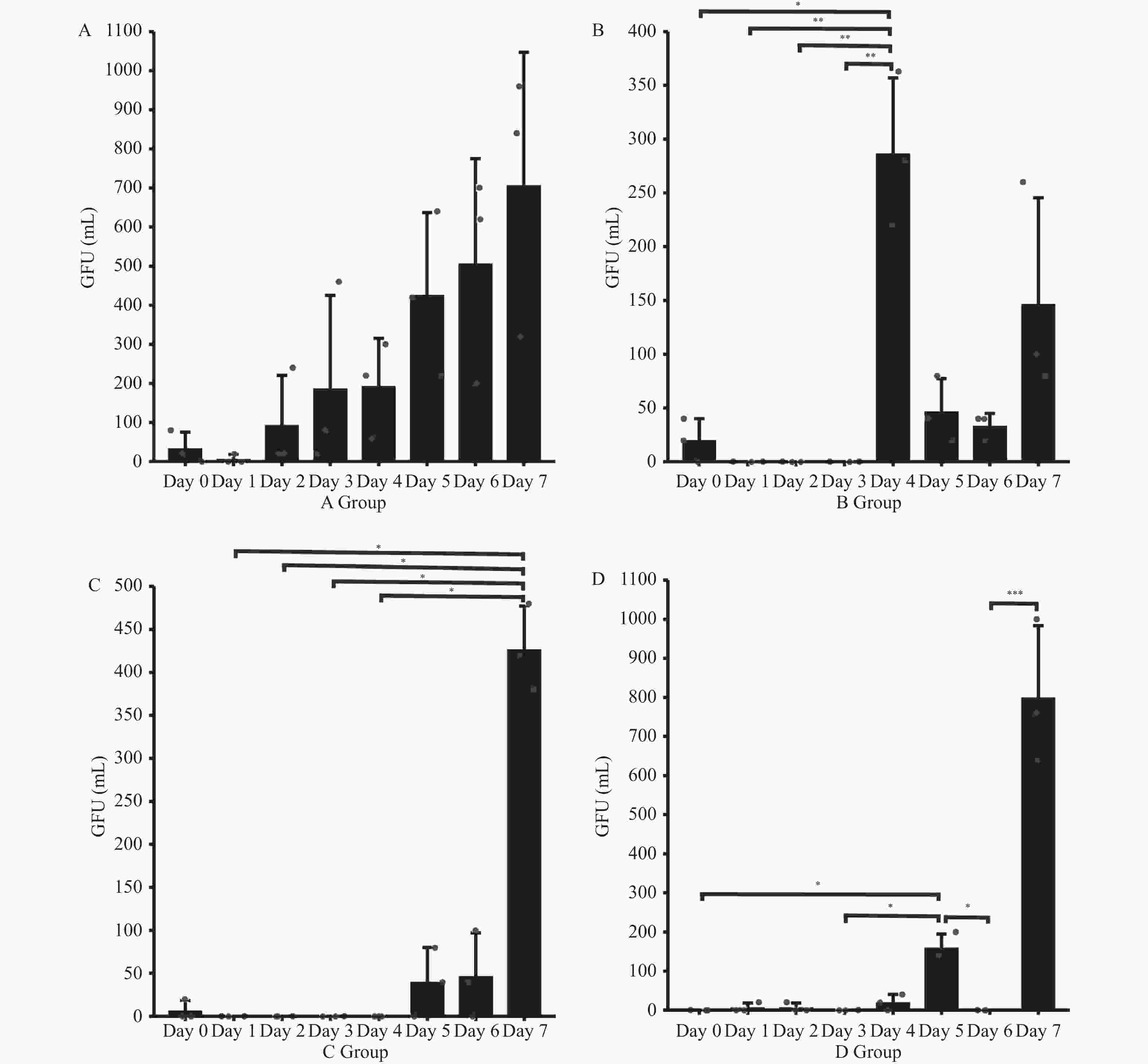
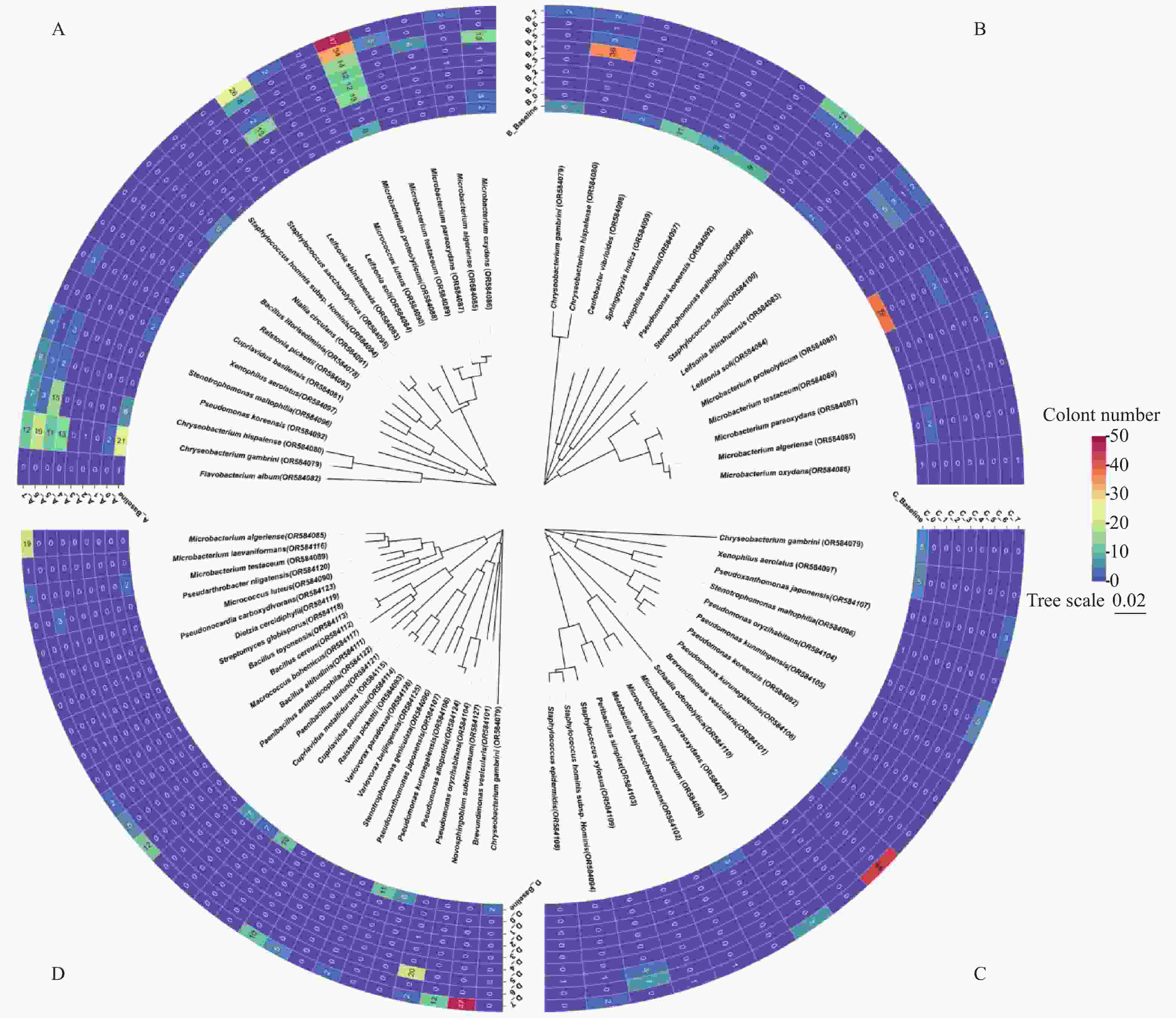
目的 评估3种临床常用消毒剂对超声洁牙机单元水路的消毒效果。 方法 将12台牙周超声洁牙机按消毒方案随机分为4组:A组蒸馏水、B组3%过氧化氢(H2O2)、C组500 mg/L含氯消毒剂和D组5%聚维酮碘。每台洁牙机用于日常牙周治疗,每天有效工作6 h。分别在基线、消毒后即刻和消毒后1~7 d收集水样。对可培养细菌进行计数、分离和纯化,通过16S rRNA 基因测序进行鉴定,基因扩增子序列按操作分类单元(OTUs)进行聚类。扫描电子显微镜(SEM)观察消毒7 d后水路内壁生物膜的形态和厚度。 结果 所有组别基线菌落总数均超过了100 CFU/mL。但在消毒后,各组菌落总数均显著降低(P < 0.05)。3% H2O2消毒后3 d内、500 mg/L含氯消毒剂消毒后6 d内和5%聚维酮碘消毒后4 d内,菌落总数保持在100 CFU/mL以下(P < 0.05)。在超声洁牙机单元水路中检测到了嗜麦芽窄食单胞菌(Stenotrophomonas maltophilia)等病原体。扫描电镜显示,与对照组相比,3% H2O2组和5%聚维酮碘组生物膜厚度更薄(P < 0.05)。 结论 牙科超声洁牙机单元水路中存在致病菌污染,对患者和医务人员构成潜在的感染风险。在日益严峻的感控背景下应根据临床实际制定适宜的消毒方案。 Abstract:Objective To assess the efficacy of three disinfectants in disinfecting the ultrasonic scaling unit waterline. Methods Twelve periodontal ultrasonic scalers were randomly divided into four groups based on the disinfection protocol: distilled water (A group), 3% hydrogen peroxide (B group), 500 mg/L chlorine disinfectant (C group) and 5% povidone-iodine (D group). Each scaler was used for daily periodontal treatment and worked effectively for 6 hours per day. Water samples were collected at baseline, after disinfection, and 1-7 days post-disinfection. Scanning electron microscopy (SEM) was used to observe biofilm formation. Culturable bacteria were counted, isolated and purified, then identified through 16S rRNA gene sequencing. Sequences of 16S rRNA gene amplicons were clustered by operational taxonomic units (OTUs). Results Baseline colony counts exceeded 100 CFU/mL in all groups. However, after disinfection, the colony counts decreased significantly in all groups (P < 0.05). The colony counts remained below 100 CFU/mL within 3 d after disinfection with 3% H2O2, within 6 days after disinfection with 500 mg/L chlorine-containing disinfectant, and within 4 d after disinfection with 5% povidone-iodine (P < 0.05).Pathogens such as Stenotrophomonas maltophilia were detected in the ultrasonic scaler unit water circuit. SEM showed thinner biofilm thickness in the 3% H2O2 and 5% povidone-iodine groups compared to the control group (P < 0.05). Conclusions Bacterial contamination is present in the ultrasonic scaling unit waterline, posing a potential infection risk to periodontal staff and patients. In the context of increasingly severe nosocomial infections, appropriate disinfection protocols should be developed based on clinical realities. -
Key words:
- Ultrasonic scaler /
- Dental unit waterline /
- Plaque biofilms /
- Hydrogen peroxide /
- Sodium hypochlorite /
- Povidone iodine
-
表 1 A、B、C、D 组不同时间点可培养菌落的描述性分析[ M(P25,P75),CFU/mL]
Table 1. Descriptive analysis of culturable colonies at different time points in groups A,B,C,D [ M(P25,P75),CFU/mL]
时间 A组 B组 C组 D组 基线 200(160,660) 360(240,940) 120(100,140) 280(160,400) 消毒后即刻 20(0,80) 20(0,40) 0(0,20) 0(0,0) 消毒后第1天 0(0,20) 0(0,0) 0(0,0) 0(0,20) 消毒后第2天 20(20,240) 0(0,0) 0(0,0) 0(0,20) 消毒后第3天 80(20,460) 0(0,0) 0(0,0) 0(0,0) 消毒后第4天 220(60,300) 280(220,360) 0(0,0) 20(0,40) 消毒后第5天 420(220,640) 40(20,80) 40(0,80) 140(140,200) 消毒后第6天 620(200,700) 40(20,40) 40(0,100) 0(0,0) 消毒后第7天 840(320,960) 100(80,260) 420(380,480) 760(640, 1000 )表 2 不同组别消毒后7 d的重复测量秩和检验
Table 2. Repeated-measures rank-sum test 7 d after disinfection in different groups
组别 χ2 df P A组 18.657 7 0.009 B组 20.079 7 0.005 C组 15.621 7 0.029 D组 17.604 7 0.014 -
[1] Cui Y,Tian G,Li R,et al. Epidemiological and sociodemographic transitions of severe periodontitis incidence,prevalence,and disability-adjusted life years for 21 world regions and globally from 1990 to 2019: An age-period-cohort analysis[J]. Journal of Periodontology,2023,94(2):193-203. doi: 10.1002/JPER.22-0241 [2] Sanz M,Herrera D,Kebschull M,et al. Treatment of stage I-III periodontitis-The EFP S3 level clinical practice guideline [J]. Journal of Clinical Periodontology,2020,47 Suppl(22): 4-60. [3] Herrera D,Sanz M,Kebschull M,et al. Treatment of stage IV periodontitis: The EFP S3 level clinical practice guideline[J]. Journal of Clinical Periodontology,2022,49(Suppl 24):4-71. [4] 孟焕新. 牙周病学 [M]. 北京: 人民卫生出版社,2020: 36. [5] Yu Y,Mahmud M,Vyas N,et al. Cavitation in a periodontal pocket by an ultrasonic dental scaler: A numerical investigation[J]. Ultrasonics Sonochemistry,2022,90(1):1-9. [6] Dang Y,Zhang Q,Wang J,et al. Assessment of microbiota diversity in dental unit waterline contamination[J]. Peer J,2022,6(10):1-16 [7] Dahlen G. Biofilms in dental unit water lines[J]. Monographs in Oral Science,2021,29(1):12-18. [8] Kumbargere Nagraj S,Eachempati P,Paisi M,et al. Interventions to reduce contaminated aerosols produced during dental procedures for preventing infectious diseases[J]. The Cochrane Database of Systematic Reviews,2020,10(12):1-88. [9] Marino F,Mazzotta M,Pascale M R,et al. First water safety plan approach applied to a Dental Clinic complex: identification of new risk factors associated with Legionella and P. aeruginosa contamination,using a novel sampling,maintenance and management program[J]. Journal of Oral Microbiology,2023,15(1):1-14. [10] Bayani M,Raisolvaezin K,Almasi-Hashiani A,et al. Bacterial biofilm prevalence in dental unit waterlines: A systematic review and meta-analysis[J]. BMC Oral Health,2023,23(1):158-172. doi: 10.1186/s12903-023-02885-4 [11] 国家市场监督管理局,国家标准化管理委员会. GB 5749-2022. 生活饮用水卫生标准[S]. 北京: 中国标准出版社,2022. [12] Wirthlin M R,Marshall G J. Evaluation of ultrasonic scaling unit waterline contamination after use of chlorine dioxide mouthrinse lavage[J]. Journal of Periodontology,2001,72(3):401-410. doi: 10.1902/jop.2001.72.3.401 [13] 侯雅蓉,倪佳,周俏怡,等. 2种消毒剂对牙周超声洁牙机独立水路消毒效果的比较[J]. 口腔疾病防治,2023,31(12):855-862. [14] 国家卫生和计划生育委员会,国家食品药品监督管理局. GB4789.2-2016. 食品安全国家标准食品微生物学检验菌落总数测定[S]. 北京: 中国标准出版社,2017. [15] Standard Methods Committee of the American Public Health Association, American Water Works Association, and Water Environment Federation. 9215. Standard Methods for the Examination of Water and Waste Water. 23rd ed: 9215 Heterotrophic Plate Count[M]. Washington, DC: American Public Health Association, 2017:2-8. [16] Rice E W,Rich W K,Johnson C H,et al. The role of flushing dental water lines for the removal of microbial contaminants [J]. Public Health Reports (Washington,DC: 1974),2006,121(3): 270-274. [17] Montebugnoli L,Chersoni S,Prati C,et al. A between-patient disinfection method to control water line contamination and biofilm inside dental units[J]. The Journal of Hospital Infection,2004,56(4):297-304. doi: 10.1016/j.jhin.2004.01.015 [18] Urban M V,Rath T,Radtke C. Hydrogen peroxide (H2O2): A review of its use in surgery [J]. Wiener Medizinische Wochenschrift (1946),2019,169(9-10): 222-225. [19] Scarano A, Inchingolo F, Lorusso F, et al. Environmental disinfection of a dental clinic during the covid-19 pandemic: A narrative insight[J]. BioMed research international,2020,9(4):1-15. [20] da Cruz Nizer W S,Inkovskiy V,Overhage J. Surviving reactive chlorine stress: Responses of gram-negative bacteria to hypochlorous acid[J]. Microorganisms,2020,8(8):1-27. [21] Slots J. Low-cost periodontal therapy[J]. Periodontology 2000,2012,60(1):110-137. [22] Dioguardi M,Gioia G D,Illuzzi G,et al. Endodontic irrigants: Different methods to improve efficacy and related problems[J]. European Journal of Dentistry,2018,12(3):459-466. doi: 10.4103/ejd.ejd_56_18 [23] Haapasalo M,Shen Y,Wang Z,et al. Irrigation in endodontics[J]. Br Dent J,2014,216(6):299-303. doi: 10.1038/sj.bdj.2014.204 [24] 郭婉晴,王卫. 不同浓度含氯消毒液对口腔综合治疗台水路消毒效果研究[J]. 全科口腔医学电子杂志,2016,3(9):100-101. [25] Hoang T,Jorgensen M G,Keim R G,et al. Povidone-iodine as a periodontal pocket disinfectant[J]. Journal of Periodontal Research,2003,38(3):311-317. doi: 10.1034/j.1600-0765.2003.02016.x [26] Eggers M. Infectious disease management and control with povidone iodine[J]. Infectious Diseases and Therapy,2019,8(4):581-593. doi: 10.1007/s40121-019-00260-x [27] Fidler A,Steyer A,Manevski D,et al. Virus transmission by ultrasonic scaler and its prevention by antiviral agent: An in vitro study[J]. Journal of Periodontology,2022,93(7):116-124. [28] Baker M A,Rhee C,Tucker R,et al. Ralstonia pickettii and pseudomonas aeruginosa bloodstream infections associated with contaminated extracorporeal membrane oxygenation water heater devices[J]. Clinical Infectious Diseases,2022,75(10):1838-1840. doi: 10.1093/cid/ciac379 [29] Sun X,Li M,Xia L,et al. Alteration of salivary microbiome in periodontitis with or without type-2 diabetes mellitus and metformin treatment[J]. Scientific Reports,2020,10(1):1-14. doi: 10.1038/s41598-019-56847-4 [30] Inkster T,Weinbren M. Xenophilus aerolatus: What's in a name?[J]. The Journal of Hospital Infection,2023,139(9):238-239. [31] Li L H,Shih Y L,Huang J Y,et al. Protection from hydrogen peroxide stress relies mainly on AhpCF and KatA2 in Stenotrophomonas maltophilia[J]. Journal of Biomedical Science,2020,27(1):37. doi: 10.1186/s12929-020-00631-4 [32] An S Q,Berg G. Stenotrophomonas maltophilia[J]. Trends in Microbiology,2018,26(7):637-638. doi: 10.1016/j.tim.2018.04.006 [33] Olwal C O,Ang'ienda P O,Onyango D M,et al. Susceptibility patterns and the role of extracellular DNA in Staphylococcus epidermidis biofilm resistance to physico-chemical stress exposure[J]. BMC Microbiology,2018,18(1):40. doi: 10.1186/s12866-018-1183-y [34] Koksal F,Yasar H,Samasti M. Antibiotic resistance patterns of coagulase-negative staphylococcus strains isolated from blood cultures of septicemic patients in Turkey[J]. Microbiological Research,2009,164(4):404-410. doi: 10.1016/j.micres.2007.03.004 [35] Ryan M P,Pembroke J T. Brevundimonas spp: Emerging global opportunistic pathogens[J]. Virulence,2018,9(1):480-493. doi: 10.1080/21505594.2017.1419116 [36] Lee M R,Huang Y T,Liao C H,et al. Bacteremia caused by Brevundimonas species at a tertiary care hospital in Taiwan,2000-2010[J]. European Journal of Clinical Microbiology & Infectious Diseases,2011,30(10):1185-1191. [37] Yan J,Bassler B L. Surviving as a community: Antibiotic tolerance and persistence in bacterial biofilms[J]. Cell Host & Microbe,2019,26(1):15-21. [38] Hall C W,Mah T F. Molecular mechanisms of biofilm-based antibiotic resistance and tolerance in pathogenic bacteria[J]. FEMS Microbiology Reviews,2017,41(3):276-301. doi: 10.1093/femsre/fux010 [39] Høiby N,Bjarnsholt T,Givskov M,et al. Antibiotic resistance of bacterial biofilms[J]. International Journal of Antimicrobial Agents,2010,35(4):322-332. doi: 10.1016/j.ijantimicag.2009.12.011 [40] Spagnolo A M,Sartini M,Cristina M L. Microbial contamination of dental unit waterlines and potential risk of infection: A narrative review[J]. Pathogens (Basel,Switzerland),2020,9(8):1-16 [41] Fan C,Gu H,Liu L,et al. Distinct microbial community of accumulated biofilm in dental unit waterlines of different specialties[J]. Frontiers in Cellular and Infection Microbiology,2021,11(6):1-14. -


 下载:
下载:


