

Mechanism of Rosiglitazone in Reducing Liver Ischemia Reperfusion Injury in Rats by Inducing HO-1
-
摘要:
目的 观察过氧化物酶体增殖物激活受体γ(peroxisome proliferator-activated receptor,PPAR-γ)激动剂罗格列酮是否通过调控血红素加氧酶1(heme oxygenase 1,HO-1)活性来减轻大鼠肝缺血再灌注损伤(ischemia reperfusion injury,IRI)。 方法 建立大鼠70%肝脏热缺血再灌注(ischemia/reperfusion,I/R)模型和缺氧缺糖/复氧复糖(oxygen-glucose deprivation/reperfusion,OGD/R)诱导的大鼠肝窦内皮细胞(liver sinusoidal endothelial cells,LSECs)损伤模型,随机分为假手术组、模型组、罗格列酮预处理组和锌原卟啉(zinc protoporphyrin,ZnPP)组(n = 6)。全自动生化分析仪检测大鼠血清ALT、AST水平;HE染色评估肝组织病理学损伤;Western blot检测PPAR-γ和HO-1蛋白表达水平;CCK8法测定LSECs的细胞活力,流式细胞仪测定LSECs中活性氧(reactive oxygen species,ROS)含量。 结果 与I/R组相比,罗格列酮预处理能显著降低肝IRI大鼠ALT、AST水平(P < 0.01),减少肝细胞凋亡并减轻肝组织IRI (P < 0.01)。Western blot结果显示,罗格列酮能上调PPAR-γ和HO-1蛋白的表达(P < 0.01)。此外,罗格列酮预处理(10,30 μmol/L)能改善OGD/R诱导的LSECs存活率,显著降低细胞ROS含量,并呈剂量反应相关性(P < 0.01)。使用ZnPP阻断HO-1活性后,罗格列酮对大鼠肝IRI的保护作用均消失。 结论 罗格列酮通过上调HO-1活性介导抗氧化和抗炎作用,减轻大鼠肝IRI。 Abstract:Objective To observe whether rosiglitazone, a peroxisome proliferator-activated receptor (PPAR-γ) agonist, can attenuate hepatic ischemia-reperfusion injury (IRI) in rats by modulating heme oxygenase 1 (HO-1) activity. Methods A rat 70% hepatic thermal ischemia/reperfusion (I/R) model and hypoxia-glucose deprivation/reperfusion (OGD/R)-induced liver sinusoidal endothelial cells (LSECs) injury model in rats were established. The rats were randomly divided into sham operation group, model group, rosiglitazone pretreatment group and zinc protoporphyrin (ZnPP) group (n = 6/group). The serum ALT and AST levels of rats were detected by fully automatic biochemical analyzer. HE staining was used to assess liver histopathological injury. PPAR-γ and HO-1 protein expression levels were detected by Western Blot. The cell viability of LSECs was determined by CCK8 method, and reactive oxygen species (ROS) content in LSECs was determined by flow cytometry. Results Compared with the I/R model group, rosiglitazone pretreatment significantly decreased ALT and AST levels, reduced liver cell apoptosis, and alleviated liver tissue IRI (P < 0.01). WB results showed that rosiglitazone upregulated the expression of PPAR-γ and HO-1protein (P < 0.01). In addition, compared with the OGD/R model group, rosiglitazone pretreatment (10, 30 μmol/L) improved the survival rate of OGD/ R-induced LSECs and significantly reduced the ROS content in cells in a dose-responsive manner (P < 0.01). When ZnPP blocked HO-1 activity, the protective effect of rosiglitazone on hepatic IRI in rats disappeared. Conclusion Rosiglitazone can mediate antioxidant and anti-inflammatory effects through up-regulation of HO-1 activity and reduce hepatic IRI in rats. -
Key words:
- Hepatic ischemia reperfusion injury /
- PPAR-γ /
- HO-1 /
- Liver sinusoidal endothelial cell
-

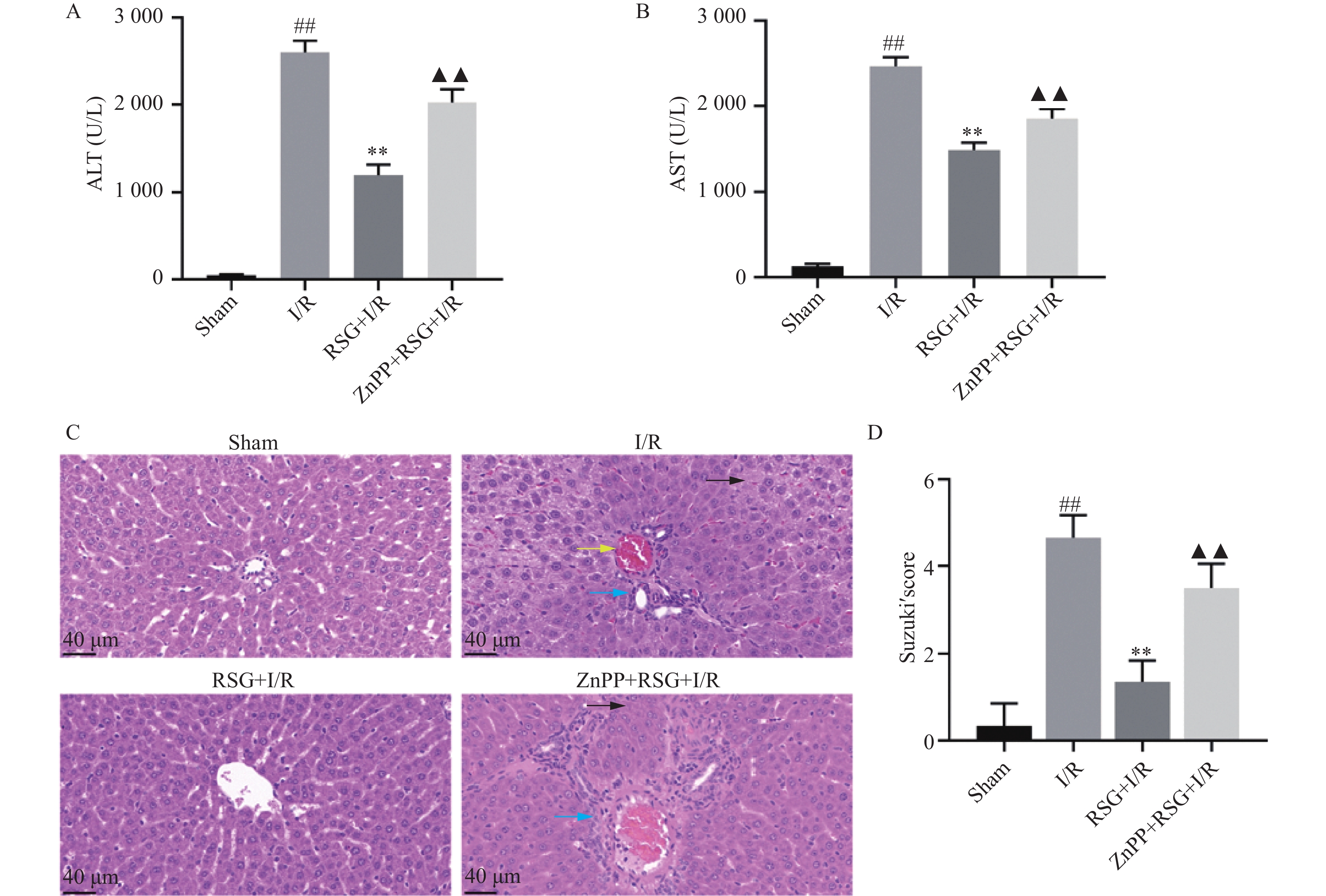
图 1 罗格列酮对I/R诱导的肝损伤的影响
A:各组大鼠血清中ALT水平;B:各组大鼠血清中AST水平;C:大鼠肝脏HE染色 (×400),
Figure 1. Effect of rosiglitazone on I/ R-induced liver injury
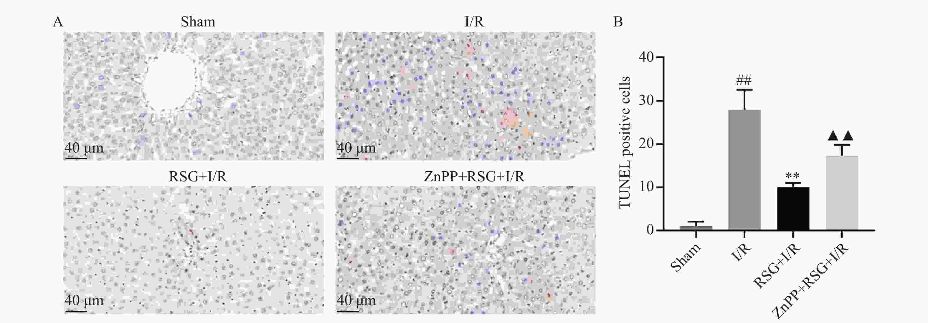
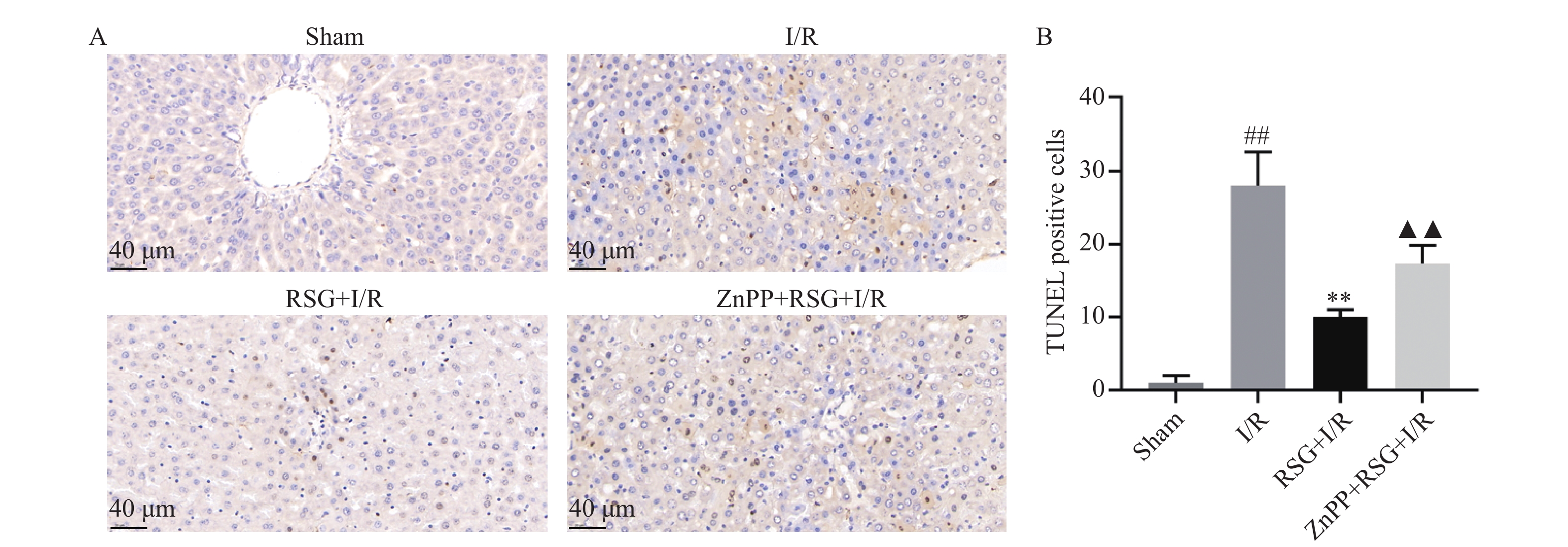
图 2 罗格列酮对大鼠IRI肝脏中TUNEL表达的影响
A:大鼠肝组织TUNEL染色图(×400);B:大鼠肝组织中TUNEL阳性细胞数统计图。与假手术组比较,##P < 0.01;与I/R模型组比较,**P < 0.01;与罗格列酮组比较,▲▲P < 0.01。
Figure 2. Effect of rosiglitazone on TUNEL expression in rat IRI liver
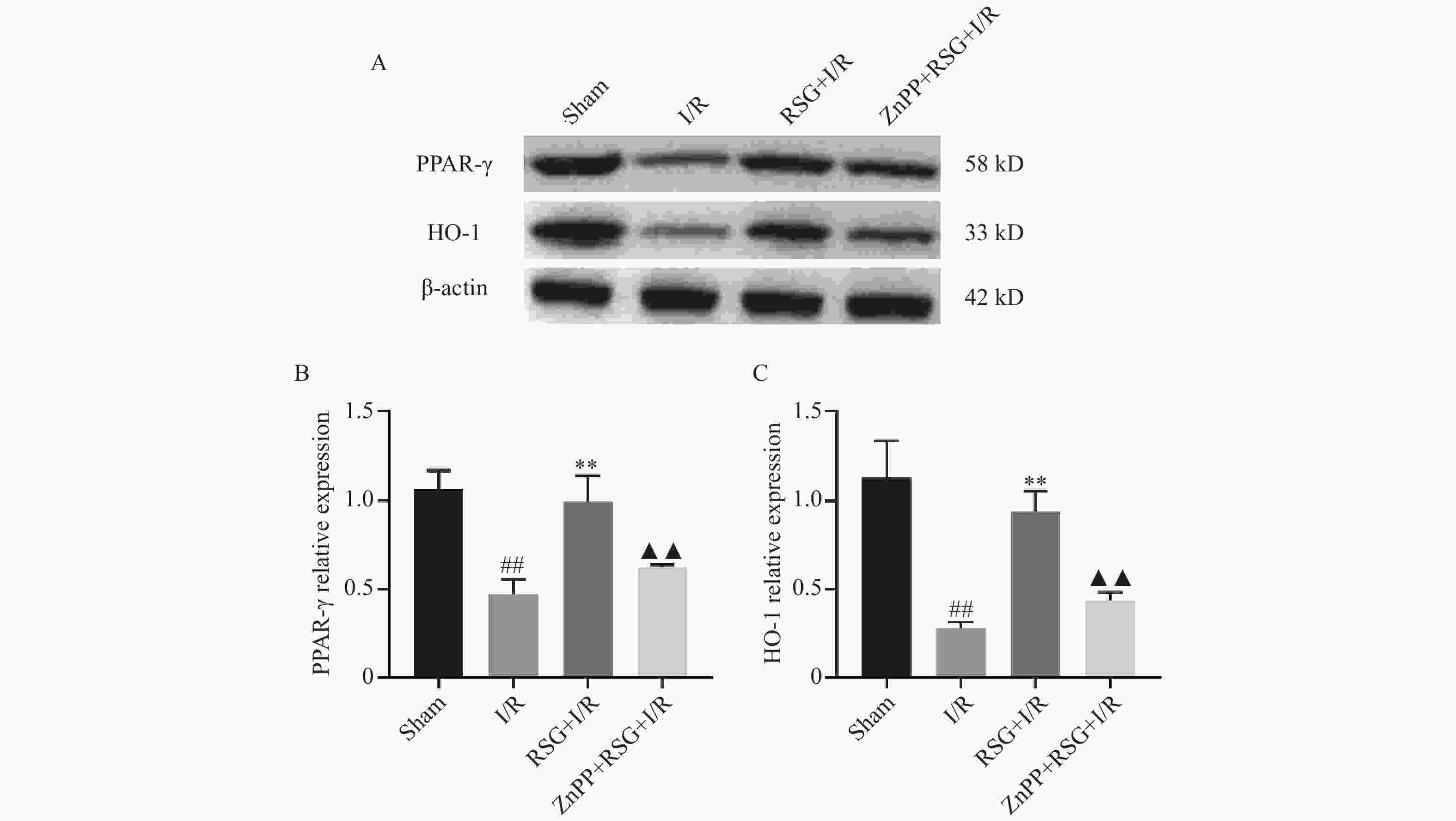
图 3 罗格列酮对大鼠IRI肝脏中PPAR-γ和HO-1蛋白表达的影响
A:肝组织中PPAR-γ、HO-1蛋白表达条带图;B:肝组织中PPAR-γ蛋白表达统计图;C:肝组织中HO-1蛋白表达统计图。与假手术组比较,##P < 0.05;与I/R模型组比较,**P < 0.01;与罗格列酮组比较,▲▲P < 0.01。
Figure 3. Effect of rosiglitazone on the expression of PPAR-γ and HO-1 proteins in the liver of rat IRI
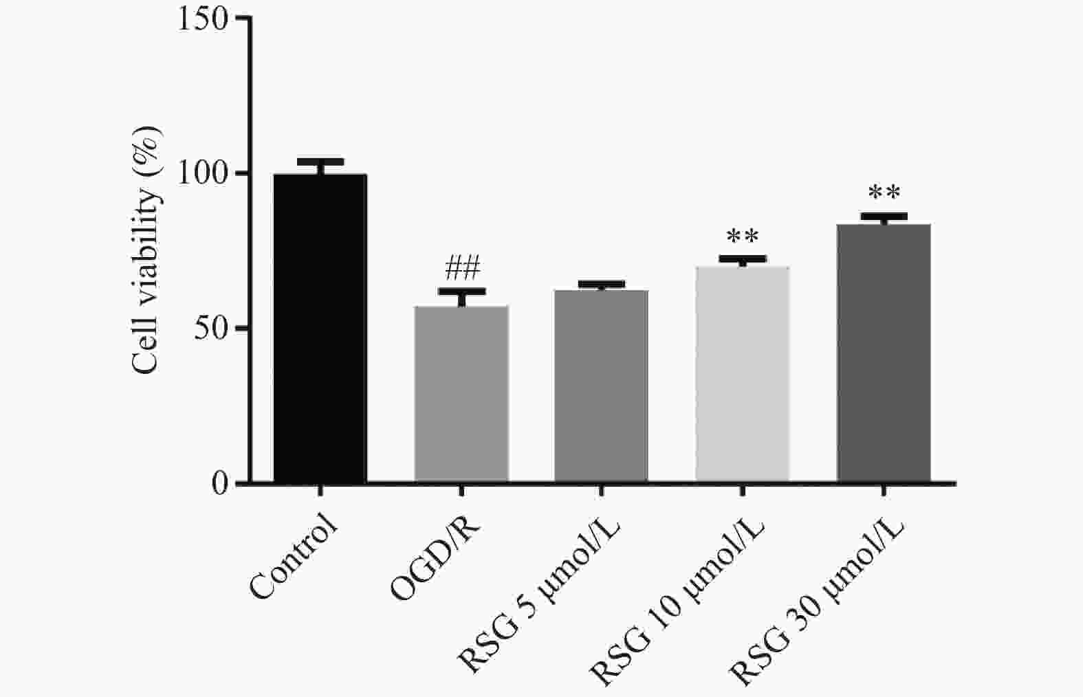
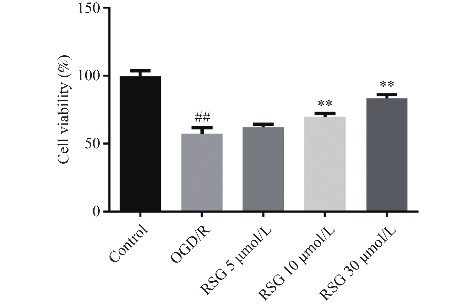
图 4 罗格列酮对LSECs细胞活力的影响
与正常对照组比较,##P < 0.01;与OGD/R模型组比较,**P < 0.01。
Figure 4. Effect of rosiglitazone on the viability of LSECs cells
-
[1] Ling S,Jiang G,Que Q,et al. Liver transplantation in patients with liver Failure: Twenty years of experience from China[J]. Liver International : Official Journal of the International Association for the Study of the Liver,2022,42(9) : 2110-2116. [2] Guan Y,Yao W,Yi K,et al. Nanotheranostics for the management of hepatic ischemia-reperfusion injury[J]. Small (Weinheim an der Bergstrasse,Germany),2021,17(23):e2007727. doi: 10.1002/smll.202007727 [3] Gao F,Qiu X,Wang K,Shao C,et al. Targeting the hepatic microenvironment to improve ischemia/reperfusion injury: New insights into the immune and metabolic compartments[J]. Aging and Disease,2022,13(4):1196-1214. doi: 10.14336/AD.2022.0109 [4] Tang S P,Mao X L,Chen Y H,et al. Reactive oxygen species induce fatty liver and ischemia-reperfusion injury by promoting inflammation and cell death[J]. Frontiers in Immunology,2022,13:870239. doi: 10.3389/fimmu.2022.870239 [5] Takada I,Makishima M. Peroxisome proliferator-activated receptor agonists and antagonists: A patent review (2014-Present)[J]. Expert Opinion on Therapeutic Patents,2020,30(1):1-13. doi: 10.1080/13543776.2020.1703952 [6] Sauer S. Ligands for the nuclear peroxisome proliferator-activated receptor gamma[J]. Trends in Pharmacological Sciences,2015,36(10):688-704. doi: 10.1016/j.tips.2015.06.010 [7] Katori M,Busuttil R W,Kupiec-Weglinski J W. Heme oxygenase-1 system in organ transplantation[J]. Transplantation,2002,74(7):905-912. doi: 10.1097/00007890-200210150-00001 [8] Sano N,Tamura T,Toriyabe N,et al. New drug delivery system for liver sinusoidal endothelial cells for ischemia-reperfusion injury[J]. World Journal of Gastroenterology,2015,21(45):12778-12786. doi: 10.3748/wjg.v21.i45.12778 [9] Yuan B,Ming T,Qu S,et al. The effect of heme oxygenase-1 on liver sinusoidal endothelial cells proliferation and pro-regeneration[J]. Chinese Journal of Hepatobiliary Surgery,2022,28(7):536-541. [10] Shen X,Wang M,Bi X,et al. Resveratrol prevents endothelial progenitor cells from senescence and reduces the oxidative reaction via PPAR-γ/HO-1 pathways[J]. Molecular Medicine Reports,2016,14(6):5528-5534. doi: 10.3892/mmr.2016.5929 [11] Abdalla H B,Napimoga M H,Lopes A H,et al. Activation of PPAR-γ induces macrophage polarization and reduces neutrophil migration mediated by heme oxygenase 1[J]. International Immunopharmacology,2020,84(7):106565. doi: 10.1016/j.intimp.2020.106565 [12] Lhuillier F,Parmantier P,Goudable J,et al. Hepatic ischemia is associated with an increase in liver parenchyma nitric oxide that is in part enzyme-independent[J]. Anesthesiology,2003,98(2):373-378. doi: 10.1097/00000542-200302000-00017 [13] Jaeschke H. Molecular mechanisms of hepatic ischemia-reperfusion injury and preconditioning[J]. American Journal of Physiology Gastrointestinal and Liver Physiology,2003,284(1):G15-26. doi: 10.1152/ajpgi.00342.2002 [14] Roushansarai N S,Pascher A,Becker F. Innate immune cells during machine perfusion of liver grafts-the janus face of hepatic macrophages[J]. Journal of Clinical Medicine,2022,11(22):6669. [15] Kotlinowski J,Jozkowicz A. PPAR-gamma and angiogenesis: endothelial cells perspective[J]. Journal of Diabetes Research,2016,2016:8492353. [16] Han X,Wu Y,Yang Q,et al. Peroxisome proliferator-activated receptors in the pathogenesis and therapies of liver fibrosis[J]. Pharmacology & Therapeutics,2021,222(6):107791. [17] Huang R,Zhang C,Wang X,et al. PPAR-γ in ischemia-reperfusion injury: Overview of the biology and therapy[J]. Frontiers in Pharmacology,2021,12(4):600618. doi: 10.3389/fphar.2021.600618 [18] Hirao H,Nakamura K,Kupiec-Weglinski J W. Liver ischaemia-reperfusion injury: A new understanding of the role of innate immunity[J]. Nature Reviews Gastroenterology & Hepatology,2022,19(4):239-256. [19] Abu-Amara M,Yang S Y,Tapuria N,et al. Liver ischemia/reperfusion injury: Processes in inflammatory networks-a review[J]. Liver transplantation : Official publication of the American Association for the Study of Liver Diseases and the International Liver Transplantation Society,2010,16(9): 1016-1032. [20] Forman HJ,Zhang H. Targeting oxidative stress in disease: Promise and limitations of antioxidant therapy[J]. Nature Reviews Drug Discovery,2021,20(9):689-709. doi: 10.1038/s41573-021-00233-1 [21] Linares I,Farrokhi K,Echeverri J,et al. PPAR-gamma activation is associated with reduced liver ischemia-reperfusion injury and altered tissue-resident macrophages polarization in a mouse model[J]. PloS One,2018,13(4):e0195212. doi: 10.1371/journal.pone.0195212 [22] Zingarelli B,Chima R,O'Connor M,et al. Liver apoptosis is age dependent and is reduced by activation of peroxisome proliferator-activated receptor-gamma in hemorrhagic shock[J]. American Journal of Physiology Gastrointestinal and Liver Physiology,2010,298(1):G133-141. doi: 10.1152/ajpgi.00262.2009 [23] R G B,Panisello-Roselló A,Sanchez-Nuno S,et al. Nrf2 and oxidative stress in liver ischemia/reperfusion injury[J]. The FEBS Journal,2022,289(18):5463-5479. doi: 10.1111/febs.16336 [24] Zhan X,Zhang Z,Huang H,et al. Effect of heme oxygenase-1 on the protection of ischemia reperfusion injury of bile duct in rats after liver transplantation[J]. Clinics and Research in Hepatology and Gastroenterology,2018,42(3):245-254. doi: 10.1016/j.clinre.2017.09.008 [25] Qu S,Yuan B,Zhang H,et al. Heme oxygenase 1 attenuates hypoxia-reoxygenation injury in mice liver sinusoidal endothelial cells[J]. Transplantation,2018,102(3):426-432. doi: 10.1097/TP.0000000000002028 [26] Shen X,Wang M,Bi X,et al. Resveratrol prevents endothelial progenitor cells from senescence and reduces the oxidative reaction via PPAR-γ/HO-1 pathways[J]. Molecular Medicine Reports,2016,14(6):5528-5534. doi: 10.3892/mmr.2016.5929 [27] Cho R L,Yang C C,Tseng H C,et al. Heme oxygenase-1 up-regulation by rosiglitazone via ROS-dependent Nrf2-antioxidant response elements axis or PPAR-γ attenuates LPS-mediated lung inflammation[J]. British Journal of Pharmacology,2018,175(20):3928-3946. doi: 10.1111/bph.14465 [28] Yang Y,Li X,Zhang L,et al. Ginsenoside Rg1 suppressed inflammation and neuron apoptosis by activating PPAR/HO-1 in hippocampus in rat model of cerebral ischemia-reperfusion injury[J]. International Journal of Clinical and Experimental Pathology,2015,8(3):2484-2494. [29] Yang H,Zhang L,Chen J,et al. Heme oxygenase-1 inhibits the poliferation of hepatic stellate cells by activating PPAR-γ and suppressing Nf-κB[J]. Computational and Mathematical Methods in Medicine,2022,2022:8920861. [30] Jang H Y,Hong O Y,Youn H J,et al. 15d-Pgj2 inhibits Nf-κB and AP-1-mediated MMP-9 expression and invasion of breast cancer cell by means of a heme oxygenase-1-dependent mechanism[J]. BMB Reports,2020,53(4):212-217. doi: 10.5483/BMBRep.2020.53.4.164 -

 下载:
下载:

 点击查看大图
计量
- 文章访问数: 1988
- HTML全文浏览量: 1028
- PDF下载量: 19
- 被引次数: 0
