




Mechanism of Ginsenoside Rg1 in Alleviating Alcohol-Induced Liver Injury in Tree Shrews via Activating of Nrf2 Pathway and Inhibiting of Mitochondrial Apoptosis
-
摘要:
目的 探讨人参皂苷Rg1对酒精诱导树鼩肝损伤的保护作用,并基于Nrf2信号轴与线粒体凋亡途径揭示其分子机制,为酒精性肝病的治疗提供新思路。 方法 将树鼩随机分为对照组、酒精模型组及人参皂苷Rg1给药组(每组6只)。后两组通过灌胃酒精建立肝损伤模型,Rg1给药组同时灌服人参皂苷Rg1,连续干预12周。采用肝脏超声评估肝实质变化;检测血清肝功能指标(ALT、AST)、氧化应激指标丙二醛(MDA)水平,以及乳酸脱氢酶(LDH)与谷胱甘肽过氧化物酶(GSH-Px)的活性;肝组织切片经HE染色后观察脂肪变性、炎症细胞浸润及气球样变程度;透射电镜用于观察线粒体超微结构改变;Western Blot法检测Nrf2通路蛋白(Nrf2、Keap1)及凋亡有关蛋白(Bcl-2、Bax、Cytochrome c、总Caspase-9、总Caspase-3)的表达。免疫组化检测肝组织中Keap1,HO-1及总Caspase-3的蛋白表达定位。 结果 与模型组相比,人参皂苷Rg1组树鼩肝脏超声影像学改善,血清ALT、AST、MDA含量及LDH活性显著降低(P < 0.05,F值分别为327.57、218.70、510.07、331.59),GSH-Px活性显著上升(P < 0.05);肝组织内的脂肪变性、炎症浸润及气球样病变均显著减轻;透射电镜显示线粒体肿胀等超微结构损伤得到修复。在分子表达层面,人参皂苷Rg1显著上调Nrf2蛋白表达,下调其负调控因子Keap1表达(P < 0.05);免疫组化证实人参皂苷Rg1干预后Keap1阳性信号减弱,HO-1阳性信号显著增强(P < 0.05)。同时,人参皂苷Rg1干预提高抗凋亡蛋白Bcl-2表达,降低促凋亡蛋白Bax表达,抑制Cytochrome c释放(P < 0.05);总Caspase-9(前体形式)表达水平回升,总Caspase-3(前体形式)表达下调趋势被逆转(P < 0.05);免疫组化显示总Caspase-3阳性信号在人参皂苷Rg1组显著减弱(P < 0.05)。 结论 人参皂苷Rg1对酒精诱导的树鼩肝损伤具有明确的保护作用,其机制可能涉及激活Nrf2/Keap1/HO-1抗氧化通路、改善线粒体结构、调控Bcl-2/Bax平衡及抑制Caspase级联反应,从而阻断线粒体依赖性凋亡途径。 Abstract:Objective To explore the protective effect of ginsenoside Rg1 on alcohol-induced liver injury in tree shrews, and to reveal its molecular mechanism based on the Nrf2 signaling axis and mitochondrial apoptosis pathway, so as to provide new insights for the treatment of alcoholic liver disease. Methods Tree shrews were randomly divided into a control group, an alcohol model group and a ginsenoside Rg1 treatment group(6 animals per group). The latter two groups were given alcohol by gavage to establish a liver injury model, while the Rg1 group was simultaneously administered ginsenoside Rg1 by gavage for 12 consecutive weeks. Hepatic ultrasonography was used to assess parenchymal changes; serum liver function indicators (ALT, AST), oxidative stress indicator malondialdehyde (MDA) levels, and the activities of lactate dehydrogenase (LDH) and glutathione peroxidase (GSH-Px) were measured; liver tissue sections were stained with HE to observe the degree of steatosis, inflammatory cell infiltration, and ballooning degeneration; transmission electron microscopy was used to observe changes in mitochondrial ultrastructure; Western Blot was used to detect the expression of Nrf2 pathway proteins (Nrf2, Keap1) and apoptosis-related proteins (Bcl-2, Bax, Cytochrome c, total Caspase-9, total Caspase-3). Immunohistochemistry was used to detect the protein expression and localization of Keap1, HO-1 and total Caspase-3 in liver tissue. Results Compared with the model group, the ginsenoside Rg1 group showed improved hepatic ultrasound imaging, significantly reduced serum ALT, AST, MDA levels and LDH activity (P < 0.05, F values were 327.57, 218.70, 510.07, and 331.59, respectively), and significantly increased GSH-Px activity (P < 0.05). Hepatic steatosis, inflammatory infiltration, and ballooning degeneration were all significantly attenuated; transmission electron microscopy showed repair of mitochondrial ultrastructural damage such as swelling. At the molecular level, ginsenoside Rg1 significantly upregulated Nrf2 expression and downregulated its negative regulator Keap1 (P < 0.05); immunohistochemistry confirmed that after Rg1 intervention, Keap1-positive signals were weakened and HO-1-positive signals were significantly enhanced (P < 0.05). Meanwhile, ginsenoside Rg1 increased the expression of the anti-apoptotic protein Bcl-2, decreased the expression of the pro-apoptotic protein Bax, and inhibited Cytochrome c release (P < 0.05); the expression level of total Caspase-9 (precursor form) increased, and the downward trend in total Caspase-3 (precursor form) expression was reversed (P < 0.05); immunohistochemistry showed that total Caspase-3-positive signals were significantly weakened in the ginsenoside Rg1 group (P < 0.05). Conclusion Ginsenoside Rg1 has a definite protective effect against alcohol-induced liver injury in tree shrews. Its mechanism may involve activation of the Nrf2/Keap1/HO-1 antioxidant pathway, improvement of mitochondrial structure, regulation of Bcl-2/Bax balance, and inhibition of the caspase cascade, there by blocking the mitochondrial-dependent apoptosis pathway. -
Key words:
- Ginsenoside Rg1 /
- Alcoholic liver disease /
- Tree shrew /
- Nrf2 /
- Mitochondrial apoptosis /
- Oxidative stress
-
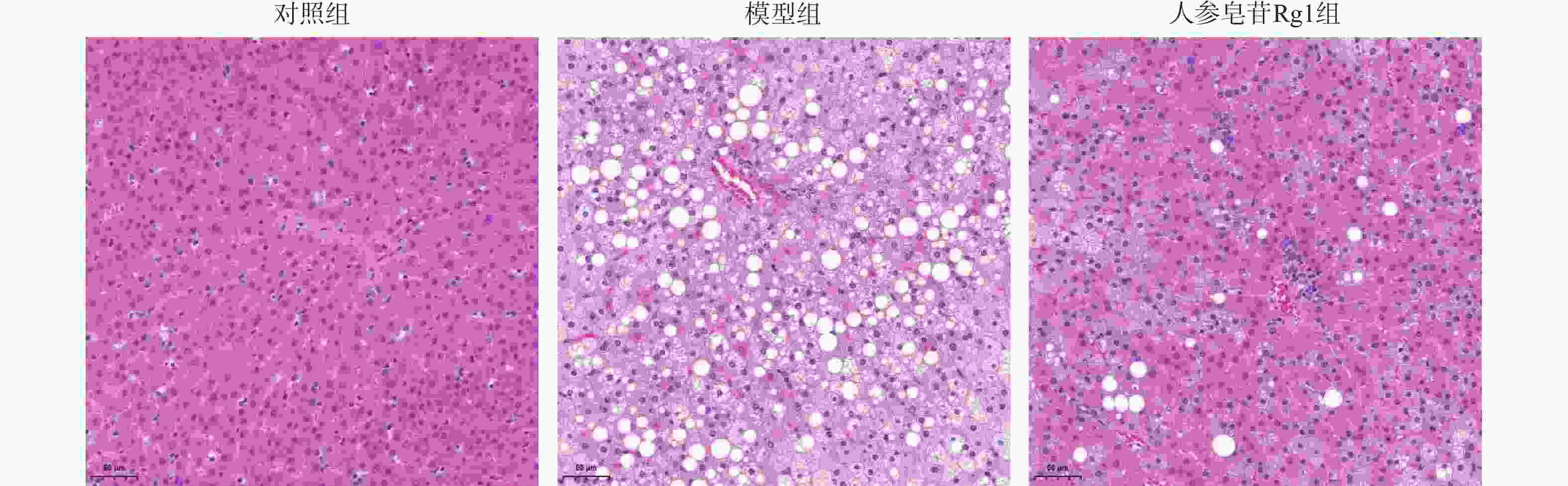
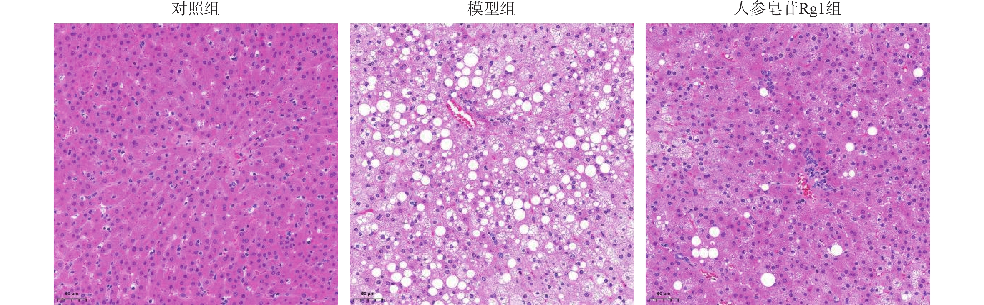
图 2 苏木素-伊红染色观察树鼩肝组织形态学(×200)
Figure 2. Histomorphology of tree shrew liver tissue observed by hematoxylin and eosin staining (×200)
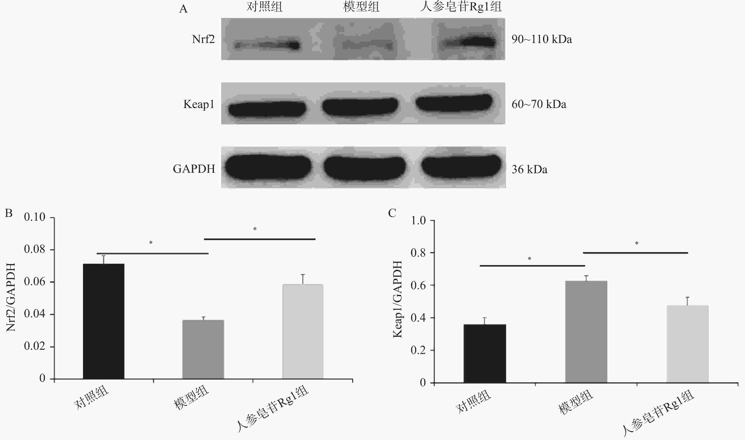
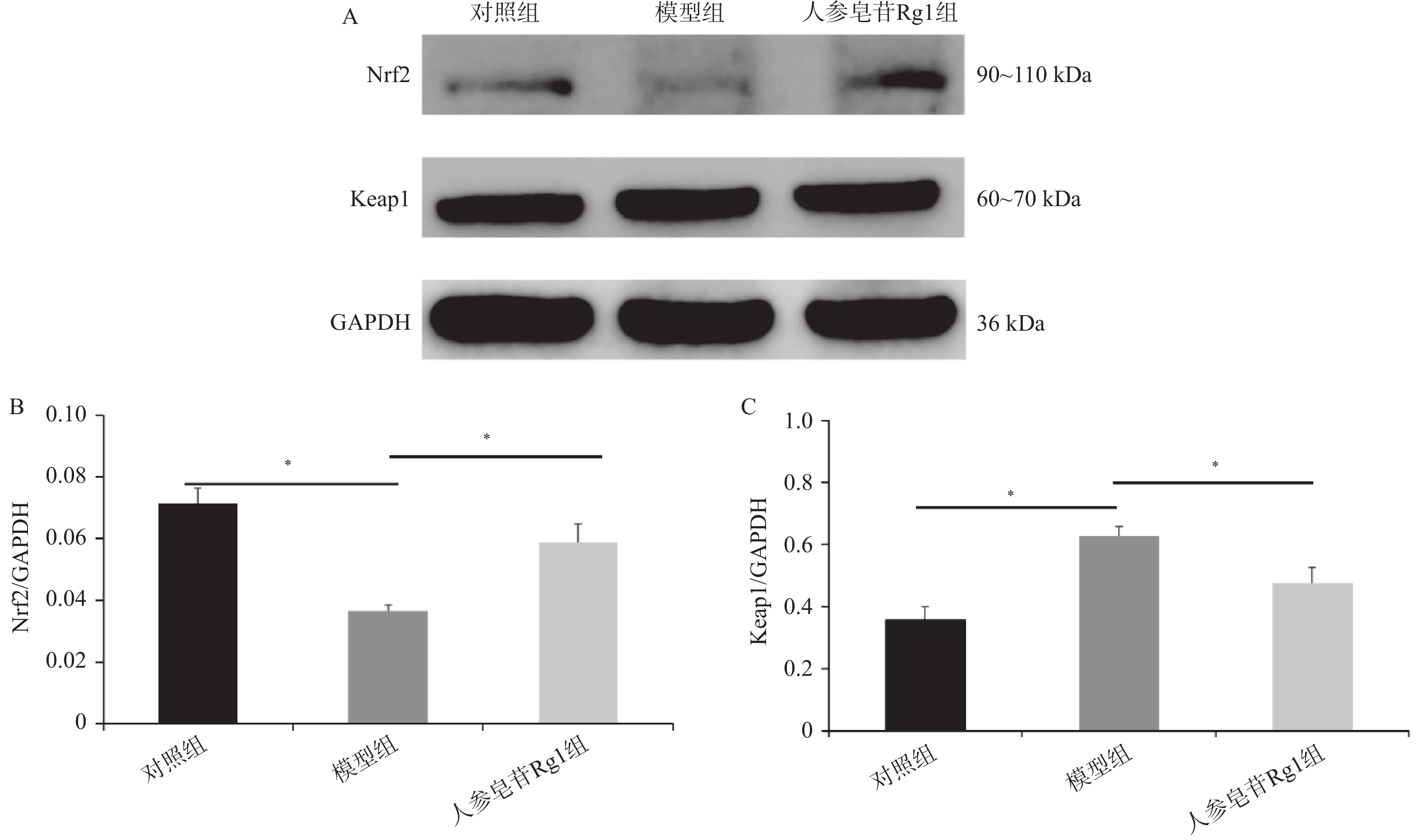
图 3 人参皂苷Rg1对树鼩肝脏Nrf2和Keap1蛋白表达的影响(n = 6,$ \bar x \pm s $)
A:各组树鼩肝脏Nrf2、Keap1蛋白表达条带;B~C:各组树鼩肝脏Nrf2、Keap1蛋白表达水平定量分析;Nrf2:F = 42.36,Keap1:F = 38.72,*P < 0.05。
Figure 3. Effect of ginsenoside Rg1 on the expression of Nrf2 and Keap1 proteins in the liver of tree shrews(n = 6,$ \bar x \pm s $)
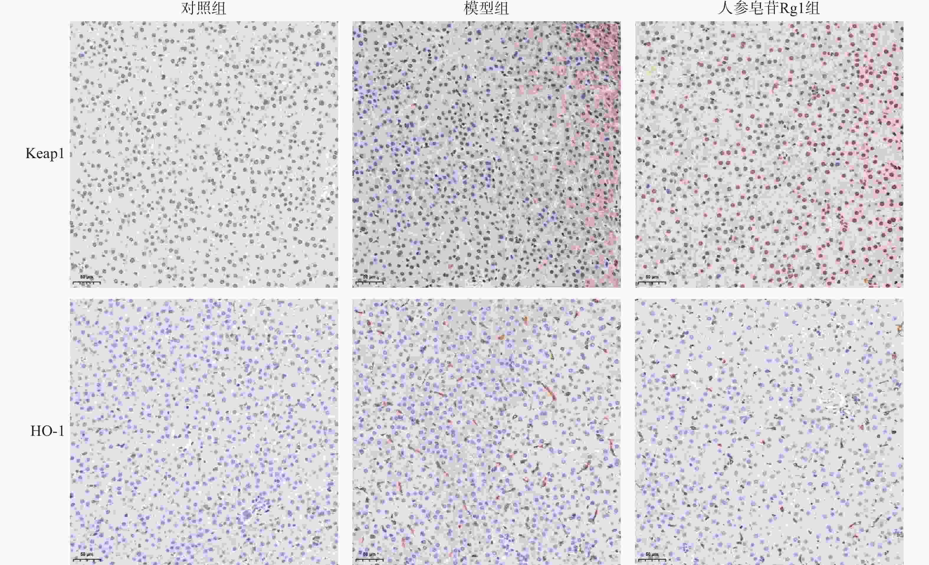
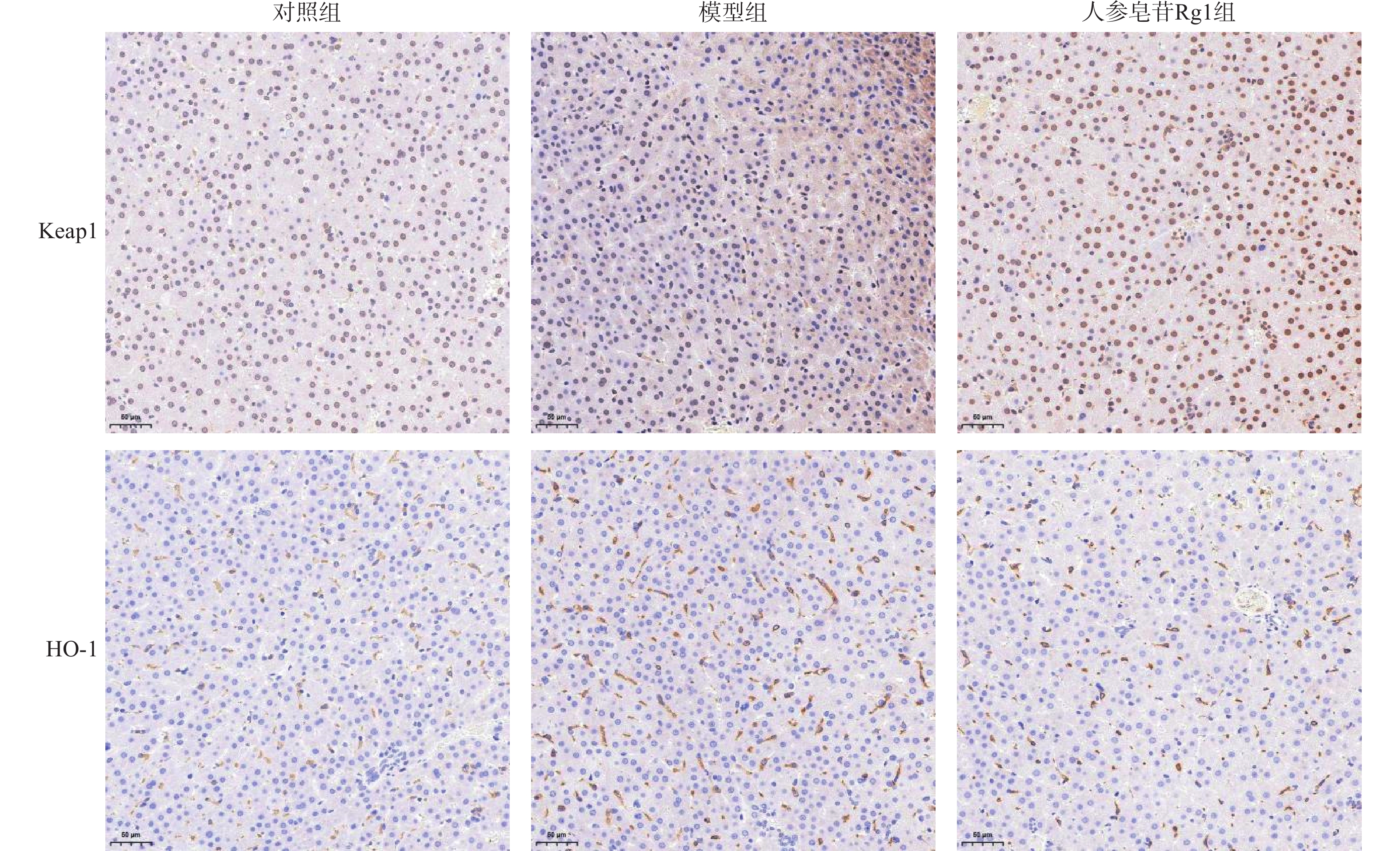
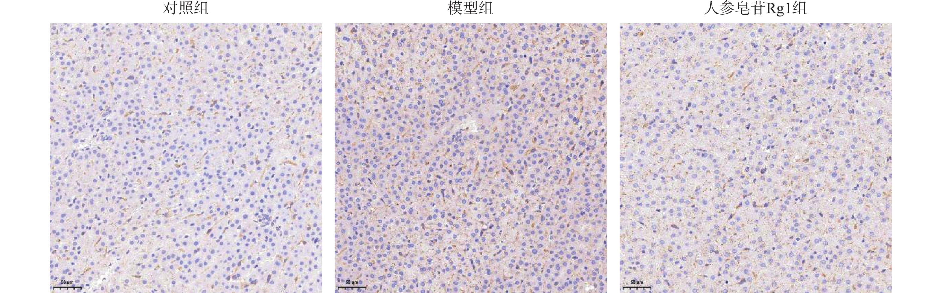
图 4 人参皂苷Rg1对树鼩肝脏Keap1和HO-1免疫组化染色的影响(×200)
Figure 4. Effects of Ginsenoside Rg1 on the immunohistochemical staining of Keap1 and HO-1 in the liver of tree shrews (×200)
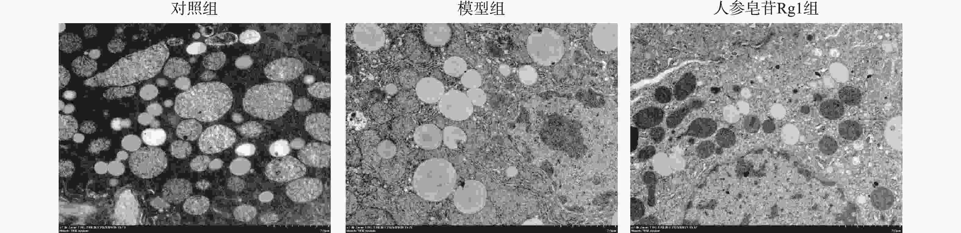
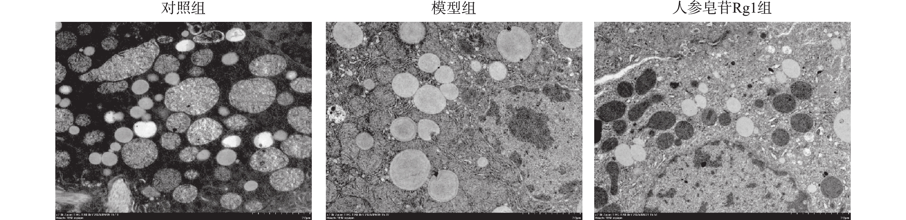
图 5 透射电镜观察树鼩肝细胞线粒体超微结构(×
7000 )Figure 5. Transmission electron microscopy observation of the ultrastructure of mitochondria in tree shrew hepatocytes (×
7000 )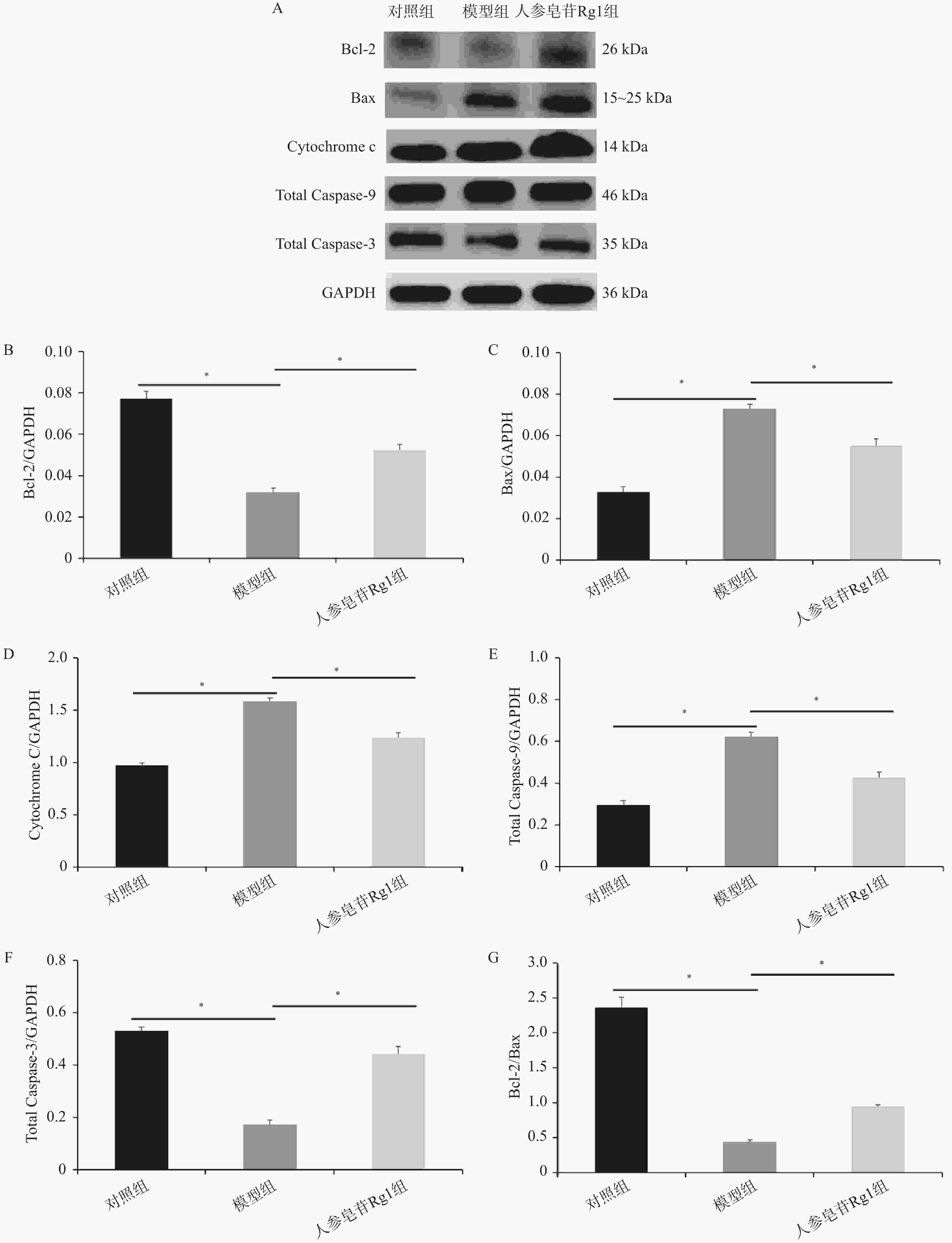
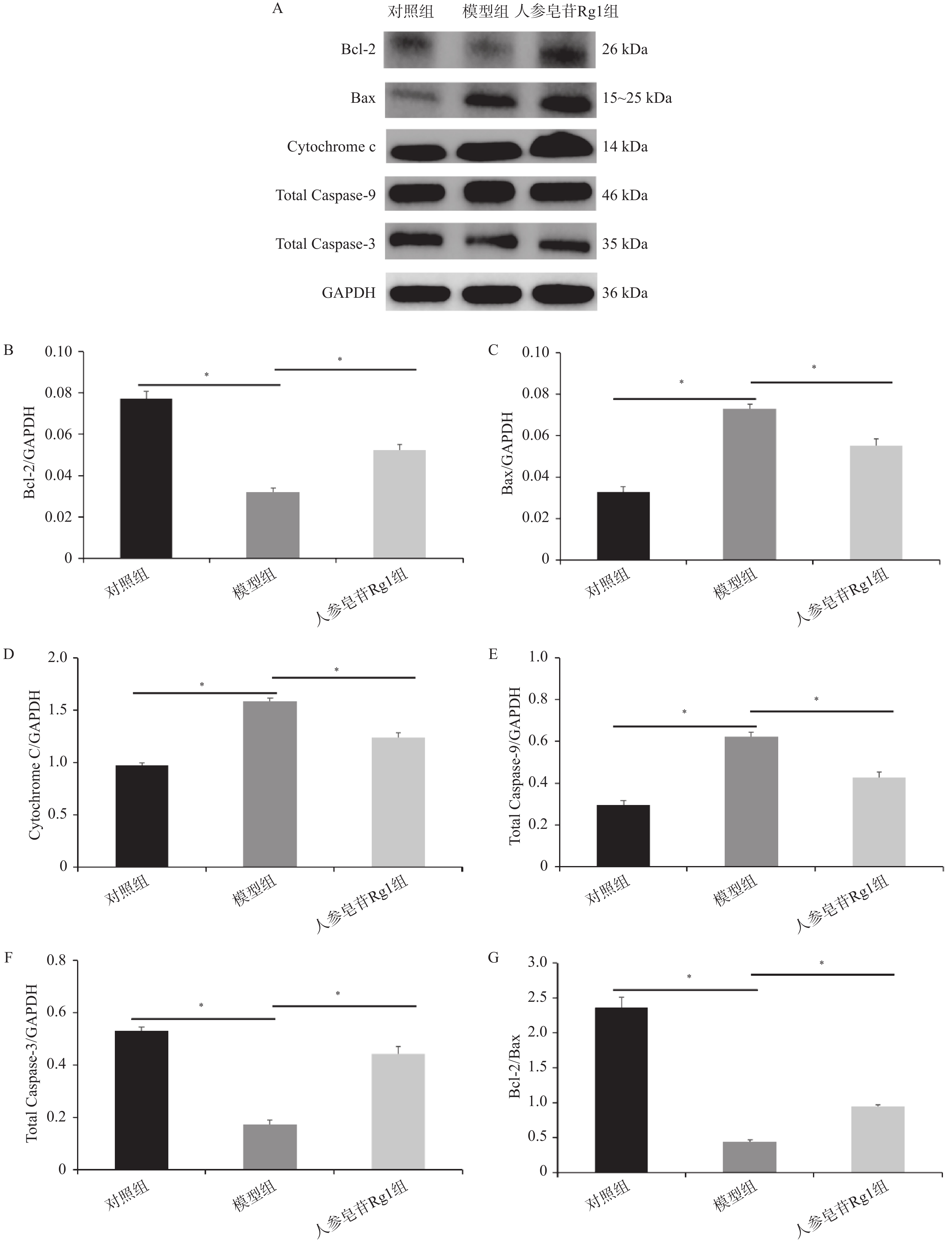
图 6 人参皂苷Rg1对树鼩肝脏Bcl-2、Bax、Cytochrome C、Total Caspase-9和Total Caspase-3蛋白表达的影响(n = 6,$ \bar x \pm s $)
A:各组树鼩肝脏Bcl-2、Bax、Cytochrome C、Total Caspase-9和Total Caspase-3蛋白表达条带;B~G:各组树鼩肝脏Bcl-2、Bax、Cytochrome C、Total Caspase-9和Total Caspase-3蛋白表达水平定量分析及Bcl-2/Bax比值;Bcl-2:F = 35.18,Bax:F = 29.64,Cytochrome C:F = 48.52,Total Caspase-9:F = 27.83,Total Caspase-3:F = 31.47,*P < 0.05。
Figure 6. Effects of Ginsenoside Rg1 on the expression of Bcl-2,Bax,cytochrome c,total caspase-9 and total caspase-3 proteins in the liver of tree shrews(n = 6,$ \bar x \pm s $)
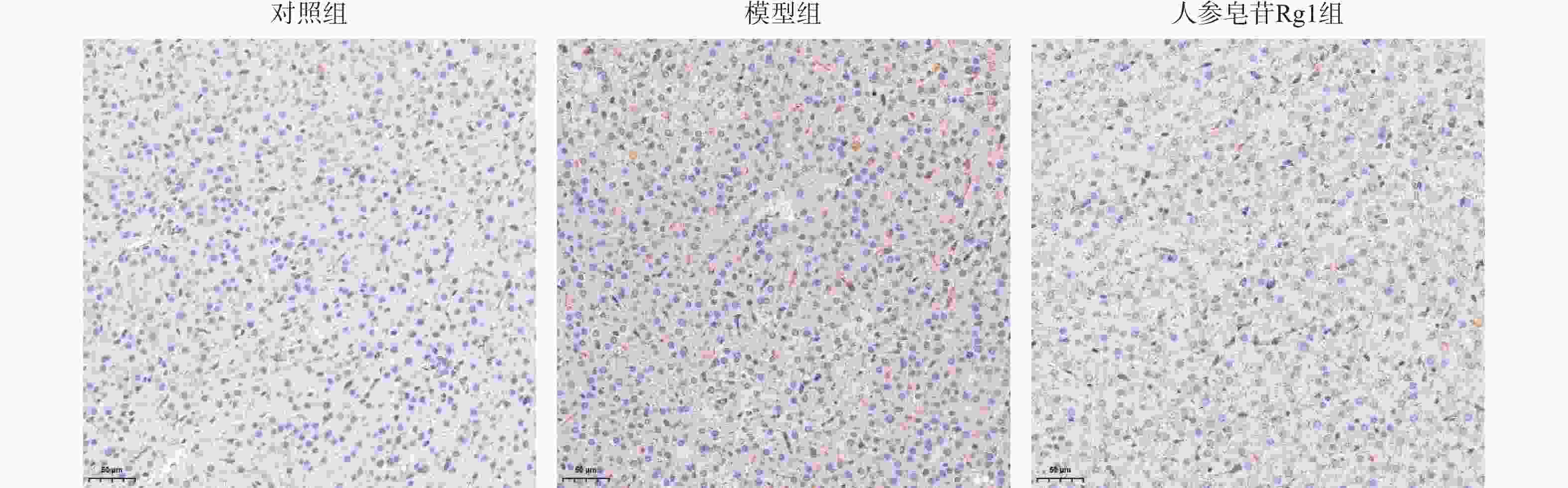
图 7 人参皂苷Rg1对树鼩肝脏总Caspase-3免疫组化染色的影响(×200)
Figure 7. Effect of Ginsenoside Rg1 on total caspase-3 immunohistochemical staining in tree shrew liver(×200)
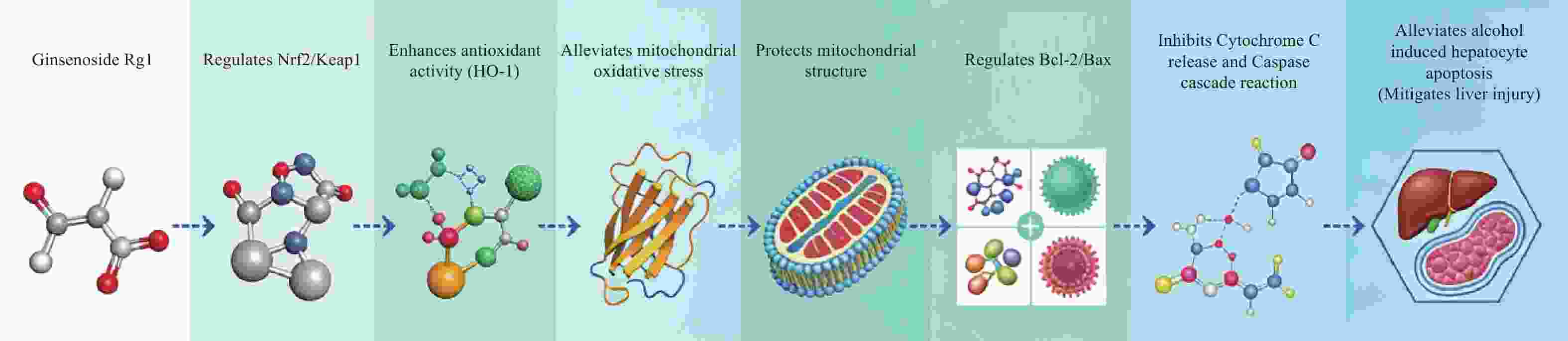
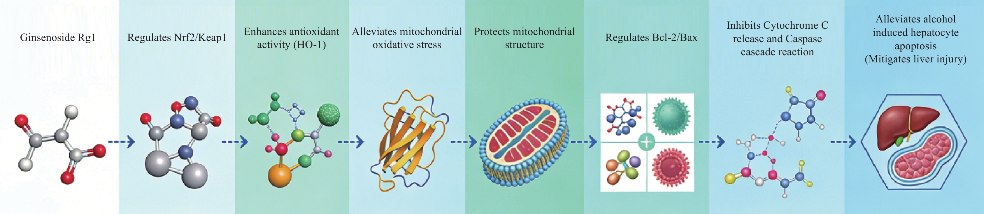
图 8 人参皂苷Rg1肝脏保护机制假设示意图
注:人参皂苷Rg1可能通过调控Nrf2/Keap1通路,增强HO-1等抗氧化酶活性,减轻线粒体氧化应激,保护线粒体结构完整性,调节Bcl-2/Bax平衡,抑制Cytochrome c释放与Caspase级联反应,最终减轻酒精诱导的肝细胞凋亡。图中箭头表示各环节间的调控关系。本图基于本研究结果提出的机制假设,尚需Nrf2特异性抑制剂或基因敲低实验进一步验证。
Figure 8. Schematic diagram of the proposed mechanism hypothesis of Ginsenoside Rg1 liver protection
表 1 各组树鼩血清肝功能、氧化应激及LDH、GSH-Px活性比较(n = 6,$ \bar x \pm s $)
Table 1. Comparison of serum liver function,oxidative stress,LDH and GSH PX activities in each group of tree shrews (n = 6,$ \bar x \pm s $)
组别 AST(U/L) ALT(U/L) MDA(μmol/L) LDH(U/L) GSH-Px(U/mL) 对照组 182.20 ± 6.42 85.00 ± 5.96 13.54 ± 0.57 1768.86 ± 59.04214.92 ± 7.12 模型组 331.80 ± 12.60a 172.40 ± 6.47a 26.32 ± 0.57a 3235.30 ± 90.16a170.88 ± 4.96a 人参皂苷Rg1组 238.20 ± 7.85b 127.80 ± 7.33b 19.79 ± 0.74b 2510.53 ± 112.72b183.12 ± 5.45b F 327.57 218.70 510.07 331.59 73.56 P <0.001* <0.001* <0.001* <0.001* <0.001* *P < 0.05;与对照组比较,aP < 0.05;与模型组比较,bP < 0.05。  下载: 导出CSV
表 2 各组树鼩肝组织病理学评分(n = 6,$ \bar x \pm s $,分)
Table 2. Histopathological scores of tree shrew liver tissue by group(n = 6,$ \bar x \pm s $,score)
组别 脂肪变性 炎性细胞浸润 气球样变 总分 对照组 0.00 ± 0.00 0.00 ± 0.00 0.00 ± 0.00 0.00 ± 0.00 模型组 3.00 ± 0.00a 1.00 ± 0.00a 1.40 ± 0.55a 5.20 ± 0.84a 人参皂苷Rg1组 1.40 ± 0.55b 0.80 ± 0.45b 0.60 ± 0.55b 2.80 ± 0.45b H 15.24 12.86 9.42 15.38 P <0.001* <0.001* <0.001* <0.001* *P < 0.05;与对照组比较,aP < 0.05;与模型组比较,bP < 0.05;多组间比较采用Kruskal-Wallis H检验,两两比较采用Dunn-Bonferroni检验。
下载: 导出CSV
表 3 各组树鼩Keap1和HO-1免疫组化阳性面积比较(n = 6,$ \bar x \pm s $)
Table 3. Comparison of the positive areas of Keap1 and HO-1 immunohistochemistry in each group of tree shrews (n = 6,$ \bar x \pm s $)
组别 Keap1阳性面积(%) HO-1阳性面积(%) 对照组 7.61 ± 0.72 1.56 ± 0.21 模型组 20.15 ± 1.61a 2.37 ± 0.29 人参皂苷Rg1组 13.90 ± 1.09b 5.44 ± 0.28b F 136.94 306.45 P <0.001* <0.001* *P < 0.05;与对照组比较,aP < 0.05;与模型组比较,bP < 0.05。
下载: 导出CSV
表 4 各组树鼩总Caspase-3免疫组化阳性面积与阳性细胞比例的比较(n = 6,$ \bar x \pm s $)
Table 4. Comparison of total Caspase-3 immunohistochemical positive area and positive cell proportion in each group of tree shrews (n = 6,$ \bar x \pm s $)
组别 总Caspase-3
阳性面积 (%)阳性细胞
比例 (%)对照组 1.76 ± 0.31 13.79 ± 1.82 模型组 6.85 ± 0.91a 36.54 ± 6.08a 人参皂苷Rg1组 2.62 ± 0.22b 18.31 ± 1.94b F 113.60 49.46 P <0.001* <0.001* *P < 0.05;与对照组比较,aP < 0.05;与模型组比较,bP < 0.05。
下载: 导出CSV
-
[1] Louvet A, Mathurin P. Alcoholic liver disease: Mechanisms of injury and targeted treatment[J]. Nat Rev Gastroenterol Hepatol, 2015, 12(4): 231-242. doi: 10.1038/nrgastro.2015.35 [2] Gao B, Bataller R. Alcoholic liver disease: Pathogenesis and new therapeutic targets[J]. Gastroenterology, 2011, 141(5): 1572-1585. doi: 10.1053/j.gastro.2011.09.002 [3] Wu X, Fan X, Miyata T, et al. Recent advances in understanding of pathogenesis of alcohol-associated liver disease[J]. Annu Rev Pathol Mech Dis, 2023, 18: 411-438. doi: 10.1146/annurev-pathmechdis-031521-030435 [4] Chen M, Zhong W, Xu W. Alcohol and the mechanisms of liver disease[J]. J Gastroenterol Hepatol, 2023, 38(8): 1233-1240. doi: 10.1111/jgh.16282 [5] Seitz H K, Bataller R, Cortez-Pinto H, et al. Alcoholic liver disease[J]. Nat Rev Dis Primers, 2018, 4: 16. doi: 10.1038/s41572-018-0014-7 [6] Xia T, Fang B, Kang C, et al. Hepatoprotective mechanism of ginsenoside Rg1 against alcoholic liver damage based on gut microbiota and network pharmacology[J]. Oxid Med Cell Longev, 2022, 2022(1): 5025237. doi: 10.1155/2022/5025237 [7] Zhou H, Liu Y, Su Y, et al. Ginsenoside Rg1 attenuates lipopolysaccharide-induced chronic liver damage by activating Nrf2 signaling and inhibiting inflammasomes in hepatic cells[J]. J Ethnopharmacol, 2024, 324: 117794. doi: 10.1016/j.jep.2024.117794 [8] Yang C, He X, Zhao J, et al. Hepatoprotection by Ginsenoside Rg1 in alcoholic liver disease[J]. Int Immunopharmacol, 2021, 92: 107327. doi: 10.1016/j.intimp.2020.107327 [9] Wu M, Li K, Wu J, et al. Ginsenoside Rg1: A bioactive therapeutic agent for diverse liver diseases[J]. Pharmacol Res, 2025, 212: 107571. doi: 10.1016/j.phrs.2024.107571 [10] Park J S, Rustamov N, Roh Y S. The roles of NFR2-regulated oxidative stress and mitochondrial quality control in chronic liver diseases[J]. Antioxidants, 2023, 12(11): 1928. doi: 10.3390/antiox12111928 [11] Sun J, Hong Z, Shao S, et al. Liver-specific Nrf2 deficiency accelerates ethanol-induced lethality and hepatic injury in vivo[J]. Toxicol Appl Pharmacol, 2021, 426: 115617. doi: 10.1016/j.taap.2021.115617 [12] Bellezza I, Giambanco I, Minelli A, et al. Nrf2-Keap1 signaling in oxidative and reductive stress[J]. Biochim Biophys Acta BBA Mol Cell Res, 2018, 1865(5): 721-733. doi: 10.1016/j.bbamcr.2018.02.010 [13] Panda H, Rowland N G, Krall C M, et al. NRF2 immunobiology in cancer: Implications for immunotherapy and therapeutic targeting[J]. Oncogene, 2025, 44(39): 3641-3651. doi: 10.1038/s41388-025-03560-4 [14] Czabotar P E, Garcia-Saez A J. Mechanisms of BCL-2 family proteins in mitochondrial apoptosis[J]. Nat Rev Mol Cell Biol, 2023, 24(10): 732-748. doi: 10.1038/s41580-023-00629-4 [15] King L E, Hohorst L, García-Sáez A J. Expanding roles of BCL-2 proteins in apoptosis execution and beyond[J]. J Cell Sci, 2023, 136(22): jcs260790. doi: 10.1242/jcs.260790 [16] Green D R, Reed J C. Mitochondria and apoptosis[J]. Science, 1998, 281(5381): 1309-1312. doi: 10.1126/science.281.5381.1309 [17] Liu X, Kim C N, Yang J, et al. Induction of apoptotic program in cell-free extracts: Requirement for dATP and cytochrome c[J]. Cell, 1996, 86(1): 147-157. doi: 10.1016/S0092-8674(00)80085-9 [18] Li P, Nijhawan D, Budihardjo I, et al. Cytochrome c and dATP-dependent formation of apaf-1/caspase-9 complex initiates an apoptotic protease cascade[J]. Cell, 1997, 91(4): 479-489. doi: 10.1016/S0092-8674(00)80434-1 [19] Shi Z, Xing H, Qi C, et al. Chinese tree shrews as a primate experimental animal eligible for the study of alcoholic liver disease: Characterization and confirmation by MRI[J]. Exp Anim, 2020, 69(1): 110-118. doi: 10.1538/expanim.19-0073 [20] Xing H, Jia K, He J, et al. Establishment of the tree shrew as an alcohol-induced fatty liver model for the study of alcoholic liver diseases[J]. PLoS One, 2015, 10(6): e0128253. doi: 10.1371/journal.pone.0128253 [21] Fan Y, Huang Z Y, Cao C C, et al. Tree shrew as an animal model for alcohol-related liver disease[J]. Frontiers in Pharmacology, 2022, 13: 891903. [22] Yao Y G. Creating animal models, why not use the Chinese tree shrew (Tupaia belangeri Chinensis)[J]. Zool Res, 2017, 38(3): 118-126. [23] 李进涛, 马怡, 郑红, 等. 滇西亚种树鼩实验性酒精肝损伤研究[J]. 云南农业大学学报(自然科学), 2017, 32(6): 1155-1161. doi: 10.16211/j.issn.1004-390X(n).2017.06.028 [24] 张钰雯, 李进丹, 郑红, 等. 人参皂苷Rg1对树鼩酒精性肝炎的疗效观察[J]. 养殖与饲料, 2018(11): 19-20. doi: 10.3969/j.issn.1671-427X.2018.11.008 [25] Lackner C, Gouw A S H, Alves VAF, et al. Consensus position statements for the standardized application of histological grading and staging systems in MASH clinical trials[J]. Journal of Hepatology, 2026, 84(4): 693-701. doi: 10.1016/j.jhep.2025.09.019 -

 点击查看大图

计量
- 文章访问数: 2
- HTML全文浏览量: 3
- PDF下载量: 0
- 被引次数: 0
