

Research Progress on the Mechanisms of Action of Artemisinin and Its Derivatives in Cancer Therapy
-
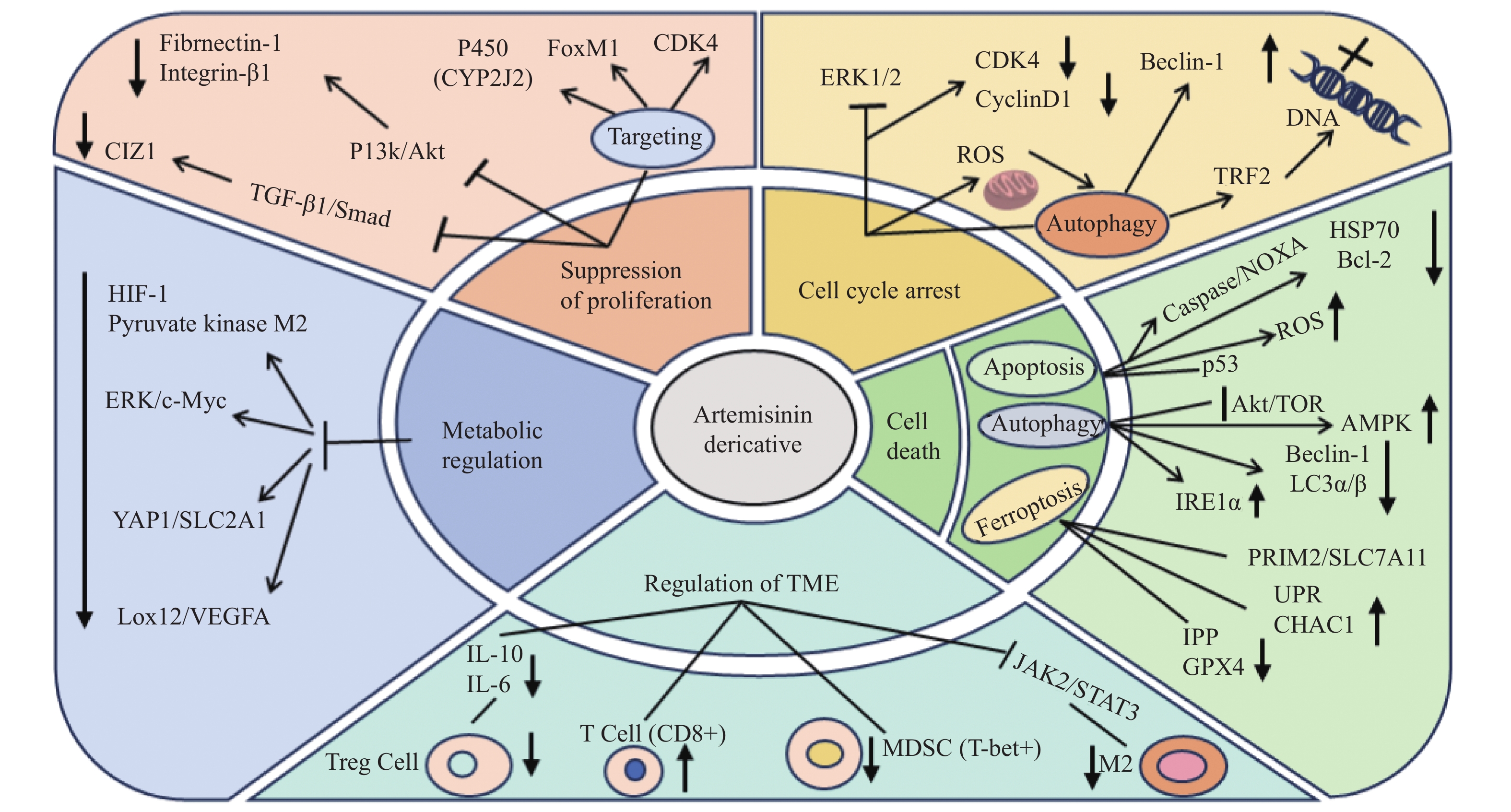
摘要: 青蒿素及其衍生物由于其独特的过氧化桥结构,不仅对恶性疟疾显示出显著的疗效,也展现了强大的抗肿瘤能力。这些化合物通过多效机制干扰癌细胞生长,包括抑制细胞增殖、引起细胞周期阻滞、诱导多种细胞死亡模式、调节肿瘤微环境和调节肿瘤代谢等。临床试验已证实青蒿素类化合物具有显著的抗癌活性。综述近年来关于青蒿素及其衍生物作为潜在抗癌药物的作用机制的最新进展和发现,为未来青蒿素类化合物的抗癌机制研究提供参考。Abstract: Due to their unique peroxide bridge structure, artemisinin and its derivatives have demonstrated the remarkable efficacy in treating severe malaria and exhibited the potent anticancer properties. These compounds interfere with the cancer cell growth through the multiple mechanisms, including the inhibition of cell proliferation, induction of various forms of cell death, cell cycle arrest, suppression of angiogenesis and modulation of the tumor microenvironment. Clinical trials have confirmed the significant anticancer activity of artemisinin-based compounds. This paper summarizes the recent advances and findings regarding the mechanisms of action of artemisinin and its derivatives as the potential anticancer agents, providing the valuable insights for future researches on the anticancer mechanisms of these compounds.
-
Key words:
- Artemisinin /
- Artemisinin derivatives /
- Anticancer /
- Mechanisms of action
-
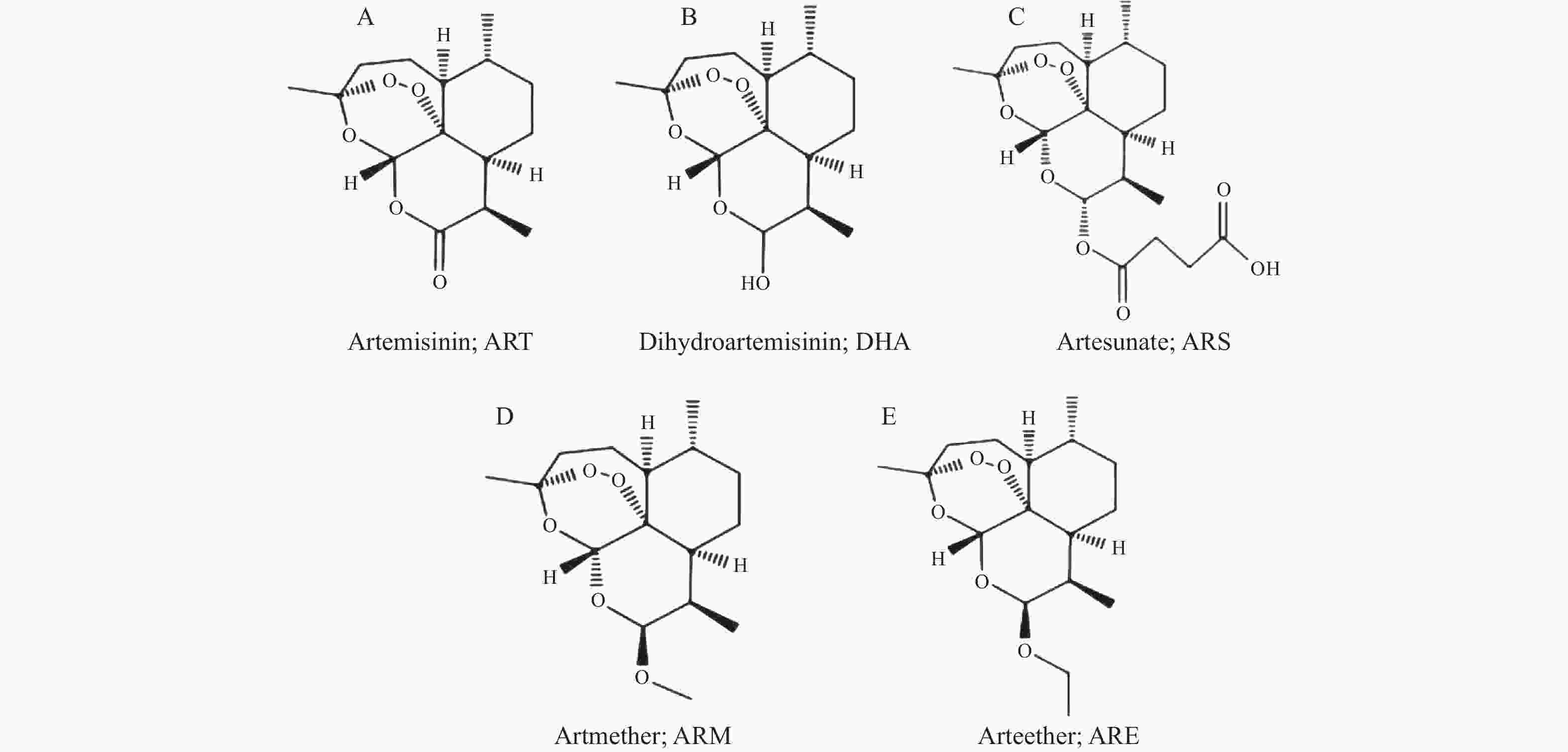
图 1 青蒿素及其衍生物的化学结构
A:青蒿素;B:双氢青蒿素;C:青蒿琥酯;D:蒿甲醚;E:蒿乙醚。
Figure 1. Chemical structures of artemisinin and its derivatives
-
[1] Ma L,Zhang M,Zhao R,et al. Plant natural products: Promising resources for cancer chemoprevention[J]. Molecules (Basel,Switzerland),2021,26(4):933-955. [2] Sonkin D,Thomas A,Teicher B A. Cancer treatments: Past,present,and future[J]. Cancer Genetics,2024,286-287(5):18-24. [3] Guo M,Jin J,Zhao D,et al. Research advances on anti-cancer natural products[J]. Frontiers in Oncology,2022,12(5):866154-866170. [4] Marchesi E,Perrone D,Navacchia M L. Molecular hybridization as a strategy for developing artemisinin-derived anticancer candidates[J]. Pharmaceutics,2023,15(9):2185-2224. [5] Zeng Z W,Chen D,Chen L,et al. A comprehensive overview of Artemisinin and its derivatives as anticancer agents[J]. European Journal of Medicinal Chemistry,2023,247(4):115000-115030. [6] Trimble C L,Levinson K,Maldonado L,et al. A first-in-human proof-of-concept trial of intravaginal artesunate to treat cervical intraepithelial neoplasia 2/3 (CIN2/3)[J]. Gynecologic Oncology,2020,157(1):188-194. [7] Ferrall L,Lin K Y,Roden R B S,et al. Cervical cancer immunotherapy: Facts and hopes[J]. Clinical Cancer Research: An Official Journal of the American Association for Cancer Research,2021,27(18):4953-4973. [8] Dong J,Chen Y,Yang W,et al. Antitumor and anti-angiogenic effects of artemisinin on breast tumor xenografts in nude mice[J]. Research in Veterinary Science,2020,129(2):66-69. [9] Li Y,Zhou X,Liu J,et al. Dihydroartemisinin inhibits the tumorigenesis and metastasis of breast cancer via downregulating CIZ1 expression associated with TGF-β1 signaling[J]. Life Sciences,2020,248(9):117454-117463. [10] Zhang Q,Yi H,Yao H,et al. Artemisinin derivatives inhibit non-small cell lung cancer cells through induction of ROS-dependent apoptosis/ferroptosis[J]. Journal of Cancer,2021,12(13):4075-4085. [11] Zhang Y,Wang Y,Li Y,et al. Dihydroartemisinin and artesunate inhibit aerobic glycolysis via suppressing c-myc signaling in non-small cell lung cancer[J]. Biochemical Pharmacology,2022,198(4):114941-114949. [12] Xiao X,Li Y,Wang Y,et al. Dihydroartemisinin inhibits Lewis Lung carcinoma progression by inducing macrophages M1 polarization via AKT/mTOR pathway[J]. International Immunopharmacology,2022,103(2):108427-108429. [13] Jia J,Qin Y,Zhang L,et al. Artemisinin inhibits gallbladder cancer cell lines through triggering cell cycle arrest and apoptosis[J]. Molecular Medicine Reports,2016,13(5):4461-4468. [14] Kandoth C,McLellan M D,Vandin F,et al. Mutational landscape and significance across 12 major cancer types[J]. Nature,2013,502(7471):333-339. [15] Zielińska K A,Katanaev V L. Information theory: New look at oncogenic signaling pathways[J]. Trends in Cell Biology,2019,29(11):862-875. [16] Tin A S,Sundar S N,Tran K Q,et al. Antiproliferative effects of artemisinin on human breast cancer cells requires the downregulated expression of the E2F1 transcription factor and loss of E2F1-target cell cycle genes[J]. Anti-Cancer Drugs,2012,23(4):370-379. [17] Nandi D,Cheema P S,Singal A,et al. Artemisinin mediates its tumor-suppressive activity in hepatocellular carcinoma through targeted inhibition of FoxM1[J]. Frontiers in Oncology,2021,11(1):751271-751289. [18] Liang R,Chen W,Chen X Y,et al. Dihydroartemisinin inhibits the tumorigenesis and invasion of gastric cancer by regulating STAT1/KDR/MMP9 and P53/BCL2L1/CASP3/7 pathways[J]. Pathology,Research and Practice,2021,218(2):153318-153330. [19] Wu R,Gao Y,Wu J,et al. Semi-synthetic product dihydroartemisinin inhibited fibronectin-1 and integrin-β1 and interfered with the migration of HCCLM6 cells via PI3K-AKT pathway[J]. Biotechnology Letters,2020,42(6):917-926. [20] Zhu X,Yang M,Song Z,et al. Artemether inhibits proliferation,invasion and migration of hepatocellular carcinoma cells via targeting of CYP2J2[J]. Oncology Letters,2022,23(6):180-188. [21] Slezakova S,Ruda-Kucerova J. Anticancer activity of Artemisinin and its derivatives[J]. Anticancer Research,2017,37(11):5995-6003. [22] Willoughby J A,Sundar S N,Cheung M,et al. Artemisinin blocks prostate cancer growth and cell cycle progression by disrupting Sp1 interactions with the cyclin-dependent kinase-4 (CDK4) promoter and inhibiting CDK4 gene expression[J]. Journal of Biological Chemistry,2009,284(4):2203-2213. [23] Ma Q,Liao H,Xu L,et al. Autophagy-dependent cell cycle arrest in esophageal cancer cells exposed to dihydroartemisinin[J]. Chinese Medicine,2020,15(9):37-49. [24] Li B,Bu S,Sun J,et al. Artemisinin derivatives inhibit epithelial ovarian cancer cells via autophagy-mediated cell cycle arrest[J]. Acta Biochimica et Biophysica Sinica,2018,50(12):1227-1235. [25] Greenshields A L,Shepherd T G,Hoskin D W. Contribution of reactive oxygen species to ovarian cancer cell growth arrest and killing by the anti-malarial drug artesunate[J]. Molecular Carcinogenesis,2017,56(1):75-93. [26] Xu C,Zhang H,Mu L,et al. Artemisinins as anticancer drugs: Novel therapeutic approaches,molecular mechanisms,and clinical trials[J]. Frontiers in Pharmacology,2020,11(10):529881-529895. [27] Eskandari E,Eaves C J. Paradoxical roles of caspase-3 in regulating cell survival,proliferation,and tumorigenesis[J]. Journal of Cell Biology,2022,221(6):e202201159-e202201172. [28] Efferth T,Rücker G,Falkenberg M,et al. Detection of apoptosis in KG-1a leukemic cells treated with investigational drugs[J]. Arzneimittel Forschung,1996,46(2):196-200. [29] Lang S J,Schmiech M,Hafner S,et al. Antitumor activity of an artemisia annua herbal preparation and identification of active ingredients[J]. Phytomedicine: International Journal of Phytotherapy and Phytopharmacology,2019,62(12):152962-152971. [30] Noori S,Hassan Z M,Farsam V. Artemisinin as a Chinese medicine,selectively induces apoptosis in pancreatic tumor cell line[J]. Chinese Journal of Integrative Medicine,2014,20(8):618-623. [31] Mondal A,Chatterji U. Artemisinin represses telomerase subunits and induces apoptosis in HPV-39 infected human cervical cancer cells[J]. Journal of Cellular Biochemistry,2015,116(9):1968-1981. [32] Pirali M,Taheri M,Zarei S,et al. Artesunate,as a HSP70 ATPase activity inhibitor,induces apoptosis in breast cancer cells[J]. International Journal of Biological Macromolecules,2020,164(5):3369-3375. [33] Cabello C M,Lamore S D,Bair W B,et al. The redox antimalarial dihydroartemisinin targets human metastatic melanoma cells but not primary melanocytes with induction of NOXA-dependent apoptosis[J]. Investigational New Drugs,2012,30(4):1289-1301. [34] Chatterjee R,Shukla A,Chakrabarti K,et al. CLEC12A sensitizes differentially responsive breast cancer cells to the anti-cancer effects of artemisinin by repressing autophagy and inflammation[J]. Frontiers in Oncology,2023,13(12):1242432-1242449. [35] Huang Z,Gan S,Zhuang X,et al. Artesunate inhibits the cell growth in colorectal cancer by promoting ROS-dependent cell senescence and autophagy[J]. Cells,2022,11(16):2472-2492. [36] Zhou X,Chen Y,Wang F,et al. Artesunate induces autophagy dependent apoptosis through upregulating ROS and activating AMPK-mTOR-ULK1 axis in human bladder cancer cells[J]. Chemico-Biological Interactions,2020,331(17):109273-109284. [37] Chen X,He L Y,Lai S,et al. Dihydroartemisinin inhibits the migration of esophageal cancer cells by inducing autophagy[J]. Oncology Letters,2020,20(4):94-102. [38] Zhou Q,Meng Y,Li D,et al. Ferroptosis in cancer: From molecular mechanisms to therapeutic strategies[J]. Signal Transduction and Targeted Therapy,2024,9(1):55-84. [39] Chen F,Fan Y,Hou J,et al. Integrated analysis identifies TfR1 as a prognostic biomarker which correlates with immune infiltration in breast cancer[J]. Aging,2021,13(17):21671-21699. [40] Yuan B,Liao F,Shi Z Z,et al. Dihydroartemisinin inhibits the proliferation,colony formation and induces ferroptosis of lung cancer cells by inhibiting PRIM2/SLC7A11 axis[J]. OncoTargets and Therapy,2020,13(1):10829-10840. [41] Wang Z,Li M,Liu Y,et al. Dihydroartemisinin triggers ferroptosis in primary liver cancer cells by promoting and unfolded protein response-induced upregulation of CHAC1 expression[J]. Oncology Reports,2021,46(5):240-253. [42] Roh J L,Kim E H,Jang H,et al. Nrf2 inhibition reverses the resistance of cisplatin-resistant head and neck cancer cells to artesunate-induced ferroptosis[J]. Redox Biology,2017,11(1):254-262. [43] Liang L,Liu Y,Wu X,et al. Artesunate induces ferroptosis by inhibiting the nuclear localization of SREBP2 in myeloma cells[J]. International Journal of Medical Sciences,2023,20(12):1535-1550. [44] Hartupee C,Nagalo B M,Chabu C Y,et al. Pancreatic cancer tumor microenvironment is a major therapeutic barrier and target[J]. Frontiers in Immunology,2024,15(2):1287459-1287475. [45] Zhang Q,Sioud M. Tumor-associated macrophage subsets: Shaping polarization and targeting[J]. International Journal of Molecular Sciences,2023,24(8):7493-7524. [46] Yu R,Jin L,Li F,et al. Dihydroartemisinin inhibits melanoma by regulating CTL/treg anti-tumor immunity and STAT3-mediated apoptosis via IL-10 dependent manner[J]. Journal of Dermatological Science,2020,99(3):193-202. [47] Wang C Z,Wan C,Luo Y,et al. Effects of dihydroartemisinin,a metabolite of artemisinin,on colon cancer chemoprevention and adaptive immune regulation[J]. Molecular Biology Reports,2022,49(4):2695-2709. [48] Mancuso R I,Olalla Saad S T,Azambuja J H. Artesunate switches monocytes to an inflammatory phenotype with the ability to kill leukemic cells[J]. International Journal of Molecular Sciences,2021,22(2):608-621. [49] Cao Y,Feng Y H,Gao L W,et al. Artemisinin enhances the anti-tumor immune response in 4T1 breast cancer cells in vitro and in vivo[J]. International Immunopharmacology,2019,70(5):110-116. [50] Li Z,Munim M B,Sharygin D A,et al. Understanding the Warburg effect in cancer[J]. Cold Spring Harbor Perspectives in Medicine,2024,14(9):a041532. [51] Wong Y K,Xu C,Kalesh K A,et al. Artemisinin as an anticancer drug: Recent advances in target profiling and mechanisms of action[J]. Medicinal Research Reviews,2017,37(6):1492-1517. [52] Wang M,Chen H,He X,et al. Artemisinin inhibits the development of esophageal cancer by targeting HIF-1α to reduce glycolysis levels[J]. Journal of Gastrointestinal Oncology,2022,13(5):2144-2153. [53] Peng Q,Hao L,Guo Y,et al. Dihydroartemisinin inhibited the Warburg effect through YAP1/SLC2A1 pathway in hepatocellular carcinoma[J]. Journal of Natural Medicines,2023,77(1):28-40. [54] Ding X,Zhang Y,Liang J,et al. Dihydroartemisinin potentiates VEGFR-TKIs antitumorigenic effect on osteosarcoma by regulating Loxl2/VEGFA expression and lipid metabolism pathway[J]. Journal of Cancer,2023,14(5):809-820. -

 下载:
下载:
 点击查看大图

图(2)
计量
- 文章访问数: 1245
- HTML全文浏览量: 611
- PDF下载量: 81
- 被引次数: 0
